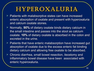
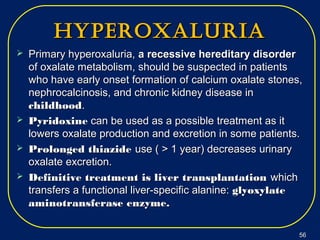
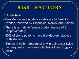
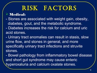
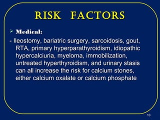
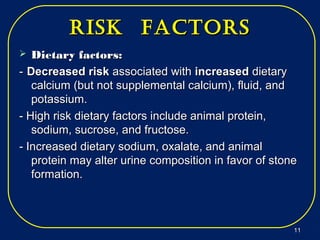
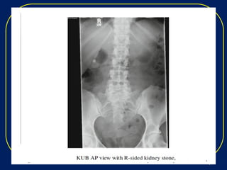
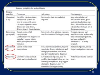
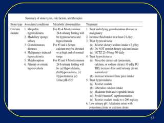
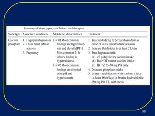
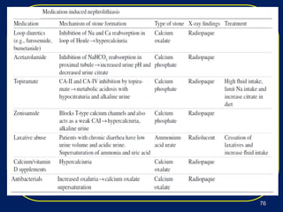
Nephrolithiasis, or kidney stones, is a common condition where stones form in the kidneys or urinary tract. The prevalence increases with age, affecting 11% of men and 5.6% of women in the US by age 70. Calcium oxalate and calcium phosphate are the most common stone types. Risk factors include genetics, medical conditions like obesity and hyperparathyroidism, dietary factors high in sodium and animal protein, and low urine volume. Patients typically present with sudden severe flank or abdominal pain known as renal colic. Diagnosis involves urinalysis, blood tests, abdominal imaging like CT, and stone analysis if a sample can be obtained. Treatment depends on factors like stone size,
Urolithiasis is a common disease that is estimated to
produce medical costs of $2.1 billion per year in the United States alone.
Renal colic affects approximately 1.2 million people
each year in USA and accounts for approximately 1% of
all hospital admissions.
Most active emergency departments (EDs) manage
patients with acute renal colic every day.
Pyelonephritis
It is the inflammation of the kidney & upper urinary tract that usually results from the bacterial infection of the bladder.
Pyelonephritis can be classified in several different catagories:
-acute pyelonephritis
-chronic pyelonephritis
-xanthogranulomatous pyelonephritis
definition of hydronephrosis,
causes and types of hydronephrosis
pathophysiology of hydronephrosis
clinical manifestation and diagnostic test for hydronephrosis
management
Nephrolithiasis, commonly known as kidney stones, refers to the formation of hard mineral and salt deposits within the kidneys or urinary tract. These stones can vary in size, ranging from tiny grains to larger, more substantial formations. Nephrolithiasis is a relatively common condition and can affect people of all ages, although it is more prevalent in adults.
RENAL STONES & STONES IN PREGNANCY .pptxBipul Thakur
THis presentation discusses about the formation of kidney stones the different theories related to formation of stones, thdifferent types of stones and management of kidney stones in case of pregnant female and various considerations required regarding the fetus and the pregnant female.
Title: Sense of Smell
Presenter: Dr. Faiza, Assistant Professor of Physiology
Qualifications:
MBBS (Best Graduate, AIMC Lahore)
FCPS Physiology
ICMT, CHPE, DHPE (STMU)
MPH (GC University, Faisalabad)
MBA (Virtual University of Pakistan)
Learning Objectives:
Describe the primary categories of smells and the concept of odor blindness.
Explain the structure and location of the olfactory membrane and mucosa, including the types and roles of cells involved in olfaction.
Describe the pathway and mechanisms of olfactory signal transmission from the olfactory receptors to the brain.
Illustrate the biochemical cascade triggered by odorant binding to olfactory receptors, including the role of G-proteins and second messengers in generating an action potential.
Identify different types of olfactory disorders such as anosmia, hyposmia, hyperosmia, and dysosmia, including their potential causes.
Key Topics:
Olfactory Genes:
3% of the human genome accounts for olfactory genes.
400 genes for odorant receptors.
Olfactory Membrane:
Located in the superior part of the nasal cavity.
Medially: Folds downward along the superior septum.
Laterally: Folds over the superior turbinate and upper surface of the middle turbinate.
Total surface area: 5-10 square centimeters.
Olfactory Mucosa:
Olfactory Cells: Bipolar nerve cells derived from the CNS (100 million), with 4-25 olfactory cilia per cell.
Sustentacular Cells: Produce mucus and maintain ionic and molecular environment.
Basal Cells: Replace worn-out olfactory cells with an average lifespan of 1-2 months.
Bowman’s Gland: Secretes mucus.
Stimulation of Olfactory Cells:
Odorant dissolves in mucus and attaches to receptors on olfactory cilia.
Involves a cascade effect through G-proteins and second messengers, leading to depolarization and action potential generation in the olfactory nerve.
Quality of a Good Odorant:
Small (3-20 Carbon atoms), volatile, water-soluble, and lipid-soluble.
Facilitated by odorant-binding proteins in mucus.
Membrane Potential and Action Potential:
Resting membrane potential: -55mV.
Action potential frequency in the olfactory nerve increases with odorant strength.
Adaptation Towards the Sense of Smell:
Rapid adaptation within the first second, with further slow adaptation.
Psychological adaptation greater than receptor adaptation, involving feedback inhibition from the central nervous system.
Primary Sensations of Smell:
Camphoraceous, Musky, Floral, Pepperminty, Ethereal, Pungent, Putrid.
Odor Detection Threshold:
Examples: Hydrogen sulfide (0.0005 ppm), Methyl-mercaptan (0.002 ppm).
Some toxic substances are odorless at lethal concentrations.
Characteristics of Smell:
Odor blindness for single substances due to lack of appropriate receptor protein.
Behavioral and emotional influences of smell.
Transmission of Olfactory Signals:
From olfactory cells to glomeruli in the olfactory bulb, involving lateral inhibition.
Primitive, less old, and new olfactory systems with different path
Flu Vaccine Alert in Bangalore Karnatakaaddon Scans
As flu season approaches, health officials in Bangalore, Karnataka, are urging residents to get their flu vaccinations. The seasonal flu, while common, can lead to severe health complications, particularly for vulnerable populations such as young children, the elderly, and those with underlying health conditions.
Dr. Vidisha Kumari, a leading epidemiologist in Bangalore, emphasizes the importance of getting vaccinated. "The flu vaccine is our best defense against the influenza virus. It not only protects individuals but also helps prevent the spread of the virus in our communities," he says.
This year, the flu season is expected to coincide with a potential increase in other respiratory illnesses. The Karnataka Health Department has launched an awareness campaign highlighting the significance of flu vaccinations. They have set up multiple vaccination centers across Bangalore, making it convenient for residents to receive their shots.
To encourage widespread vaccination, the government is also collaborating with local schools, workplaces, and community centers to facilitate vaccination drives. Special attention is being given to ensuring that the vaccine is accessible to all, including marginalized communities who may have limited access to healthcare.
Residents are reminded that the flu vaccine is safe and effective. Common side effects are mild and may include soreness at the injection site, mild fever, or muscle aches. These side effects are generally short-lived and far less severe than the flu itself.
Healthcare providers are also stressing the importance of continuing COVID-19 precautions. Wearing masks, practicing good hand hygiene, and maintaining social distancing are still crucial, especially in crowded places.
Protect yourself and your loved ones by getting vaccinated. Together, we can help keep Bangalore healthy and safe this flu season. For more information on vaccination centers and schedules, residents can visit the Karnataka Health Department’s official website or follow their social media pages.
Stay informed, stay safe, and get your flu shot today!
Ethanol (CH3CH2OH), or beverage alcohol, is a two-carbon alcohol
that is rapidly distributed in the body and brain. Ethanol alters many
neurochemical systems and has rewarding and addictive properties. It
is the oldest recreational drug and likely contributes to more morbidity,
mortality, and public health costs than all illicit drugs combined. The
5th edition of the Diagnostic and Statistical Manual of Mental Disorders
(DSM-5) integrates alcohol abuse and alcohol dependence into a single
disorder called alcohol use disorder (AUD), with mild, moderate,
and severe subclassifications (American Psychiatric Association, 2013).
In the DSM-5, all types of substance abuse and dependence have been
combined into a single substance use disorder (SUD) on a continuum
from mild to severe. A diagnosis of AUD requires that at least two of
the 11 DSM-5 behaviors be present within a 12-month period (mild
AUD: 2–3 criteria; moderate AUD: 4–5 criteria; severe AUD: 6–11 criteria).
The four main behavioral effects of AUD are impaired control over
drinking, negative social consequences, risky use, and altered physiological
effects (tolerance, withdrawal). This chapter presents an overview
of the prevalence and harmful consequences of AUD in the U.S.,
the systemic nature of the disease, neurocircuitry and stages of AUD,
comorbidities, fetal alcohol spectrum disorders, genetic risk factors, and
pharmacotherapies for AUD.
MANAGEMENT OF ATRIOVENTRICULAR CONDUCTION BLOCK.pdfJim Jacob Roy
Cardiac conduction defects can occur due to various causes.
Atrioventricular conduction blocks ( AV blocks ) are classified into 3 types.
This document describes the acute management of AV block.
These lecture slides, by Dr Sidra Arshad, offer a quick overview of physiological basis of a normal electrocardiogram.
Learning objectives:
1. Define an electrocardiogram (ECG) and electrocardiography
2. Describe how dipoles generated by the heart produce the waveforms of the ECG
3. Describe the components of a normal electrocardiogram of a typical bipolar leads (limb II)
4. Differentiate between intervals and segments
5. Enlist some common indications for obtaining an ECG
Study Resources:
1. Chapter 11, Guyton and Hall Textbook of Medical Physiology, 14th edition
2. Chapter 9, Human Physiology - From Cells to Systems, Lauralee Sherwood, 9th edition
3. Chapter 29, Ganong’s Review of Medical Physiology, 26th edition
4. Electrocardiogram, StatPearls - https://www.ncbi.nlm.nih.gov/books/NBK549803/
5. ECG in Medical Practice by ABM Abdullah, 4th edition
6. ECG Basics, http://www.nataliescasebook.com/tag/e-c-g-basics
New Directions in Targeted Therapeutic Approaches for Older Adults With Mantl...i3 Health
i3 Health is pleased to make the speaker slides from this activity available for use as a non-accredited self-study or teaching resource.
This slide deck presented by Dr. Kami Maddocks, Professor-Clinical in the Division of Hematology and
Associate Division Director for Ambulatory Operations
The Ohio State University Comprehensive Cancer Center, will provide insight into new directions in targeted therapeutic approaches for older adults with mantle cell lymphoma.
STATEMENT OF NEED
Mantle cell lymphoma (MCL) is a rare, aggressive B-cell non-Hodgkin lymphoma (NHL) accounting for 5% to 7% of all lymphomas. Its prognosis ranges from indolent disease that does not require treatment for years to very aggressive disease, which is associated with poor survival (Silkenstedt et al, 2021). Typically, MCL is diagnosed at advanced stage and in older patients who cannot tolerate intensive therapy (NCCN, 2022). Although recent advances have slightly increased remission rates, recurrence and relapse remain very common, leading to a median overall survival between 3 and 6 years (LLS, 2021). Though there are several effective options, progress is still needed towards establishing an accepted frontline approach for MCL (Castellino et al, 2022). Treatment selection and management of MCL are complicated by the heterogeneity of prognosis, advanced age and comorbidities of patients, and lack of an established standard approach for treatment, making it vital that clinicians be familiar with the latest research and advances in this area. In this activity chaired by Michael Wang, MD, Professor in the Department of Lymphoma & Myeloma at MD Anderson Cancer Center, expert faculty will discuss prognostic factors informing treatment, the promising results of recent trials in new therapeutic approaches, and the implications of treatment resistance in therapeutic selection for MCL.
Target Audience
Hematology/oncology fellows, attending faculty, and other health care professionals involved in the treatment of patients with mantle cell lymphoma (MCL).
Learning Objectives
1.) Identify clinical and biological prognostic factors that can guide treatment decision making for older adults with MCL
2.) Evaluate emerging data on targeted therapeutic approaches for treatment-naive and relapsed/refractory MCL and their applicability to older adults
3.) Assess mechanisms of resistance to targeted therapies for MCL and their implications for treatment selection
The prostate is an exocrine gland of the male mammalian reproductive system
It is a walnut-sized gland that forms part of the male reproductive system and is located in front of the rectum and just below the urinary bladder
Function is to store and secrete a clear, slightly alkaline fluid that constitutes 10-30% of the volume of the seminal fluid that along with the spermatozoa, constitutes semen
A healthy human prostate measures (4cm-vertical, by 3cm-horizontal, 2cm ant-post ).
It surrounds the urethra just below the urinary bladder. It has anterior, median, posterior and two lateral lobes
It’s work is regulated by androgens which are responsible for male sex characteristics
Generalised disease of the prostate due to hormonal derangement which leads to non malignant enlargement of the gland (increase in the number of epithelial cells and stromal tissue)to cause compression of the urethra leading to symptoms (LUTS
1. Nephrolithiasis
Tarek M. El Tantawy
Nephrology Consultant
MD, MSc Nephrology – Ain Shams UniversityD, MSc Nephrology – Ain Shams University
Egyptian Nephrology Fellowship Trainer – MNGHEgyptian Nephrology Fellowship Trainer – MNGH
Secretary-General of the Dakhlia Nephrology GroupSecretary-General of the Dakhlia Nephrology Group
HQM – CambridgeHQM – Cambridge
2. iNtroductioN
Nephrolithiasis is a common, often recurrent,
and occasionally morbid condition.
The prevalence of kidney stones increases with
age such that 11% of men and 5.6% of women in
the USA will have had a symptomatic kidney stone
by age 70.
The first episode of renal colic related to
nephrolithiasis is most likely to occur between the
ages of 20 and 30 years for women and 30–60
years for men.
22
3. iNtroductioNiNtroductioN
Urological intervention is required inUrological intervention is required in
approximately 20% of episodes of nephrolithiasis.approximately 20% of episodes of nephrolithiasis.
Recurrence rates are as high as 50% within 5Recurrence rates are as high as 50% within 5
years and 80% within 20 years.years and 80% within 20 years.
Many health care providers will encounterMany health care providers will encounter
nephrolithiasis in their clinical practice, includingnephrolithiasis in their clinical practice, including
primary care physicians, nephrologists,primary care physicians, nephrologists,
urologists, interventional radiologists, andurologists, interventional radiologists, and
emergency department physicians.emergency department physicians.
33
4. types of stoNestypes of stoNes
Major stone types frequency in adults
1 Calcium oxalate 40-60%
2 Calcium phosphate 10-20%
3 Uric acid 10-15%
4 Magnesium ammonium phosphate
(struvite(
10-15%
5 Cystine 1%<
6 Other: indinavir, triamterene, xanthine 1%<
44
5. pathophysiologypathophysiology
Calcium stonesCalcium stones are thought to start in the medullaryare thought to start in the medullary
interstitium asinterstitium as calcium phosphatecalcium phosphate stones that erodestones that erode
through the renal papilla.through the renal papilla.
Calcium oxalateCalcium oxalate is then deposited around the calciumis then deposited around the calcium
phosphate nidus.phosphate nidus.
Urinary citrateUrinary citrate inhibits calcium stones formation byinhibits calcium stones formation by
chelating calcium, decreasing the free calcium availablechelating calcium, decreasing the free calcium available
for crystal formation with oxalate and phosphate.for crystal formation with oxalate and phosphate.
Primary hyperparathyroidism and idiopathic hypercalciuriaPrimary hyperparathyroidism and idiopathic hypercalciuria
increase urinary calcium and the risk of calcium oxalateincrease urinary calcium and the risk of calcium oxalate
and calcium phosphate stones.and calcium phosphate stones.
55
6. pathophysiologypathophysiology
Crohn’s diseaseCrohn’s disease and other diseases ofand other diseases of malabsorptionmalabsorption
increase fecal fat which binds luminal calcium, increasingincrease fecal fat which binds luminal calcium, increasing
the delivery of oxalate to the colon since calcium bindthe delivery of oxalate to the colon since calcium bind
oxalate and prevents its absorption.oxalate and prevents its absorption.
An intact colon is needed to absorb the extra oxalate,An intact colon is needed to absorb the extra oxalate,
producing enteric hyperoxaluria and predisposing toproducing enteric hyperoxaluria and predisposing to
calcium oxalate stones.calcium oxalate stones.
Distal (orDistal (or type 2) renal tubular acidosistype 2) renal tubular acidosis resultsresults
increased citrate absorption (as a potential source ofincreased citrate absorption (as a potential source of
bicarbonate) andbicarbonate) and decreaseddecreased urinary citrate to bind calcium.urinary citrate to bind calcium.
Metabolic acidosisMetabolic acidosis from diarrhea and gastrointestinalfrom diarrhea and gastrointestinal
bicarbonate losses will have a similar effect.bicarbonate losses will have a similar effect.
66
7. pathophysiologypathophysiology
Uric acid stonesUric acid stones form in acidic urine (pH < 5.5).form in acidic urine (pH < 5.5).
Dietary purine intake and endogenous uric acidDietary purine intake and endogenous uric acid
production from purine turnover will increase urinary uricproduction from purine turnover will increase urinary uric
acid.acid.
Cystine stonesCystine stones form in patients with cystinuria, anform in patients with cystinuria, an
autosomal recessive condition.autosomal recessive condition.
Struvite stonesStruvite stones form as a result of urinary tractform as a result of urinary tract
infections with urea splitting bacteria, such asinfections with urea splitting bacteria, such as Proteus ,Proteus ,
KlebsiellaKlebsiella thatthat produce the enzyme urease.produce the enzyme urease.
The increased urine pH increases the risk of struviteThe increased urine pH increases the risk of struvite
stones.stones.
77
8. risk factorsrisk factors
Genetics:Genetics:
- Prevalence and incidence rates are highest for- Prevalence and incidence rates are highest for
whites, followed by Hispanics, blacks, and Asians.whites, followed by Hispanics, blacks, and Asians.
- There is a male to female predominance of 3:1.- There is a male to female predominance of 3:1.
ApproximatelyApproximately
- 50% of stone patients have first-degree relatives- 50% of stone patients have first-degree relatives
with stones.with stones.
- Stones in both members of a twin pair occur twice- Stones in both members of a twin pair occur twice
as frequently in monozygotic twins than dizygoticas frequently in monozygotic twins than dizygotic
twins.twins.
88
9. Medical:
- Stones are associated with weight gain, obesity,
diabetes, gout, and the metabolic syndrome.
- Diabetes increases the risk for calcium and uric
acid stones.
- Urinary tract anomalies can result in stasis, slow
urine flow, and stones in general, and more
specifically urinary tract infections and struvite
stones.
- Bowel pathology from inflammatory bowel disease
and short gut syndrome may cause enteric
hyperoxaluria and calcium oxalate stones.
risk factorsrisk factors
99
10. risk factorsrisk factors
Medical:Medical:
- Ileostomy, bariatric surgery, sarcoidosis, gout,- Ileostomy, bariatric surgery, sarcoidosis, gout,
RTA, primary hyperparathyroidism, idiopathicRTA, primary hyperparathyroidism, idiopathic
hypercalciuria, myeloma, immobilization,hypercalciuria, myeloma, immobilization,
untreated hyperthyroidism, and urinary stasisuntreated hyperthyroidism, and urinary stasis
can all increase the risk for calcium stones,can all increase the risk for calcium stones,
either calcium oxalate or calcium phosphateeither calcium oxalate or calcium phosphate
1010
11. risk factorsrisk factors
Dietary factors:Dietary factors:
-- Decreased riskDecreased risk associated withassociated with increasedincreased dietarydietary
calcium (but not supplemental calcium), fluid, andcalcium (but not supplemental calcium), fluid, and
potassium.potassium.
- High risk dietary factors include animal protein,- High risk dietary factors include animal protein,
sodium, sucrose, and fructose.sodium, sucrose, and fructose.
- Increased dietary sodium, oxalate, and animal- Increased dietary sodium, oxalate, and animal
protein may alter urine composition in favor of stoneprotein may alter urine composition in favor of stone
formation.formation.
1111
12. risk factorsrisk factors
Low urine volume:Low urine volume:
Increases the urine concentration of calciumIncreases the urine concentration of calcium
salts and oxalate. Patients in hot climates andsalts and oxalate. Patients in hot climates and
others with increased insensible losses fromothers with increased insensible losses from
sweating may have decreased urine volumessweating may have decreased urine volumes
and increased stone risk.and increased stone risk.
Anatomic abnormalities:Anatomic abnormalities:
Such as ureteropelvic junction obstruction,Such as ureteropelvic junction obstruction,
horseshoe kidney, and polycystic kidney canhorseshoe kidney, and polycystic kidney can
cause urinary stasis, promoting stone formation.cause urinary stasis, promoting stone formation.
1212
13. cliNicalcliNical
preseNtatioNpreseNtatioN
Nephrolithiasis presents with renal colic, a paroxysmalNephrolithiasis presents with renal colic, a paroxysmal
pain that begins as the stone enters the ureter.pain that begins as the stone enters the ureter.
The onset of pain is typically sudden.The onset of pain is typically sudden.
The location of the pain will vary depending on theThe location of the pain will vary depending on the
location of the stone within the ureter:location of the stone within the ureter:
-- Proximal ureteral stonesProximal ureteral stones may cause flank pain ormay cause flank pain or
upper abdominal pain.upper abdominal pain.
-- Distal stonesDistal stones may cause groin, pelvic,may cause groin, pelvic,
testicular, or labial pain.testicular, or labial pain.
-- Ureterovesical junctionUreterovesical junction lodged stoneslodged stones
may cause suprapubic pain, urinary urgency, andmay cause suprapubic pain, urinary urgency, and
frequency.frequency.
1313
14. cliNicalcliNical
preseNtatioNpreseNtatioN
Nausea and vomiting are often present, butNausea and vomiting are often present, but
fever is not unless there is a superimposedfever is not unless there is a superimposed
urinary tract infection or pyelonephritis.urinary tract infection or pyelonephritis.
Patients with stones often have microscopic orPatients with stones often have microscopic or
gross hematuria.gross hematuria.
Other less common presentations includeOther less common presentations include
painless hematuria or persistent urinary tractpainless hematuria or persistent urinary tract
infections.infections.
1414
15. differeNtialdiffereNtial
diagNosisdiagNosis
• PyelonephritisPyelonephritis
•• Renal infarctRenal infarct
•• Renal papillary necrosisRenal papillary necrosis
with a sloughed papillawith a sloughed papilla
causing obstructioncausing obstruction
•• Renal cell carcinomaRenal cell carcinoma
(e.g., renal colic from(e.g., renal colic from
blood clots in the ureter)blood clots in the ureter)
•• Ureteral obstruction fromUreteral obstruction from
non-stone pathology,non-stone pathology,
e.g., a sloughed papilla ore.g., a sloughed papilla or
thrombusthrombus
• Ureteral strictureUreteral stricture
•• Pelvic inflammatoryPelvic inflammatory
disease, ovarian torsion,disease, ovarian torsion,
ectopic pregnancyectopic pregnancy
•• Prostatitis, prostateProstatitis, prostate
cancer, testicular torsioncancer, testicular torsion
•• Cholecystitis, e.g., a rightCholecystitis, e.g., a right
sided stone at thesided stone at the
ureteropelvic junctionureteropelvic junction
•• Appendicitis, e.g., a rightAppendicitis, e.g., a right
ureteral stone crossingureteral stone crossing
the pelvic brimthe pelvic brim
1515
16. differeNtialdiffereNtial
diagNosisdiagNosis
•• Cystitis, e.g., a stone atCystitis, e.g., a stone at
the ureterovesicalthe ureterovesical
Junction.Junction.
• PeritonitisPeritonitis
•• Intestinal obstructionIntestinal obstruction
•• Duodenal ulcerDuodenal ulcer
•• Abdominal aorticAbdominal aortic
aneurysmaneurysm
•• Musculoskeletal painMusculoskeletal pain
•• Herpes zosterHerpes zoster
1616
17. diagNosisdiagNosis
The physical examinationThe physical examination is nonspecific, but mayis nonspecific, but may
exclude other pathology.exclude other pathology.
The patient is typically in pain and uncomfortable.The patient is typically in pain and uncomfortable.
There may be ipsilateral costovertebral angle tenderness.There may be ipsilateral costovertebral angle tenderness.
With superimposed infection as such as pyelonephritis,With superimposed infection as such as pyelonephritis,
there may be signs of sepsis including fever, tachycardia,there may be signs of sepsis including fever, tachycardia,
and hypotension.and hypotension.
Pelvic examination may be necessary in female patientsPelvic examination may be necessary in female patients
to rule out obstetrical or gynecological pathology, whileto rule out obstetrical or gynecological pathology, while
rectal examination may reveal prostate pathology in men.rectal examination may reveal prostate pathology in men.
1717
18. diagNosisdiagNosis
Urinalysis with microscopy (and urine culture if evidenceUrinalysis with microscopy (and urine culture if evidence
of infection).of infection).
Serum electrolytes, BUN, creatinine, and calcium.Serum electrolytes, BUN, creatinine, and calcium.
Complete blood count in cases of suspected infection.Complete blood count in cases of suspected infection.
Abdominal imaging (most commonly spiral abdominalAbdominal imaging (most commonly spiral abdominal
CT without contrast) it is critical in the care of patientsCT without contrast) it is critical in the care of patients
with nephrolithiasis.with nephrolithiasis.
Stone analysis (if a specimen can be obtained).Stone analysis (if a specimen can be obtained).
Tests to rule out alternate diagnoses: amylase/ lipaseTests to rule out alternate diagnoses: amylase/ lipase
(pancreatitis), urine pregnancy test (pregnancy(pancreatitis), urine pregnancy test (pregnancy
complications), liver function tests (biliary disease).complications), liver function tests (biliary disease).
1818
19. diagNosisdiagNosis
Complicated cases such as recurrent nephrolithiasis,Complicated cases such as recurrent nephrolithiasis,
solitary kidney, transplant kidney, heavy stone burdensolitary kidney, transplant kidney, heavy stone burden
(e.g., staghorn calculus), chronic kidney disease,(e.g., staghorn calculus), chronic kidney disease,
obstruction, and superimposed infection warrant a moreobstruction, and superimposed infection warrant a more
thorough evaluationthorough evaluation
Patients who present with their first episode of renalPatients who present with their first episode of renal
colic, but have multiple stones should be considered tocolic, but have multiple stones should be considered to
have recurrent disease.have recurrent disease.
1919
21. Urine analysisUrine analysis
Urine that is positive for hemoglobin on dipstick analysisUrine that is positive for hemoglobin on dipstick analysis
and erythrocytes on microscopic examination supportsand erythrocytes on microscopic examination supports
the diagnosis of kidney stones. However, the lack ofthe diagnosis of kidney stones. However, the lack of
hematuria does not definitively rule out nephrolithiasis.hematuria does not definitively rule out nephrolithiasis.
Leukocytes in the urine suggest a urinary tract infection.Leukocytes in the urine suggest a urinary tract infection.
If pyuria is present, a urine culture should also beIf pyuria is present, a urine culture should also be
obtained.obtained. Nephrolithiasis with associatedNephrolithiasis with associated
pyelonephritis is an indication for urgentpyelonephritis is an indication for urgent
urological evaluation and possible intervention.urological evaluation and possible intervention.
2121
22. Urine analysis andUrine analysis and
sediment examinationsediment examination
Microscopic evaluation of urine for crystals may revealMicroscopic evaluation of urine for crystals may reveal
the composition of the stone,the composition of the stone, although the absence ofalthough the absence of
crystals does not rule out nephrolithiasiscrystals does not rule out nephrolithiasis..
Examination may yield crystals of calcium oxalate,Examination may yield crystals of calcium oxalate,
calcium phosphate, uric acid, cystine, magnesiumcalcium phosphate, uric acid, cystine, magnesium
ammonium phosphate (struvite), or drug crystals.ammonium phosphate (struvite), or drug crystals.
2222
24. Urine analysisUrine analysis
Urinary pH may also be helpful in determining the causeUrinary pH may also be helpful in determining the cause
of nephrolithiasisof nephrolithiasis::
Alkaline urine (pH > 7.5) or pyuria can be caused byAlkaline urine (pH > 7.5) or pyuria can be caused by
urinary tract infections with urea-splitting organisms; theurinary tract infections with urea-splitting organisms; the
alkaline urine promotes struvite stones.alkaline urine promotes struvite stones.
Acidic urine (pH < 5.5) is associated with uric acid stones.Acidic urine (pH < 5.5) is associated with uric acid stones.
An inappropriately high urine pH in the setting ofAn inappropriately high urine pH in the setting of
metabolic acidosis may suggest RTA, which is a potentialmetabolic acidosis may suggest RTA, which is a potential
cause of kidney stones.cause of kidney stones.
2424
25. stone analysisstone analysis
The patient should be given a urine strainer withThe patient should be given a urine strainer with
the instructions to strain all urine until the stonethe instructions to strain all urine until the stone
has passed.has passed.
A 4 × 4 gauze pad placed over a cup can beA 4 × 4 gauze pad placed over a cup can be
used to collect stone specimens if a urineused to collect stone specimens if a urine
strainer is not available.strainer is not available.
Collected fragments or stones can be sent forCollected fragments or stones can be sent for
chemical analysis.chemical analysis.
2525
26. electrolytes, BUnelectrolytes, BUn
and creatinineand creatinine
An acutely elevated creatinine in the setting ofAn acutely elevated creatinine in the setting of
nephrolithiasis could be a sign of urinarynephrolithiasis could be a sign of urinary
obstruction and warrants urgent urologicalobstruction and warrants urgent urological
consultation.consultation.
A low serum bicarbonate could suggest RTA, which canA low serum bicarbonate could suggest RTA, which can
cause nephrolithiasis. Distal RTA is the type mostcause nephrolithiasis. Distal RTA is the type most
commonly associated with nephrolithiasis.commonly associated with nephrolithiasis.
hypokalemia and an inappropriately high urinary pHhypokalemia and an inappropriately high urinary pH
would also be suggestive of distal RTA.would also be suggestive of distal RTA.
Nausea and vomiting may produce a metabolic alkalosisNausea and vomiting may produce a metabolic alkalosis
from volume contraction.from volume contraction.
2626
27. serUm calciUmserUm calciUm
Hypercalcemia may precipitate stone formation.Hypercalcemia may precipitate stone formation.
If the serum calcium is elevated, a parathyroid hormoneIf the serum calcium is elevated, a parathyroid hormone
level and serum phosphorus should be obtained tolevel and serum phosphorus should be obtained to
detect disorders such as primary hyperparathyroidism.detect disorders such as primary hyperparathyroidism.
The serum calcium is often only modestly elevated or atThe serum calcium is often only modestly elevated or at
the high end of the normal range in primarythe high end of the normal range in primary
hyperparathyroidism; the threshold for checking a serumhyperparathyroidism; the threshold for checking a serum
PTH should be relatively low.PTH should be relatively low.
Other etiologies of hypercalcemia include malignancy,Other etiologies of hypercalcemia include malignancy,
vitamin D intoxication, and granulomatous diseasesvitamin D intoxication, and granulomatous diseases
(e.g., sarcoidosis and tuberculosis).(e.g., sarcoidosis and tuberculosis).
2727
28. imagingimaging
modalitiesmodalities
Spiral computed tomography (CT) of the abdomen withoutSpiral computed tomography (CT) of the abdomen without
intravenous contrast is the imaging modality of choice forintravenous contrast is the imaging modality of choice for
confirming nephrolithiasis.confirming nephrolithiasis.
CT can detect stones as small as 1 mm in diameter.CT can detect stones as small as 1 mm in diameter.
The sensitivity and specificity of noncontrast spiral CT areThe sensitivity and specificity of noncontrast spiral CT are
98% and 100%, respectively, for the diagnosis of stones.98% and 100%, respectively, for the diagnosis of stones.
CT can detect radiolucent stones such as uric acid andCT can detect radiolucent stones such as uric acid and
indinavir stones.indinavir stones.
CT provides information regarding location, size, numberCT provides information regarding location, size, number
of stones, and any structural abnormalities predisposingof stones, and any structural abnormalities predisposing
to stone formation.to stone formation.
2828
29. imagingimaging
modalitiesmodalities
Intravenous pyelography (IVP) should be rarely used inIntravenous pyelography (IVP) should be rarely used in
the evaluation of nephrolithiasis since it requires contrastthe evaluation of nephrolithiasis since it requires contrast
administration and may miss small stones.administration and may miss small stones.
IVP also takes more time to perform than CT.IVP also takes more time to perform than CT.
Some medical centers do not routinely perform IVP in theSome medical centers do not routinely perform IVP in the
era of CT.era of CT.
IVP remains the gold standard for the diagnosis forIVP remains the gold standard for the diagnosis for
medullary sponge kidneymedullary sponge kidney, a condition which may cause, a condition which may cause
calcium oxalate and calcium phosphate stones andcalcium oxalate and calcium phosphate stones and
nephrocalcinosisnephrocalcinosis..
Spiral CT and CT urography can be used in lieu of IVP forSpiral CT and CT urography can be used in lieu of IVP for
the diagnosis of medullary sponge kidney.the diagnosis of medullary sponge kidney.
2929
30. imagingimaging
modalitiesmodalities
Ultrasound is inferior to CT for visualization of stones,Ultrasound is inferior to CT for visualization of stones,
compared to spiral CT as the gold standard .compared to spiral CT as the gold standard .
Ultrasound can only image the kidney and proximalUltrasound can only image the kidney and proximal
ureter and may miss distal stones.ureter and may miss distal stones.
Although a suboptimal imaging modality forAlthough a suboptimal imaging modality for
nephrolithiasis, ultrasound does not use radiation andnephrolithiasis, ultrasound does not use radiation and
should be used in cases where radiation isshould be used in cases where radiation is
contraindicated, such as pregnant patients.contraindicated, such as pregnant patients.
3030
31. imaging modalitiesimaging modalities
Abdominal X-ray of the kidneys, ureters, and bladderAbdominal X-ray of the kidneys, ureters, and bladder
(KUB) is inadequate since it may miss radiolucent stones(KUB) is inadequate since it may miss radiolucent stones
and stones less than 5 mm in diameter.and stones less than 5 mm in diameter.
Calcium stones are radiopaque, while cystine andCalcium stones are radiopaque, while cystine and
struvite stones are often, but not consistently,struvite stones are often, but not consistently,
radiopaque.radiopaque.
Uric acid stones are radiolucent unless they contain aUric acid stones are radiolucent unless they contain a
calcium component.calcium component.
KUB also does not provide information about obstructionKUB also does not provide information about obstruction
and hydronephrosis and yields limited information aboutand hydronephrosis and yields limited information about
surrounding anatomy.surrounding anatomy.
3131
33. nephrocalcinosisnephrocalcinosis
Nephrocalcinosis refers to the deposition of calciumNephrocalcinosis refers to the deposition of calcium
oxalate and calcium phosphate in the renal parenchyma,oxalate and calcium phosphate in the renal parenchyma,
while oxalosis refers specifically to calcium oxalatewhile oxalosis refers specifically to calcium oxalate
deposition.deposition.
Nephrocalcinosis may be acute or chronic and associatedNephrocalcinosis may be acute or chronic and associated
with normal or reduced kidney function.with normal or reduced kidney function.
Often noted incidentally on radiological imaging, includingOften noted incidentally on radiological imaging, including
plain films, CT, or ultrasound, nephrocalcinosis may beplain films, CT, or ultrasound, nephrocalcinosis may be
diagnosed during the evaluation of nephrolithiasis.diagnosed during the evaluation of nephrolithiasis.
Although associated with some of the nephrolithiasis riskAlthough associated with some of the nephrolithiasis risk
factors, nephrocalcinosis may occur withoutfactors, nephrocalcinosis may occur without
nephrolithiasis, and vice versa.nephrolithiasis, and vice versa.
3333
34. nephrocalcinosisnephrocalcinosis
Risk factors for nephrocalcinosis include:Risk factors for nephrocalcinosis include:
- Hypercalciuria with hypercalcemia (primary- Hypercalciuria with hypercalcemia (primary
hyperparathyroidism, vitamin D therapy, sarcoidosis),hyperparathyroidism, vitamin D therapy, sarcoidosis),
hypercalciuria without hypercalcemia (distal / type I RTA,hypercalciuria without hypercalcemia (distal / type I RTA,
medullary sponge kidney, loop diuretics, neonatalmedullary sponge kidney, loop diuretics, neonatal
nephrocalcinosis, congenital tubulopathies, and chronicnephrocalcinosis, congenital tubulopathies, and chronic
hypokalemia).hypokalemia).
- Hyperphosphaturia- Hyperphosphaturia (e.g., tumor lysis syndrome, oral(e.g., tumor lysis syndrome, oral
sodiumsodium phosphate bowel preparations).phosphate bowel preparations).
- Hyperoxaluria (i.e., primary, secondary, and enteric).- Hyperoxaluria (i.e., primary, secondary, and enteric).
3434
35. nephrocalcinosisnephrocalcinosis
Treatment of nephrocalcinosis involves:Treatment of nephrocalcinosis involves:
Treating the underlying cause, but maintenance ofTreating the underlying cause, but maintenance of
adequate urine output (>2 L/day) may benefit all patientsadequate urine output (>2 L/day) may benefit all patients
with nephrocalcinosis.with nephrocalcinosis.
Hypercalciuric patients may benefit from oral potassiumHypercalciuric patients may benefit from oral potassium
citrate to increase solubility of calcium oxalate andcitrate to increase solubility of calcium oxalate and
dietary restriction of animal protein (<0.7 g/kg/day) anddietary restriction of animal protein (<0.7 g/kg/day) and
sodium (<2.3 g/day) along with liberalized potassiumsodium (<2.3 g/day) along with liberalized potassium
intake.intake.
Calcium deposition is typically irreversible, even if theCalcium deposition is typically irreversible, even if the
underlying cause is corrected.underlying cause is corrected.
3535
36. acUte medicalacUte medical
managementmanagement
In general, stones >10 mm typically do not passIn general, stones >10 mm typically do not pass
spontaneously, while stones < 5 mm will. Intermediatespontaneously, while stones < 5 mm will. Intermediate
stones between 5 and 10 mm have variable outcomes.stones between 5 and 10 mm have variable outcomes.
Distal ureter stones are more likely to pass than proximalDistal ureter stones are more likely to pass than proximal
stones.stones.
Patients with ureteral stones <10 mm in diameter in thePatients with ureteral stones <10 mm in diameter in the
absence of fever, infection, kidney injury, or otherabsence of fever, infection, kidney injury, or other
complications are candidates for conservativecomplications are candidates for conservative
management with analgesia and hydration.management with analgesia and hydration.
3636
37. acUte medicalacUte medical
managementmanagement
Nonsteroidal anti-infl ammatory drugsNonsteroidal anti-infl ammatory drugs (NSAIDs) and(NSAIDs) and
opiates are the two most commonly used classes ofopiates are the two most commonly used classes of
analgesics used for renal colic.analgesics used for renal colic.
NSAIDs have been shown to be at least as effectiveNSAIDs have been shown to be at least as effective
as opiates in controlling pain. Intravenous ketorolac isas opiates in controlling pain. Intravenous ketorolac is
commonly used in the emergency department settingcommonly used in the emergency department setting
for effective analgesia with less sedation than opiates.for effective analgesia with less sedation than opiates.
The choice between NSAIDs and opiates largelyThe choice between NSAIDs and opiates largely
depends on the side effect profile of the medicationdepends on the side effect profile of the medication
and the patient’s comorbidities.and the patient’s comorbidities.
3737
38. acUte medicalacUte medical
managementmanagement
Hydration can correct hypovolemia associated withHydration can correct hypovolemia associated with
nausea and vomiting from nephrolithiasis.nausea and vomiting from nephrolithiasis.
Forced intravenous hydration does not reduce painForced intravenous hydration does not reduce pain
medication requirements or facilitate stone passagemedication requirements or facilitate stone passage
compared to regular IV hydration.compared to regular IV hydration.
Increased oral fluid intake is recommended to increaseIncreased oral fluid intake is recommended to increase
urine flow and hasten stone passage.urine flow and hasten stone passage.
Patients are usually instructed to drink at least 2–3 L ofPatients are usually instructed to drink at least 2–3 L of
fluid per day to maintain a urine output of at least 2.5 Lfluid per day to maintain a urine output of at least 2.5 L
per day.per day.
Increased urine output will decrease the soluteIncreased urine output will decrease the solute
supersaturation that lead to stone formation and growth.supersaturation that lead to stone formation and growth.
3838
39. expUlsive medicalexpUlsive medical
therapytherapy
Alpha 1 - adrenergic blockers and calcium channelAlpha 1 - adrenergic blockers and calcium channel
blockers have been used to relax the ureter and increaseblockers have been used to relax the ureter and increase
hydrostatic pressure proximal to the stone, resulting inhydrostatic pressure proximal to the stone, resulting in
stone passage. They reduce time to stone passage, painstone passage. They reduce time to stone passage, pain
episodes, pain scores, and analgesic requirements.episodes, pain scores, and analgesic requirements.
Both medications are generally well tolerated and mayBoth medications are generally well tolerated and may
be considered for patients with smaller stones < 5 mmbe considered for patients with smaller stones < 5 mm
and/ or stones located in the distal ureter.and/ or stones located in the distal ureter.
Tamsulosin may cause less hypotension and lightTamsulosin may cause less hypotension and light
headedness than nifedipine. The usual doses areheadedness than nifedipine. The usual doses are
tamsulosin 0.4 mg daily and extended-release nifedipinetamsulosin 0.4 mg daily and extended-release nifedipine
30 mg daily.30 mg daily.
3939
40. acUte medicalacUte medical
managementmanagement
Complicated cases including larger stones (> 5 mm),Complicated cases including larger stones (> 5 mm),
proximal stones, superimposed infection, pyelonephritis,proximal stones, superimposed infection, pyelonephritis,
obstruction (either unilateral with a solitary kidney orobstruction (either unilateral with a solitary kidney or
bilateral), acute kidney injury, and significant comorbiditiesbilateral), acute kidney injury, and significant comorbidities
may requiremay require hospitalizationhospitalization..
Patients with larger stones requiring IV analgesics mayPatients with larger stones requiring IV analgesics may
requirerequire hospitalizationhospitalization for pain control.for pain control.
Consultation byConsultation by urologyurology oror interventional radiologyinterventional radiology
for stone removal or definitive drainage should befor stone removal or definitive drainage should be
considered.considered.
NephrologyNephrology consultation may be necessary for risk factorconsultation may be necessary for risk factor
identification, risk reduction for recurrent nephrolithiasis,identification, risk reduction for recurrent nephrolithiasis,
and any concomitant acute and/or chronic kidney disease.and any concomitant acute and/or chronic kidney disease.
4040
41. DietaryDietary
MoDificationMoDification Specific dietary recommendations depend on theSpecific dietary recommendations depend on the
patient’s risk factors, type of stone, and results of a 24 hpatient’s risk factors, type of stone, and results of a 24 h
urine collection.urine collection.
Increased fluid intake is useful in decreasing the urinaryIncreased fluid intake is useful in decreasing the urinary
concentration of solutes should reduce supersaturationconcentration of solutes should reduce supersaturation
and stone formation and/or growth.and stone formation and/or growth.
Patients should drink 2–3 L of water per day, titratingPatients should drink 2–3 L of water per day, titrating
oral fluid intake to keep urine output greater than 2.5 Loral fluid intake to keep urine output greater than 2.5 L
per day.per day.
One consideration when prescribing increased fluidOne consideration when prescribing increased fluid
intake is that patients may develop increased urinaryintake is that patients may develop increased urinary
frequency which could be disruptive, particularly withfrequency which could be disruptive, particularly with
nocturia.nocturia.
4141
42. MeDicationsMeDications
Medication will be indicated, If dietary therapy does notMedication will be indicated, If dietary therapy does not
adequately modify a patient’s risk profile (i.e., normalizationadequately modify a patient’s risk profile (i.e., normalization
of 24 h urine results).of 24 h urine results).
ThiazidesThiazides such as chlorthalidone and hydrochlorothiazidesuch as chlorthalidone and hydrochlorothiazide
reduce urine calcium excretion.reduce urine calcium excretion.
Urine alkalinizationUrine alkalinization may also be helpful for uric acidmay also be helpful for uric acid
stones by increasing the solubility of uric acid, but therestones by increasing the solubility of uric acid, but there
should be adequate urine flow around the stone.should be adequate urine flow around the stone.
AllopurinolAllopurinol “xanthine oxidase enzyme inhibitor”,“xanthine oxidase enzyme inhibitor”,
preventing the formation of uric acid and decreasing urinepreventing the formation of uric acid and decreasing urine
uric acid excretion.uric acid excretion.
4242
44. calciuM oxalate anDcalciuM oxalate anD
calciuMcalciuM
phosphate stonesphosphate stones
Calcium stones tend to form in low urine volume states,Calcium stones tend to form in low urine volume states,
especially in people on high salt diets or vitamin D orespecially in people on high salt diets or vitamin D or
calcium supplements.calcium supplements.
The resultant hypercalciuria increases the supersaturationThe resultant hypercalciuria increases the supersaturation
of calcium oxalate and calcium phosphate.of calcium oxalate and calcium phosphate.
Conditions associated with urinary stasis, such asConditions associated with urinary stasis, such as
ureteropelvic junction obstruction, horseshoe kidney andureteropelvic junction obstruction, horseshoe kidney and
polycystic kidney disease increase the risk of calciumpolycystic kidney disease increase the risk of calcium
stone precipitation.stone precipitation.
Unlike calcium oxalate stones, calcium phosphate stonesUnlike calcium oxalate stones, calcium phosphate stones
tend to form intend to form in alkaline urinealkaline urine (pH > 6.3) as is seen with(pH > 6.3) as is seen with
distal RTA.distal RTA.
4343
45. calciuM oxalate anDcalciuM oxalate anD
calciuMcalciuM
phosphate stonesphosphate stones
Primary hyperparathyroidism increases urinary calciumPrimary hyperparathyroidism increases urinary calcium
and phosphate excretion, predisposing to calciumand phosphate excretion, predisposing to calcium
phosphate stones.phosphate stones.
A low calcium dietA low calcium diet has not been shown to preventhas not been shown to prevent
recurrent calcium stones. Calcium binds oxalate in therecurrent calcium stones. Calcium binds oxalate in the
gastrointestinal tract; decreasing calcium intakegastrointestinal tract; decreasing calcium intake
increases oxalate absorption and leads to hyperoxaluria,increases oxalate absorption and leads to hyperoxaluria,
which may increase calcium oxalate stones.which may increase calcium oxalate stones.
A low sodium dietA low sodium diet limits urinary calcium excretionlimits urinary calcium excretion
A low protein dietA low protein diet increases urinary citrate excretionincreases urinary citrate excretion
and decreases calcium excretion.and decreases calcium excretion.
4444
46. calciuM oxalate anDcalciuM oxalate anD
calciuMcalciuM
phosphate stonesphosphate stones
Goal oxalate intake should be < 100 mg daily andGoal oxalate intake should be < 100 mg daily and
avoid taking > 100 mg of ascorbic acid as well.avoid taking > 100 mg of ascorbic acid as well.
Oxalate-rich foodsOxalate-rich foods should be eaten in moderation,should be eaten in moderation,
including spinach, rhubarb, wheat bran, beets,including spinach, rhubarb, wheat bran, beets,
chocolate, and nuts.chocolate, and nuts.
CitrateCitrate inhibits crystallization of calcium oxalateinhibits crystallization of calcium oxalate
crystallization. Potassium citrate (20–60 mg/kg/daycrystallization. Potassium citrate (20–60 mg/kg/day
divided in three to four doses) can also be used todivided in three to four doses) can also be used to
decrease calcium absorption and calciuria.decrease calcium absorption and calciuria.
4545
47. hypercalciuriahypercalciuria
Patients withPatients with hypercalciuria tend to develop calciumhypercalciuria tend to develop calcium
oxalateoxalate and calcium phosphate stones, the latter moreand calcium phosphate stones, the latter more
notably in patients with alkaline urine.notably in patients with alkaline urine.
Causes:Causes: Idiopathic, primary hyperparathyroidism,Idiopathic, primary hyperparathyroidism,
granulomatous diseases, vitamin D excess, corticosteroidgranulomatous diseases, vitamin D excess, corticosteroid
treatment, distal RTA, hyperthyroidism, andtreatment, distal RTA, hyperthyroidism, and
malignancy(e.g., multiple myeloma).malignancy(e.g., multiple myeloma).
Idiopathic hypercalciuriaIdiopathic hypercalciuria can be further classified intocan be further classified into
three different types:three different types:
absorptive, resorptive, and renal leakabsorptive, resorptive, and renal leak..
4646
48. hypercalciuriahypercalciuria
A- Absorptive hypercalciuriaA- Absorptive hypercalciuria is thought to be due tois thought to be due to
increased gastrointestinal absorptionincreased gastrointestinal absorption of ingestedof ingested
calcium.calcium.
These patients will tend to have slightly decreased PTH,These patients will tend to have slightly decreased PTH,
slightly elevated 1,25-dihydroxyvitamin D levels, elevatedslightly elevated 1,25-dihydroxyvitamin D levels, elevated
serum calcium, and slightly low serum phosphorus.serum calcium, and slightly low serum phosphorus.
The pathophysiologyThe pathophysiology is thought to be eitheris thought to be either
overproduction of vitamin D or increased sensitivity tooverproduction of vitamin D or increased sensitivity to
vitamin D action.vitamin D action.
These patients tend to be poorly responsive to anyThese patients tend to be poorly responsive to any
dietary modifications in calcium.dietary modifications in calcium.
4747
49. hypercalciuriahypercalciuria
B- Resorptive hypercalciuriaB- Resorptive hypercalciuria occurs with increasedoccurs with increased
bone resorption and turnover (typically:bone resorption and turnover (typically:
hyperparathyroidism), leading to increased urinaryhyperparathyroidism), leading to increased urinary
calcium excretion.calcium excretion.
C- Renal leak hypercalciuriaC- Renal leak hypercalciuria is due to a primaryis due to a primary
defect in renal tubular transportdefect in renal tubular transport causingcausing
inappropriate urinary calcium losses and secondaryinappropriate urinary calcium losses and secondary
gastrointestinal calcium reabsorption and calciumgastrointestinal calcium reabsorption and calcium
mobilization from the bone. These patients have mildmobilization from the bone. These patients have mild
hypocalcemia and 2ry hyperparathyroidism.hypocalcemia and 2ry hyperparathyroidism.
4848
50. hypercalciuriahypercalciuria
Patients may requirePatients may require hydrochlorothiazidehydrochlorothiazide 50 mg50 mg
daily to achieve significant decreases in calciumdaily to achieve significant decreases in calcium
excretion; the prescribing physician shouldexcretion; the prescribing physician should
document decreased urinary calcium excretion withdocument decreased urinary calcium excretion with
24 h urine collections.24 h urine collections.
Patients onPatients on thiazidesthiazides should be monitored forshould be monitored for
hypokalemiahypokalemia sincesince hypokalemiahypokalemia can decreasecan decrease
urinary citrate excretion and increase the risk forurinary citrate excretion and increase the risk for
calcium stones.calcium stones.
Hypokalemic patients should be supplemented withHypokalemic patients should be supplemented with
potassium citratepotassium citrate or startor start potassium sparingpotassium sparing
diureticsdiuretics such as amiloride or spironolactone.such as amiloride or spironolactone.
4949
51. hypercalciuriahypercalciuria
Patients with hypercalciuria should limit dietary calcium to <Patients with hypercalciuria should limit dietary calcium to <
2 g/day. The calcium should be obtained from dietary2 g/day. The calcium should be obtained from dietary
sources.sources.
Calcium restriction in patients with hypercalciuria can resultCalcium restriction in patients with hypercalciuria can result
in decreasing bone mineral density and increased rate ofin decreasing bone mineral density and increased rate of
fractures in this patient group and should be avoided.fractures in this patient group and should be avoided.
Thiazide diuretics can decrease urinary calcium excretion byThiazide diuretics can decrease urinary calcium excretion by
> 50%. The mechanism of decreased urinary calcium> 50%. The mechanism of decreased urinary calcium
excretion with thiazides is thought to be increased absorptionexcretion with thiazides is thought to be increased absorption
of calcium in the proximal tubule due to volume contraction.of calcium in the proximal tubule due to volume contraction.
Diets high in sodium or carbohydrates increase urinaryDiets high in sodium or carbohydrates increase urinary
calcium excretion. Thereforecalcium excretion. Therefore low sodium and lowlow sodium and low
carbohydratecarbohydrate diets are recommended.diets are recommended.
5050
52. Distal rtaDistal rta
Patients that have distal RTA will usually present withPatients that have distal RTA will usually present with
nephrolithiasis due tonephrolithiasis due to excessive urinary calciumexcessive urinary calcium
excretion, decreased urinary citrate excretion andexcretion, decreased urinary citrate excretion and
persistently alkaline urine.persistently alkaline urine.
They have aThey have a chronic metabolic acidosischronic metabolic acidosis which resultswhich results
inin loss of bone calciumloss of bone calcium leading to hypercalciuria.leading to hypercalciuria.
In addition, the acidosis also contributes toIn addition, the acidosis also contributes to
hypocitraturiahypocitraturia, which is an independent risk factor that, which is an independent risk factor that
increases risk for stone recurrence.increases risk for stone recurrence.
Finally, theFinally, the chronic alkaline urinechronic alkaline urine promotes calciumpromotes calcium
phosphate precipitation.phosphate precipitation.
5151
53. hypocitraturiahypocitraturia
CitrateCitrate slows the growth of calcium crystals byslows the growth of calcium crystals by
chelating urinary calcium, preventingchelating urinary calcium, preventing
supersaturation and stone formation.supersaturation and stone formation.
HypocitraturiaHypocitraturia can be idiopathic or associatedcan be idiopathic or associated
with medical conditions, including distal RTA,with medical conditions, including distal RTA,
hypokalemia, glycogen storage disease type I, andhypokalemia, glycogen storage disease type I, and
a high protein/low carbohydrate diet.a high protein/low carbohydrate diet.
Carbonic anhydrase inhibitorsCarbonic anhydrase inhibitors (acetazolamide)(acetazolamide)
alter urinary pH and decrease urinary citratealter urinary pH and decrease urinary citrate
excretion in the urine.excretion in the urine.
5252
54. hypocitraturiahypocitraturia
Tubular reabsorption of citrate is increased inTubular reabsorption of citrate is increased in
acidemiaacidemia. Intracellular acidosis enhances proximal. Intracellular acidosis enhances proximal
tubular transport of citrate by the brush bordertubular transport of citrate by the brush border
membrane.membrane.
In patients with hypocitraturia, increase urinary citrateIn patients with hypocitraturia, increase urinary citrate
by using potassium citrate or potassium bicarbonate.by using potassium citrate or potassium bicarbonate.
Try to avoid sodium-based medications to avoid theTry to avoid sodium-based medications to avoid the
calciuric effect of sodium.calciuric effect of sodium.
Potassium citrate should be 10–20 mEq two to threePotassium citrate should be 10–20 mEq two to three
times daily. Of note, orange juice and lemonade cantimes daily. Of note, orange juice and lemonade can
also increase urinary citrate excretion.also increase urinary citrate excretion.
5353
55. hyperoxaluriahyperoxaluria
The differential diagnosisThe differential diagnosis includes high dietary oxalate,includes high dietary oxalate,
malabsorptive states, and hyperoxalosis (primary ormalabsorptive states, and hyperoxalosis (primary or
enteric).enteric).
Patients who eat a high protein diet, oxalate rich dietPatients who eat a high protein diet, oxalate rich diet
(found in nuts, soybean, spinach), or large amounts of(found in nuts, soybean, spinach), or large amounts of
ascorbic acid can also have hyperoxaluria because ofascorbic acid can also have hyperoxaluria because of
increased oxalate production.increased oxalate production.
Patients who are on a low calcium diet can also havePatients who are on a low calcium diet can also have
increased oxalate absorption and hyperoxaluria as a result.increased oxalate absorption and hyperoxaluria as a result.
Preventive measuresPreventive measures focus on a low fat and low oxalatefocus on a low fat and low oxalate
diet, increasing calcium intake with meals to bind oxalatediet, increasing calcium intake with meals to bind oxalate
and prevent absorption from the gut, increased fluid intake,and prevent absorption from the gut, increased fluid intake,
and avoiding a high protein and/or ascorbic acid-rich diet.and avoiding a high protein and/or ascorbic acid-rich diet.
5454
56. hyperoxaluriahyperoxaluria
Patients with malabsorptive states can have increasedPatients with malabsorptive states can have increased
enteric absorption of oxalate and present with hyperoxaluriaenteric absorption of oxalate and present with hyperoxaluria
and calcium oxalate stones.and calcium oxalate stones.
Normally,Normally, 90%90% of dietary oxalate binds dietary calcium inof dietary oxalate binds dietary calcium in
the small intestine and passes into the stool as calciumthe small intestine and passes into the stool as calcium
oxalate.oxalate. 10%10% of dietary oxalate is absorbed in the colon andof dietary oxalate is absorbed in the colon and
excreted in the urine.excreted in the urine.
Patients that have enteric malabsorption have increased gutPatients that have enteric malabsorption have increased gut
absorption of oxalate due to the excess enteric fat bindingabsorption of oxalate due to the excess enteric fat binding
dietary calcium and allowing free oxalate to be absorbed.dietary calcium and allowing free oxalate to be absorbed.
Chronic diarrhea, small bowel resection, ileostomy andChronic diarrhea, small bowel resection, ileostomy and
inflammatory bowel disease have been associated withinflammatory bowel disease have been associated with
enteric hyperoxaluria.enteric hyperoxaluria.
5555
57. hyperoxaluriahyperoxaluria
Primary hyperoxaluria,Primary hyperoxaluria, a recessive hereditary disordera recessive hereditary disorder
of oxalate metabolism, should be suspected in patientsof oxalate metabolism, should be suspected in patients
who have early onset formation of calcium oxalate stones,who have early onset formation of calcium oxalate stones,
nephrocalcinosis, and chronic kidney disease innephrocalcinosis, and chronic kidney disease in
childhoodchildhood..
PyridoxinePyridoxine can be used as a possible treatment as itcan be used as a possible treatment as it
lowers oxalate production and excretion in some patients.lowers oxalate production and excretion in some patients.
Prolonged thiazideProlonged thiazide use ( > 1 year) decreases urinaryuse ( > 1 year) decreases urinary
oxalate excretion.oxalate excretion.
Definitive treatment is liver transplantationDefinitive treatment is liver transplantation whichwhich
transfers a functional liver-specific alanine:transfers a functional liver-specific alanine: glyoxylateglyoxylate
aminotransferase enzyme.aminotransferase enzyme.
5656
60. hyperuricosuriahyperuricosuria
Hyperuricosuria can be associated withHyperuricosuria can be associated with calciumcalcium
oxalate stones and uric acid stonesoxalate stones and uric acid stones..
Patients that develop calcium oxalate stones havePatients that develop calcium oxalate stones have
elevated urinary uric acid levels, which promote stoneelevated urinary uric acid levels, which promote stone
formation by reducing the solubility of calcium oxalateformation by reducing the solubility of calcium oxalate
and increasing supersaturation of calcium oxalate in theand increasing supersaturation of calcium oxalate in the
urine.urine.
Patients with hyperuricosuria and idiopathic calciumPatients with hyperuricosuria and idiopathic calcium
oxalate nephrolithiasis should be started on allopurinoloxalate nephrolithiasis should be started on allopurinol
(100 mg daily which can be titrated to a maximum dose(100 mg daily which can be titrated to a maximum dose
of 300 mg daily) to decrease stone recurrence.of 300 mg daily) to decrease stone recurrence.
5959
61. Uric Acid StoneSUric Acid StoneS
Comprising 10–15% of kidney stones, uric acidComprising 10–15% of kidney stones, uric acid
stones form in the setting ofstones form in the setting of hyperuricosuria andhyperuricosuria and
decreased solubility at low urine pHdecreased solubility at low urine pH..
Uric acid nephrolithiasis is associated with obesity,Uric acid nephrolithiasis is associated with obesity,
metabolic syndrome, gout, chronic diarrhea, highmetabolic syndrome, gout, chronic diarrhea, high
protein diets, myeloproliferative disorders andprotein diets, myeloproliferative disorders and
hereditary inborn metabolism disorders such ashereditary inborn metabolism disorders such as
Lesch-Nyhan syndrome.Lesch-Nyhan syndrome.
Patients with chronic diarrhea have lower urine pHPatients with chronic diarrhea have lower urine pH
because of the loss of alkali via stool and increasedbecause of the loss of alkali via stool and increased
urinary H+ secretion.urinary H+ secretion.
6060
62. Uric Acid StoneSUric Acid StoneS
Uric acid stones are radiolucent on plain films, but canUric acid stones are radiolucent on plain films, but can
be visualized on CT.be visualized on CT.
Prevention and treatmentPrevention and treatment involves:involves:
-- AlkalinizationAlkalinization of the urine to pH 6–6.5 to increase uricof the urine to pH 6–6.5 to increase uric
acid solubility. Alkalinization consists of oral potassiumacid solubility. Alkalinization consists of oral potassium
citrate 10 – 20 mEq two to three times daily.citrate 10 – 20 mEq two to three times daily.
-- Adequate hydrationAdequate hydration to reduce supersaturation.to reduce supersaturation.
-- A low animal proteinA low animal protein diet is recommended to reducediet is recommended to reduce
acid production.acid production.
-- Dietary sodiumDietary sodium should be restricted to reduce urinaryshould be restricted to reduce urinary
uric acid excretion.uric acid excretion.
6161
63. Uric Acid StoneSUric Acid StoneS
Allopurinol should be used in hyperuricosuria or whenAllopurinol should be used in hyperuricosuria or when
urine alkalinization is difficult or not well tolerated.urine alkalinization is difficult or not well tolerated.
Before initiating therapy for recurrent stones, the urinaryBefore initiating therapy for recurrent stones, the urinary
pH and citrate levels for these patients needs to bepH and citrate levels for these patients needs to be
evaluated and if urinary citrate levels do not rise butevaluated and if urinary citrate levels do not rise but
urinary pH tends to rise, the degree of supersaturationurinary pH tends to rise, the degree of supersaturation
worsens and patients can then develop calciumworsens and patients can then develop calcium
phosphate stones and alkaline therapy is unlikely to bephosphate stones and alkaline therapy is unlikely to be
beneficial in this patient group.beneficial in this patient group.
However it is still recommended that these patientsHowever it is still recommended that these patients
should adhere toshould adhere to increasing fluid intakeincreasing fluid intake to increaseto increase
urinary output and should adhere tourinary output and should adhere to a low-salt anda low-salt and
low-protein diet with normal calcium intakelow-protein diet with normal calcium intake..
6262
65. StrUvite StoneSStrUvite StoneS
Magnesium ammonium phosphateMagnesium ammonium phosphate (struvite) stones(struvite) stones
make up 10–15% of stones.make up 10–15% of stones.
More common in women and patients withMore common in women and patients with chronicchronic
urinary obstructionurinary obstruction, struvite stones are usually, struvite stones are usually
associated withassociated with urinary tract infectionsurinary tract infections with urease-with urease-
producing organisms such as Proteus , Klebsiella ,producing organisms such as Proteus , Klebsiella ,
Providencia , Pseudomonas , enterococci,Providencia , Pseudomonas , enterococci,
Haemophilus , and Ureaplasma urealyticumHaemophilus , and Ureaplasma urealyticum..
The initial event in the pathogenesis of struvite stonesThe initial event in the pathogenesis of struvite stones
may in fact be a calcium oxalate nidus infected with amay in fact be a calcium oxalate nidus infected with a
urea-splitting organism.urea-splitting organism.
6464
66. StrUvite StoneSStrUvite StoneS
UreaseUrease hydrolyzes urea to ammonia and COhydrolyzes urea to ammonia and CO22
,,
resulting in a urine pH > 7. Ammonia combines withresulting in a urine pH > 7. Ammonia combines with
water to form ammonium, resulting in an increasedwater to form ammonium, resulting in an increased
amount of ammonium in alkaline urine. Struviteamount of ammonium in alkaline urine. Struvite
precipitates with calcium carbonate to form large stonesprecipitates with calcium carbonate to form large stones
andand staghorn calculistaghorn calculi that can fill the renal pelvis.that can fill the renal pelvis.
Struvite stones can progress rapidly over weeks toStruvite stones can progress rapidly over weeks to
months. Untreated, struvite stones can cause acute andmonths. Untreated, struvite stones can cause acute and
chronic kidney disease and end stage renal disease.chronic kidney disease and end stage renal disease.
TheThe coffin-lidcoffin-lid appearance of magnesium ammoniumappearance of magnesium ammonium
phosphate crystals on urine microscopy confirms thephosphate crystals on urine microscopy confirms the
diagnosis.diagnosis.
6565
67. StrUvite StoneSStrUvite StoneS
Treatment involves antibiotics and concomitant stoneTreatment involves antibiotics and concomitant stone
removal to eradicate the infection and to remove the nidus.removal to eradicate the infection and to remove the nidus.
Medical therapy alone is rarely successful. According toMedical therapy alone is rarely successful. According to
the American Urological Association Nephrolithiasisthe American Urological Association Nephrolithiasis
Clinical Guidelines Panel, struvite stones should beClinical Guidelines Panel, struvite stones should be
removed via percutaneous nephrolithotomy and thenremoved via percutaneous nephrolithotomy and then
patients should remain on suppressive low dose antibioticspatients should remain on suppressive low dose antibiotics
with sulfamethoxazole/trimethoprim or nitrofurantoin for 6with sulfamethoxazole/trimethoprim or nitrofurantoin for 6
months. Open nephrolithotomy is rarely indicated in themonths. Open nephrolithotomy is rarely indicated in the
modern era.modern era.
Selected patients may require shockwave lithotripsy,Selected patients may require shockwave lithotripsy,
combination therapy with percutaneous nephrolithotomycombination therapy with percutaneous nephrolithotomy
and lithotripsy, or nephrostomy tube placement.and lithotripsy, or nephrostomy tube placement.
6666
68. StrUvite StoneSStrUvite StoneS
A multidisciplinary approachA multidisciplinary approach with nephrology, awith nephrology, a
urology, and interventional radiology may be indicated.urology, and interventional radiology may be indicated.
Following successful stone removal, monitoring withFollowing successful stone removal, monitoring with
periodic CT for recurrent disease is indicated, especiallyperiodic CT for recurrent disease is indicated, especially
if an underlying urological condition predisposing toif an underlying urological condition predisposing to
recurrent urinary tract infections has not been resolved.recurrent urinary tract infections has not been resolved.
Patients should also be monitored for calcium stones asPatients should also be monitored for calcium stones as
the initial step in the pathogenesis of recurrent struvitethe initial step in the pathogenesis of recurrent struvite
stones.stones.
Any risk factors for calcium stones should be addressed.Any risk factors for calcium stones should be addressed.
6767
69. StrUvite StoneSStrUvite StoneS
If stone removal is not possible, then medical therapyIf stone removal is not possible, then medical therapy
includes antibiotics and a urease inhibitor.includes antibiotics and a urease inhibitor.
Selected on the basis of urine culture sensitivity results,Selected on the basis of urine culture sensitivity results,
antibiotics may not eradicate the infection and sterilizeantibiotics may not eradicate the infection and sterilize
the stone, but may slow stone progression.the stone, but may slow stone progression.
Acetohydroxamic acidAcetohydroxamic acid, a urease inhibitor, has been, a urease inhibitor, has been
used to slow or prevent stone growth.used to slow or prevent stone growth.
However, many patients have intolerable side effects,However, many patients have intolerable side effects,
with up to 60% experiencing headache, nausea,with up to 60% experiencing headache, nausea,
vomiting, tremor, and rash.vomiting, tremor, and rash.
Acetohydroxamic acidAcetohydroxamic acid is contraindicated in patientsis contraindicated in patients
with moderate to severe chronic kidney disease.with moderate to severe chronic kidney disease.
6868
71. cyStine StoneScyStine StoneS
Cystine stonesCystine stones are rare, comprising < 1% of stones, butare rare, comprising < 1% of stones, but
are more common in pediatric patients.are more common in pediatric patients.
Patients have anPatients have an autosomal recessive disorderautosomal recessive disorder thatthat
causes impaired reabsorption of dibasic amino acidscauses impaired reabsorption of dibasic amino acids
including cystine, ornithine, arginine, and lysine in theincluding cystine, ornithine, arginine, and lysine in the
small intestine and renal proximal tubule, resulting insmall intestine and renal proximal tubule, resulting in
increased urinary excretion.increased urinary excretion.
Cystine is insoluble in the urine, producing radiopaqueCystine is insoluble in the urine, producing radiopaque
Stones The median age at onset is 12 years, althoughStones The median age at onset is 12 years, although
some will present in infancy.some will present in infancy.
Large staghorn calculi of cystineLarge staghorn calculi of cystine can form. Patientscan form. Patients
may have decreased kidney function and diffusemay have decreased kidney function and diffuse
interstitial fibrosis and plugging of collecting ducts withinterstitial fibrosis and plugging of collecting ducts with
cystine crystals on kidney biopsy.cystine crystals on kidney biopsy.
7070
72. cyStine StoneScyStine StoneS
The diagnosis is made by family history of nephrolithiasisThe diagnosis is made by family history of nephrolithiasis
disease,disease, hexagonalhexagonal cystine crystals on urine microscopy,cystine crystals on urine microscopy,
stone analysis, and a positive cyanide - nitroprussidestone analysis, and a positive cyanide - nitroprusside
screening test for urine cystine (which indicates ascreening test for urine cystine (which indicates a
concentration > 75 mg/L; normal is < 30 mg/L).concentration > 75 mg/L; normal is < 30 mg/L).
Cystine stones are less radiopaque than calcium stonesCystine stones are less radiopaque than calcium stones
on plain films. Less common than struvite stones as theon plain films. Less common than struvite stones as the
cause of staghorn calculi, cystine stones should because of staghorn calculi, cystine stones should be
considered in pediatric patient with large branched stone.considered in pediatric patient with large branched stone.
Prevention focuses on hydration (urine volume of at leastPrevention focuses on hydration (urine volume of at least
3–3.5 L/day) and urine alkalinization to pH >7.0 to3–3.5 L/day) and urine alkalinization to pH >7.0 to
enhance solubility. Potassium citrate or potassiumenhance solubility. Potassium citrate or potassium
bicarbonate at 3–4 mEq/kg daily may be necessary forbicarbonate at 3–4 mEq/kg daily may be necessary for
adequate alkalinization,adequate alkalinization,
7171
73. cyStine StoneScyStine StoneS
Low protein and low salt dietsLow protein and low salt diets decrease urinary cystinedecrease urinary cystine
excretion. Protein is a source of methionine, the precursor toexcretion. Protein is a source of methionine, the precursor to
cystine.cystine.
Cysteine - binding medication:Cysteine - binding medication: cystine is a dimer ofcystine is a dimer of
cysteine molecules linked by disulfide bond. Cysteine -cysteine molecules linked by disulfide bond. Cysteine -
binding drugs have sulfhydryl groups that form mixedbinding drugs have sulfhydryl groups that form mixed
disulfides with cysteine that are more soluble than cystine.disulfides with cysteine that are more soluble than cystine.
Two regimens includeTwo regimens include d - penicillamined - penicillamine 1–2 g daily in 3-41–2 g daily in 3-4
divided doses anddivided doses and tiopronintiopronin at 400–1,200 mg daily in 3-4at 400–1,200 mg daily in 3-4
divided doses. Both medications have side effects includingdivided doses. Both medications have side effects including
abdominal pain, dygeusia, leukopenia, fever, proteinuria, andabdominal pain, dygeusia, leukopenia, fever, proteinuria, and
rarely nephritic syndrome.rarely nephritic syndrome.
Patients on d - penicillamine should take vitamin BPatients on d - penicillamine should take vitamin B66 50 mg50 mg
daily.daily.
7272
75. nephrolithiASiSnephrolithiASiS
relAted torelAted to
MedicAtionSMedicAtionS
Drug-induced renal calculi comprise 1–2% of all stones.Drug-induced renal calculi comprise 1–2% of all stones.
Medications can cause stone formation via twoMedications can cause stone formation via two
mechanisms:mechanisms:
-- FirstFirst, drugs can induce metabolic abnormalities that, drugs can induce metabolic abnormalities that
promote stone formation (e.g., loop diuretics, carbonicpromote stone formation (e.g., loop diuretics, carbonic
anhydrase inhibitors, laxatives).anhydrase inhibitors, laxatives).
-- SecondSecond, poorly soluble medications with high urinary, poorly soluble medications with high urinary
excretion can crystallize in urine, directly forming stonesexcretion can crystallize in urine, directly forming stones
or a nidus for subsequent stone formation (e.g.,or a nidus for subsequent stone formation (e.g.,
ciprofloxacin, sulfa drugs, triamterene, indinavir,ciprofloxacin, sulfa drugs, triamterene, indinavir,
ephedrine, and magnesium trisilicate.ephedrine, and magnesium trisilicate.
7474
76. nephrolithiASiSnephrolithiASiS
relAted torelAted to
MedicAtionSMedicAtionS
Risk factors for developing drug-induced nephrolithiasis:Risk factors for developing drug-induced nephrolithiasis:
A- Personal or family history of kidney stonesA- Personal or family history of kidney stones,,
preexisting stone, urinary stasis (e.g., from benignpreexisting stone, urinary stasis (e.g., from benign
prostatic hypertrophy or a urinary tract abnormality),prostatic hypertrophy or a urinary tract abnormality),
underlying hypercalciuria/ hypocitraturia, abnormal urineunderlying hypercalciuria/ hypocitraturia, abnormal urine
pH (high or low), urinary tract infections, low urine output,pH (high or low), urinary tract infections, low urine output,
and warm climates.and warm climates.
B- Some drug-specific risk factorsB- Some drug-specific risk factors include high dailyinclude high daily
dosing of medication, long-term treatment with thedosing of medication, long-term treatment with the
offending medication, high urinary excretion and / or lowoffending medication, high urinary excretion and / or low
solubility of the drug and / or its metabolites, short half-lifesolubility of the drug and / or its metabolites, short half-life
of the drug, and the size and morphology of drug crystals.of the drug, and the size and morphology of drug crystals.
7575
78. conclUSionconclUSion
Nephrolithiasis is a prevalent, frequently recurrent, andNephrolithiasis is a prevalent, frequently recurrent, and
occasionally morbid condition associated with increasedoccasionally morbid condition associated with increased
risk of bone disease, chronic kidney disease, andrisk of bone disease, chronic kidney disease, and
hypertension.hypertension.
Many physicians including family practitioners, internists,Many physicians including family practitioners, internists,
nephrologists, urologists, emergency room physicians,nephrologists, urologists, emergency room physicians,
and interventional radiologists will see stone patients inand interventional radiologists will see stone patients in
their routine practice.their routine practice.
Many underlying disorders have been associated withMany underlying disorders have been associated with
stone formation; recognizing these disorders is importantstone formation; recognizing these disorders is important
in stone prophylaxis.in stone prophylaxis.
7777