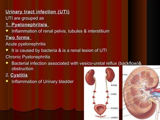
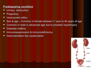
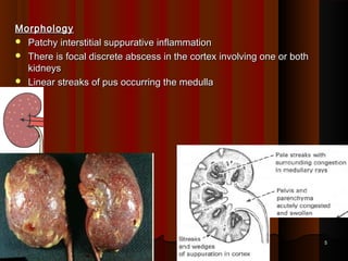
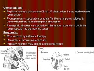
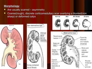
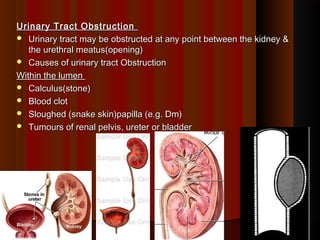
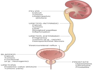
Urinary tract infection (UTI)Urinary tract infection (UTI)
UTI are grouped asUTI are grouped as
1. Pyelonephritsis1. Pyelonephritsis
 Inflammation of renal pelvis, tubules & interstitiumInflammation of renal pelvis, tubules & interstitium
Two formsTwo forms
Acute pyelonephritisAcute pyelonephritis
 It is caused by bacteria & is a renal lesion of UTIIt is caused by bacteria & is a renal lesion of UTI
Chronic PyelonephritisChronic Pyelonephritis
 Bacterial infection associated with vesico-uretal reflux (backflow)&Bacterial infection associated with vesico-uretal reflux (backflow)&
obstructionobstruction
2.2. CystitisCystitis
 Inflammation of Urinary bladderInflammation of Urinary bladder
11
Causes of Urinary tract infection (UTI)Causes of Urinary tract infection (UTI)
 Most infections are endogenousMost infections are endogenous
 Escherichia (E.) coli - 75 - 90% of UTIEscherichia (E.) coli - 75 - 90% of UTI
 Proteus species(p.vulgaris,mirabilis,penneri)Proteus species(p.vulgaris,mirabilis,penneri)
 KlebsiellaKlebsiella
 Staphylococcus epidermisStaphylococcus epidermis
22
Acute pyelonephritisAcute pyelonephritis
 Is an acute suppurative inflammation of theIs an acute suppurative inflammation of the
kidneykidney
Routes of InfectionRoutes of Infection
Ascending infectionAscending infection
 From the lower UT is the commonestFrom the lower UT is the commonest
 At first there is colonisation of the distalAt first there is colonisation of the distal
urethra .urethra .
Hematogenous infectionHematogenous infection
 Through blood stream e.g. septicaemiaThrough blood stream e.g. septicaemia
33
Predisposing conditionPredisposing condition
 Urinary obstructionUrinary obstruction
 PregnancyPregnancy
 Vesicouretic refluxVesicouretic reflux
 Sex & age – Common in female between 1Sex & age – Common in female between 1stst
year to 40 years of ageyear to 40 years of age
 Common in male in advanced age due to prostatic hypertrophyCommon in male in advanced age due to prostatic hypertrophy
 Diabetes mellitusDiabetes mellitus
 Immunosuppression & immunodeficiencyImmunosuppression & immunodeficiency
 Instrumentation like cauterizationInstrumentation like cauterization
44
MorphologyMorphology
 Patchy interstitial suppurative inflammationPatchy interstitial suppurative inflammation
 There is focal discrete abscess in the cortex involving one or bothThere is focal discrete abscess in the cortex involving one or both
kidneyskidneys
 Linear streaks of pus occurring the medullaLinear streaks of pus occurring the medulla
55
 Features of suppurative inflammation & tubular necrosisFeatures of suppurative inflammation & tubular necrosis
 Polymorphs may be present in tubular laminaPolymorphs may be present in tubular lamina
66
77
ComplicationsComplications
 Papillary necrosis particularly DM & UT obstruction it may lead to acutePapillary necrosis particularly DM & UT obstruction it may lead to acute
renal failurerenal failure
 Pyonephrosis – suppurative exudate fills the renal pelvis calyces &Pyonephrosis – suppurative exudate fills the renal pelvis calyces &
ureter when there is total complete obstructionureter when there is total complete obstruction
 Perinephric abscess – suppurative inflammation extends through thePerinephric abscess – suppurative inflammation extends through the
renal capsule into perinephric tissuerenal capsule into perinephric tissue
PrognosisPrognosis
 Most recover by antibiotic therapyMost recover by antibiotic therapy
 Recurrent - Chronic pyelonephritisRecurrent - Chronic pyelonephritis
 Papillary necrosis may lead to acute renal failurePapillary necrosis may lead to acute renal failure
88
Chronic PyelonephritisChronic Pyelonephritis
 It may be unilateral or bilateral.It may be unilateral or bilateral.
It occurs in 2 formsIt occurs in 2 forms
Reflux associated pyelonephritis (reflux nephropathy)Reflux associated pyelonephritis (reflux nephropathy)
 It starts early in childhood. There is urinary infection on congenitalIt starts early in childhood. There is urinary infection on congenital
vesicouretal refluxvesicouretal reflux
99
MorphologyMorphology
 Are usually scarred – asymmetryAre usually scarred – asymmetry
 Coarse(rough), discrete corticomedullary scar overlying a blunted(nonCoarse(rough), discrete corticomedullary scar overlying a blunted(non
sharp) or deformed calyxsharp) or deformed calyx
1010
Urinary Tract ObstructionUrinary Tract Obstruction
 Urinary tract may be obstructed at any point between the kidney &Urinary tract may be obstructed at any point between the kidney &
the urethral meatus(opening)the urethral meatus(opening)
 Causes of urinary tract ObstructionCauses of urinary tract Obstruction
Within the lumenWithin the lumen
 Calculus(stone)Calculus(stone)
 Blood clotBlood clot
 Sloughed (snake skin)papilla (e.g. Dm)Sloughed (snake skin)papilla (e.g. Dm)
 Tumours of renal pelvis, ureter or bladderTumours of renal pelvis, ureter or bladder
1111
Within the wallWithin the wall
 Congenital Intrinsic pelviureteric junction (PUJ) obstructionCongenital Intrinsic pelviureteric junction (PUJ) obstruction
Pressure from outsidePressure from outside
 Normal pregnancyNormal pregnancy
 Prostatic hypertrophyProstatic hypertrophy
 Tumours e.g. carcinoma prostate, carcinoma colon, carcinomaTumours e.g. carcinoma prostate, carcinoma colon, carcinoma
cervixcervix
 Retroperitoneal fibrosisRetroperitoneal fibrosis
1212
1313
Effects of Urinary tract obstructionEffects of Urinary tract obstruction
 Predisposes to infectionPredisposes to infection
 Predisposes to stone formationPredisposes to stone formation
 Leads to hydronephrosisLeads to hydronephrosis
1414
HydronephrosisHydronephrosis
 Hydronephrosis is an aseptic dilatation of the kidney due to a partial orHydronephrosis is an aseptic dilatation of the kidney due to a partial or
complete obstruction to the outflow of urine.complete obstruction to the outflow of urine.
It may beIt may be
 Unilateral hydronephrosisUnilateral hydronephrosis
 Bilateral hydronephrosisBilateral hydronephrosis
1515
PathologyPathology
 The dilatation first affects the pelvis alone (pelvic hydronephrosis).The dilatation first affects the pelvis alone (pelvic hydronephrosis).
 Then the calyces become increasingly dilated and the renalThen the calyces become increasingly dilated and the renal
parenchyma is progressively destroyed by pressure atrophy.parenchyma is progressively destroyed by pressure atrophy.
 In a kidney with a predominantly intrarenal pelvis, destruction of theIn a kidney with a predominantly intrarenal pelvis, destruction of the
parenchyma occurs more rapidly.parenchyma occurs more rapidly.
A kidney destroyed by long-standing hydronephrosis is a thin-walledA kidney destroyed by long-standing hydronephrosis is a thin-walled
lobulated saclobulated sac
1616
 Marked hydronephrosis with nearlyMarked hydronephrosis with nearly
complete loss of cortex.complete loss of cortex.
1717
Renal calculiRenal calculi
 Stone mostly arise in the kidney but may form anywhere in UTStone mostly arise in the kidney but may form anywhere in UT
 There is familial & hereditary predisposition to stoneThere is familial & hereditary predisposition to stone
Types/causes/pathogenesis of renal calculusTypes/causes/pathogenesis of renal calculus
Calcium stones (70%)-Calcium stones (70%)-
Most are calcium oxalate, others calcium oxalate with calciumMost are calcium oxalate, others calcium oxalate with calcium
phosphatephosphate
1818
This may be due toThis may be due to
 HypercalciuriaHypercalciuria – without hypercalcaemia in most cases– without hypercalcaemia in most cases
This may be due toThis may be due to
 More absorption of calcium ions form intestine (AbsorptiveMore absorption of calcium ions form intestine (Absorptive
hypercalciuria)hypercalciuria)
 Impairment of renal tubular reabsorb ion of calcium (renalImpairment of renal tubular reabsorb ion of calcium (renal
hypercalciuria)hypercalciuria)
 IdiopathicIdiopathic
 Hypercalcaemia & hypercalciuriaHypercalcaemia & hypercalciuria – hyperparathyroidism, diffuse bone– hyperparathyroidism, diffuse bone
diseases & other hyper calcaemic statesdiseases & other hyper calcaemic states
 With increased uric acid secretionWith increased uric acid secretion – (Hyperuricosuric calcium– (Hyperuricosuric calcium
nephrolithiasis)nephrolithiasis)
 HyperoxaluriaHyperoxaluria - Enteric disease or hereditary- Enteric disease or hereditary
 IdopathicIdopathic
1919
Magnesium Ammonium Phosphate calculus /triple phosphate stone /Magnesium Ammonium Phosphate calculus /triple phosphate stone /
struvite / infection stonestruvite / infection stone
 These are largest, smooth and dirty white & forms staghorn calculusThese are largest, smooth and dirty white & forms staghorn calculus
 Stone formation mostly follows renal infection by urea splittingStone formation mostly follows renal infection by urea splitting
bacteria – proteus species & staphylococcusbacteria – proteus species & staphylococcus
 Which convert urea to ammoniaWhich convert urea to ammonia
 Alkaline urine is formed – leads to ppt of magnesium ammoniumAlkaline urine is formed – leads to ppt of magnesium ammonium
phosphate saltphosphate salt
2020
Uric acid stone (5-10%)Uric acid stone (5-10%)
 It occurs in hyperuricaemia in goutIt occurs in hyperuricaemia in gout
 Without hyperurecimia & without increased excretion of uric acidWithout hyperurecimia & without increased excretion of uric acid
Urine pH below 5.5 may predispose to uric acid stonesUrine pH below 5.5 may predispose to uric acid stones
Cystine calculiCystine calculi
 are uncommon. They appear in the urinary tract of patients with aare uncommon. They appear in the urinary tract of patients with a
congenital error of metabolism which leads to cystinuria.congenital error of metabolism which leads to cystinuria.
Others rareOthers rare
2121
Factors that influence the formation of calculiFactors that influence the formation of calculi
 Deficiency of vitamin A causes desquamation of epitheliumDeficiency of vitamin A causes desquamation of epithelium
 Decrease urine volume - dehydrationDecrease urine volume - dehydration
 Altered urinary solutes and colloids-tends to cause them to precipitate.Altered urinary solutes and colloids-tends to cause them to precipitate.
 Change in pHChange in pH
 UTI - Infection favors the formation of urinary calculi.UTI - Infection favors the formation of urinary calculi.
 Obstruction and urinary stasis – encourages salt precipitationObstruction and urinary stasis – encourages salt precipitation
 Metabolic factors - alter the pH & increase the output of a substanceMetabolic factors - alter the pH & increase the output of a substance
e.g. Hyperparathyroidism, gout etce.g. Hyperparathyroidism, gout etc
2222
 Hence, the name "staghorn calculus". Seen here is a horn-like stoneHence, the name "staghorn calculus". Seen here is a horn-like stone
extending into a dilated calyx, with nearly unrecognizable overlyingextending into a dilated calyx, with nearly unrecognizable overlying
renal cortex from severe hydronephrosis and pyelonephritis.renal cortex from severe hydronephrosis and pyelonephritis.
2323
Congenital anomalies of kidneyCongenital anomalies of kidney
10% of all persons are born with potential significant malformations of10% of all persons are born with potential significant malformations of
urinary systemurinary system
1.1. Agenesis of kidneyAgenesis of kidney
2.2. Renal hypoplasiaRenal hypoplasia
3.3. Ectopic kidneyEctopic kidney
4.4. Horse shoe kidneyHorse shoe kidney
5.5. Cystic disease of kidneyCystic disease of kidney
2424
2525
Classification of renal cystClassification of renal cyst
 Adult polycystic disease (Autosomal dominant disease)Adult polycystic disease (Autosomal dominant disease)
 Childhood polycystic disease (Autosomal recessive disease)Childhood polycystic disease (Autosomal recessive disease)
 Cystic renal dysplasia - Disorganized development of kidneyCystic renal dysplasia - Disorganized development of kidney
characterized by cyst (primitive glomeruli, tubules & mesenchymecharacterized by cyst (primitive glomeruli, tubules & mesenchyme
 Medullary cystic disease - limited to medullary pyramids and papillae.Medullary cystic disease - limited to medullary pyramids and papillae.
 Acquired cystic disease – dialysis associated/ uraemicAcquired cystic disease – dialysis associated/ uraemic
 Simple cyst - Non neoplastic cyst lined by single cuboidal or flattenedSimple cyst - Non neoplastic cyst lined by single cuboidal or flattened
epitheliumepithelium
2626
ADULT POLYCYSTIC KIDNEY DISEASEADULT POLYCYSTIC KIDNEY DISEASE
 Common hereditary disorders characterized by expanding cysts thatCommon hereditary disorders characterized by expanding cysts that
progressively destroy the renal parenchyma of both kidneys leading toprogressively destroy the renal parenchyma of both kidneys leading to
renal failure.renal failure.
 Is generally a late-onset disorder.Is generally a late-onset disorder.
 It may be associated with other congenital anomalies e.g. polycysticIt may be associated with other congenital anomalies e.g. polycystic
liver, spleen, pancreas or lung, berry aneurysm of circle of Willisliver, spleen, pancreas or lung, berry aneurysm of circle of Willis
 APKDAPKD is a inherited autosomal dominant Disorderis a inherited autosomal dominant Disorder
 Resulting from mutations in either the PKD-1 or PKD-2 gene.Resulting from mutations in either the PKD-1 or PKD-2 gene.
2727
COMMON CLINICAL FEATURESCOMMON CLINICAL FEATURES
 Asymptomatic or may have - Haematuria (with little or noAsymptomatic or may have - Haematuria (with little or no
proteinuria), polyuria& uraemiaproteinuria), polyuria& uraemia
PathogenesisPathogenesis
 HereditaryHereditary
 Expanding cysts progressively destroy the renal parenchyma of bothExpanding cysts progressively destroy the renal parenchyma of both
kidneys, ultimately causing renal failurekidneys, ultimately causing renal failure
2828
MorphologyMorphology
 Both kidneys are enlarged to enormous sizeBoth kidneys are enlarged to enormous size
 A mass of cyst on the surface of various sizeA mass of cyst on the surface of various size
 Cyst contain clear serous fluid which may be turbid, red brown ofCyst contain clear serous fluid which may be turbid, red brown of
hemorrhagichemorrhagic
MicroscopicMicroscopic
 Functional nephrons are present between cystsFunctional nephrons are present between cysts
 Pressure ischaemia atropy of the intervening massPressure ischaemia atropy of the intervening mass
2929
Tumours of KidneyTumours of Kidney
Benign tumour – rarely have clinical significanceBenign tumour – rarely have clinical significance
 Cortical adenoma (origin in tubules)Cortical adenoma (origin in tubules)
 Renal hamartoma or fibromaRenal hamartoma or fibroma
 AngiomyolipomaAngiomyolipoma
 Renin producing juxtaglomerular tumours – rareRenin producing juxtaglomerular tumours – rare
Malignant tumourMalignant tumour
 Renal cell carcinoma (hypernephroma) – Most commonRenal cell carcinoma (hypernephroma) – Most common
 Wilm’s tumour (nephroblastoma) – childhood tumourWilm’s tumour (nephroblastoma) – childhood tumour
 Carcinoma of renal pelvisCarcinoma of renal pelvis
3030
Renal Cell Carcinoma (Adenocarcinoma ofRenal Cell Carcinoma (Adenocarcinoma of
kidney)kidney)
Malignant epithelial tumor of the kidney, composed of largeMalignant epithelial tumor of the kidney, composed of large
cells with clear or granular cytoplasm,cells with clear or granular cytoplasm,
 It arises from tubular epitheliumIt arises from tubular epithelium
 Commonest primary kidney tumor in adults.Commonest primary kidney tumor in adults.
 1-3% of all visceral tumors.1-3% of all visceral tumors.
 Over the age of 50 years; a male preponderance.Over the age of 50 years; a male preponderance.
3131
Risk factorsRisk factors
 Cigarette smokingCigarette smoking
 Chronic analgesic useChronic analgesic use
 Asbestos exposureAsbestos exposure
 Chronic renal failure and acquired cystic diseaseChronic renal failure and acquired cystic disease
 Genetic factors may play a role – VHL gene is the tumourGenetic factors may play a role – VHL gene is the tumour
supresure for Von Hippel-Lindau disease on chromosomesupresure for Von Hippel-Lindau disease on chromosome
3 – is implicated in renal carcinogenesis3 – is implicated in renal carcinogenesis
3232
MorphologyMorphology
Macroscopically:Macroscopically:
 Usually originates in the upper pole but may occur in any partUsually originates in the upper pole but may occur in any part
 Usually solitary & unilateralUsually solitary & unilateral
 Spherical mass of 3-15cmSpherical mass of 3-15cm
 Cut surface reveals a solid yellowish-grey tumor with areas ofCut surface reveals a solid yellowish-grey tumor with areas of
haemorrhage and necrosis.haemorrhage and necrosis.
 The margins of the tumor are usually well demarcated, but someThe margins of the tumor are usually well demarcated, but some
breach the renal capsule and invade the perinephric fat.breach the renal capsule and invade the perinephric fat.
3333
Macroscopically:Macroscopically:
 Extension into the renal vein is sometimes seen grossly;Extension into the renal vein is sometimes seen grossly;
occasionally, a solid mass of tumor extends into the inferior venaoccasionally, a solid mass of tumor extends into the inferior vena
cava and, rarely, into the right atrium.cava and, rarely, into the right atrium.
3434
Histologically:
Pattern of growth
• Papillary, solid, cord or tubular in some
Cell type
• Either clear - The clear cytoplasm is due to glycogen and fat
• Or Granular cells - organelles more numerous (glycogen and fat more
scanty)
3535
3636
Wilms' tumor or nephroblastomaWilms' tumor or nephroblastoma
 Most common primary renal tumor of childhood (ages 2-5 years).Most common primary renal tumor of childhood (ages 2-5 years).
 5-10% are bilateral5-10% are bilateral
 It originates from renal blastema & has epithelial & stromal derivativesIt originates from renal blastema & has epithelial & stromal derivatives
 Defect in the form of short arm of chromosome 11 may be presentDefect in the form of short arm of chromosome 11 may be present
 Mutation in tumor suppressor genes WT-1 and WT-2 genes.Mutation in tumor suppressor genes WT-1 and WT-2 genes.
 Other causes currently under investigation.Other causes currently under investigation.
3737
MorphologyMorphology
Gross:Gross:
 Large expansile solitary spherical mass with dwarf kidneyLarge expansile solitary spherical mass with dwarf kidney
3838
MicroscopicallyMicroscopically
 These are generally known as the classic triphasic combination:These are generally known as the classic triphasic combination:
1) blastmemic - Nest & sheets of primitive blastema within1) blastmemic - Nest & sheets of primitive blastema within
mesenchymemesenchyme
 2) stromal - (straited muscle, smooth muscle, cartilage, bone, fat cells,2) stromal - (straited muscle, smooth muscle, cartilage, bone, fat cells,
collagen fibrous tissue, cholesterol crystals & lipid,)collagen fibrous tissue, cholesterol crystals & lipid,)
 3) epithelial - abortive tubules, abortive glomeruli,3) epithelial - abortive tubules, abortive glomeruli,
 Spindle cell stroma gives a sarcomatoid patternSpindle cell stroma gives a sarcomatoid pattern
3939
4040
Carcinoma of renal pelvisCarcinoma of renal pelvis
 The tumor originates in the renal pelvis and has fingerlike projections.The tumor originates in the renal pelvis and has fingerlike projections.
 It can be exophytic or grow into the kidney.It can be exophytic or grow into the kidney.
 Occurs in theOccurs in the transitional epitheliumtransitional epithelium (“urothelium”).(“urothelium”).
 Occasionaly they may appear at multiple sites involving pelvis, ureter &Occasionaly they may appear at multiple sites involving pelvis, ureter &
bladderbladder
4141
Acute Renal failureAcute Renal failure
 Acute renal failure (ARF) refers to a sudden and usually reversibleAcute renal failure (ARF) refers to a sudden and usually reversible
loss of renal function, which develops over a period of days orloss of renal function, which develops over a period of days or
weeks and is usually accompanied by a reduction in urine volume.weeks and is usually accompanied by a reduction in urine volume.
 It is usually a reversible deterioration of renal function that developsIt is usually a reversible deterioration of renal function that develops
in daysin days
4242
CausesCauses
Pre-renal (kidneys are inadequately perfused / decreased GFR)Pre-renal (kidneys are inadequately perfused / decreased GFR)
 Heart failureHeart failure
 Loss of blood or plasma volumeLoss of blood or plasma volume
 Renal artery occlusionRenal artery occlusion
RenalRenal
 Glomerular diseases like crescentic GNGlomerular diseases like crescentic GN
 Acute tubular necrosisAcute tubular necrosis
 Toxins/drugsToxins/drugs
 Malignant HTNMalignant HTN
Post renalPost renal
 Obstruction in any part of UT (stones, tumors or enlarged prostate)Obstruction in any part of UT (stones, tumors or enlarged prostate)
4343
It can be divided into 3 stagesIt can be divided into 3 stages
Initiating stageInitiating stage
 Features of initiang factorFeatures of initiang factor
Maintenance stageMaintenance stage
 Oliguria (40 – 400 ml /day)Oliguria (40 – 400 ml /day)
 Progressive rise of blood urea & creatinine, hyperkalaemia,Progressive rise of blood urea & creatinine, hyperkalaemia,
metabolic acidosis with salt & water overloadmetabolic acidosis with salt & water overload
 Manifestations of uraemiaManifestations of uraemia
Recovery stageRecovery stage
 Gradual increase in urine volume maybe 3/ dayGradual increase in urine volume maybe 3/ day
 Blood urera & creatinine levels begin to return to normalBlood urera & creatinine levels begin to return to normal
 Ther is increased vulnerability to infectionTher is increased vulnerability to infection
4444
CHRONIC RENAL FAILURECHRONIC RENAL FAILURE
 Chronic renal failure (CRF) refers to an irreversible deterioration inChronic renal failure (CRF) refers to an irreversible deterioration in
renal function which classically develops over a period of years .renal function which classically develops over a period of years .
 Initially, it is manifest only as a biochemical abnormality.Initially, it is manifest only as a biochemical abnormality.
 Eventually, loss of the excretory, metabolic and endocrine functionsEventually, loss of the excretory, metabolic and endocrine functions
of the kidney leads to - the development of the uraemia.of the kidney leads to - the development of the uraemia.
 When death is likely without renal replacement therapy, it is calledWhen death is likely without renal replacement therapy, it is called
end-stage renal failure (ESRF).end-stage renal failure (ESRF).
4545
It is divided into 3 stages from normal renal function to uraemiaIt is divided into 3 stages from normal renal function to uraemia
 Diminished renal reserveDiminished renal reserve
 GFR is about 50% of normalGFR is about 50% of normal
 Asymptomatic. No azotemiaAsymptomatic. No azotemia
 Renal insufficiencyRenal insufficiency
 GFR is 20 – 50% of normal. There is azotaemia, anemia, HTN,GFR is 20 – 50% of normal. There is azotaemia, anemia, HTN,
polyuria, nocturiapolyuria, nocturia
 Renal failureRenal failure
 GFR is less than 20% of normalGFR is less than 20% of normal
 There is uraemia , hypokalaemia & metabolic acidosisThere is uraemia , hypokalaemia & metabolic acidosis
4646
Causes of end stage kidney disease(CRF)Causes of end stage kidney disease(CRF)
 Diabetes mellitusDiabetes mellitus
 HypertensionHypertension
 Glomerular diseasesGlomerular diseases
 Congenital and inherited renal diseases (e.g. PKD) 5%Congenital and inherited renal diseases (e.g. PKD) 5%
 Renal artery stenosisRenal artery stenosis
 Interstitial diseasesInterstitial diseases
 Systemic inflammatory diseases (e.g. SLE, vasculitis)Systemic inflammatory diseases (e.g. SLE, vasculitis)
 UnknownUnknown
4747

Other renal pathology

  • 1.
    Urinary tract infection(UTI)Urinary tract infection (UTI) UTI are grouped asUTI are grouped as 1. Pyelonephritsis1. Pyelonephritsis  Inflammation of renal pelvis, tubules & interstitiumInflammation of renal pelvis, tubules & interstitium Two formsTwo forms Acute pyelonephritisAcute pyelonephritis  It is caused by bacteria & is a renal lesion of UTIIt is caused by bacteria & is a renal lesion of UTI Chronic PyelonephritisChronic Pyelonephritis  Bacterial infection associated with vesico-uretal reflux (backflow)&Bacterial infection associated with vesico-uretal reflux (backflow)& obstructionobstruction 2.2. CystitisCystitis  Inflammation of Urinary bladderInflammation of Urinary bladder 11
  • 2.
    Causes of Urinarytract infection (UTI)Causes of Urinary tract infection (UTI)  Most infections are endogenousMost infections are endogenous  Escherichia (E.) coli - 75 - 90% of UTIEscherichia (E.) coli - 75 - 90% of UTI  Proteus species(p.vulgaris,mirabilis,penneri)Proteus species(p.vulgaris,mirabilis,penneri)  KlebsiellaKlebsiella  Staphylococcus epidermisStaphylococcus epidermis 22
  • 3.
    Acute pyelonephritisAcute pyelonephritis Is an acute suppurative inflammation of theIs an acute suppurative inflammation of the kidneykidney Routes of InfectionRoutes of Infection Ascending infectionAscending infection  From the lower UT is the commonestFrom the lower UT is the commonest  At first there is colonisation of the distalAt first there is colonisation of the distal urethra .urethra . Hematogenous infectionHematogenous infection  Through blood stream e.g. septicaemiaThrough blood stream e.g. septicaemia 33
  • 4.
    Predisposing conditionPredisposing condition Urinary obstructionUrinary obstruction  PregnancyPregnancy  Vesicouretic refluxVesicouretic reflux  Sex & age – Common in female between 1Sex & age – Common in female between 1stst year to 40 years of ageyear to 40 years of age  Common in male in advanced age due to prostatic hypertrophyCommon in male in advanced age due to prostatic hypertrophy  Diabetes mellitusDiabetes mellitus  Immunosuppression & immunodeficiencyImmunosuppression & immunodeficiency  Instrumentation like cauterizationInstrumentation like cauterization 44
  • 5.
    MorphologyMorphology  Patchy interstitialsuppurative inflammationPatchy interstitial suppurative inflammation  There is focal discrete abscess in the cortex involving one or bothThere is focal discrete abscess in the cortex involving one or both kidneyskidneys  Linear streaks of pus occurring the medullaLinear streaks of pus occurring the medulla 55
  • 6.
     Features ofsuppurative inflammation & tubular necrosisFeatures of suppurative inflammation & tubular necrosis  Polymorphs may be present in tubular laminaPolymorphs may be present in tubular lamina 66
  • 7.
  • 8.
    ComplicationsComplications  Papillary necrosisparticularly DM & UT obstruction it may lead to acutePapillary necrosis particularly DM & UT obstruction it may lead to acute renal failurerenal failure  Pyonephrosis – suppurative exudate fills the renal pelvis calyces &Pyonephrosis – suppurative exudate fills the renal pelvis calyces & ureter when there is total complete obstructionureter when there is total complete obstruction  Perinephric abscess – suppurative inflammation extends through thePerinephric abscess – suppurative inflammation extends through the renal capsule into perinephric tissuerenal capsule into perinephric tissue PrognosisPrognosis  Most recover by antibiotic therapyMost recover by antibiotic therapy  Recurrent - Chronic pyelonephritisRecurrent - Chronic pyelonephritis  Papillary necrosis may lead to acute renal failurePapillary necrosis may lead to acute renal failure 88
  • 9.
    Chronic PyelonephritisChronic Pyelonephritis It may be unilateral or bilateral.It may be unilateral or bilateral. It occurs in 2 formsIt occurs in 2 forms Reflux associated pyelonephritis (reflux nephropathy)Reflux associated pyelonephritis (reflux nephropathy)  It starts early in childhood. There is urinary infection on congenitalIt starts early in childhood. There is urinary infection on congenital vesicouretal refluxvesicouretal reflux 99
  • 10.
    MorphologyMorphology  Are usuallyscarred – asymmetryAre usually scarred – asymmetry  Coarse(rough), discrete corticomedullary scar overlying a blunted(nonCoarse(rough), discrete corticomedullary scar overlying a blunted(non sharp) or deformed calyxsharp) or deformed calyx 1010
  • 11.
    Urinary Tract ObstructionUrinaryTract Obstruction  Urinary tract may be obstructed at any point between the kidney &Urinary tract may be obstructed at any point between the kidney & the urethral meatus(opening)the urethral meatus(opening)  Causes of urinary tract ObstructionCauses of urinary tract Obstruction Within the lumenWithin the lumen  Calculus(stone)Calculus(stone)  Blood clotBlood clot  Sloughed (snake skin)papilla (e.g. Dm)Sloughed (snake skin)papilla (e.g. Dm)  Tumours of renal pelvis, ureter or bladderTumours of renal pelvis, ureter or bladder 1111
  • 12.
    Within the wallWithinthe wall  Congenital Intrinsic pelviureteric junction (PUJ) obstructionCongenital Intrinsic pelviureteric junction (PUJ) obstruction Pressure from outsidePressure from outside  Normal pregnancyNormal pregnancy  Prostatic hypertrophyProstatic hypertrophy  Tumours e.g. carcinoma prostate, carcinoma colon, carcinomaTumours e.g. carcinoma prostate, carcinoma colon, carcinoma cervixcervix  Retroperitoneal fibrosisRetroperitoneal fibrosis 1212
  • 13.
  • 14.
    Effects of Urinarytract obstructionEffects of Urinary tract obstruction  Predisposes to infectionPredisposes to infection  Predisposes to stone formationPredisposes to stone formation  Leads to hydronephrosisLeads to hydronephrosis 1414
  • 15.
    HydronephrosisHydronephrosis  Hydronephrosis isan aseptic dilatation of the kidney due to a partial orHydronephrosis is an aseptic dilatation of the kidney due to a partial or complete obstruction to the outflow of urine.complete obstruction to the outflow of urine. It may beIt may be  Unilateral hydronephrosisUnilateral hydronephrosis  Bilateral hydronephrosisBilateral hydronephrosis 1515
  • 16.
    PathologyPathology  The dilatationfirst affects the pelvis alone (pelvic hydronephrosis).The dilatation first affects the pelvis alone (pelvic hydronephrosis).  Then the calyces become increasingly dilated and the renalThen the calyces become increasingly dilated and the renal parenchyma is progressively destroyed by pressure atrophy.parenchyma is progressively destroyed by pressure atrophy.  In a kidney with a predominantly intrarenal pelvis, destruction of theIn a kidney with a predominantly intrarenal pelvis, destruction of the parenchyma occurs more rapidly.parenchyma occurs more rapidly. A kidney destroyed by long-standing hydronephrosis is a thin-walledA kidney destroyed by long-standing hydronephrosis is a thin-walled lobulated saclobulated sac 1616
  • 17.
     Marked hydronephrosiswith nearlyMarked hydronephrosis with nearly complete loss of cortex.complete loss of cortex. 1717
  • 18.
    Renal calculiRenal calculi Stone mostly arise in the kidney but may form anywhere in UTStone mostly arise in the kidney but may form anywhere in UT  There is familial & hereditary predisposition to stoneThere is familial & hereditary predisposition to stone Types/causes/pathogenesis of renal calculusTypes/causes/pathogenesis of renal calculus Calcium stones (70%)-Calcium stones (70%)- Most are calcium oxalate, others calcium oxalate with calciumMost are calcium oxalate, others calcium oxalate with calcium phosphatephosphate 1818
  • 19.
    This may bedue toThis may be due to  HypercalciuriaHypercalciuria – without hypercalcaemia in most cases– without hypercalcaemia in most cases This may be due toThis may be due to  More absorption of calcium ions form intestine (AbsorptiveMore absorption of calcium ions form intestine (Absorptive hypercalciuria)hypercalciuria)  Impairment of renal tubular reabsorb ion of calcium (renalImpairment of renal tubular reabsorb ion of calcium (renal hypercalciuria)hypercalciuria)  IdiopathicIdiopathic  Hypercalcaemia & hypercalciuriaHypercalcaemia & hypercalciuria – hyperparathyroidism, diffuse bone– hyperparathyroidism, diffuse bone diseases & other hyper calcaemic statesdiseases & other hyper calcaemic states  With increased uric acid secretionWith increased uric acid secretion – (Hyperuricosuric calcium– (Hyperuricosuric calcium nephrolithiasis)nephrolithiasis)  HyperoxaluriaHyperoxaluria - Enteric disease or hereditary- Enteric disease or hereditary  IdopathicIdopathic 1919
  • 20.
    Magnesium Ammonium Phosphatecalculus /triple phosphate stone /Magnesium Ammonium Phosphate calculus /triple phosphate stone / struvite / infection stonestruvite / infection stone  These are largest, smooth and dirty white & forms staghorn calculusThese are largest, smooth and dirty white & forms staghorn calculus  Stone formation mostly follows renal infection by urea splittingStone formation mostly follows renal infection by urea splitting bacteria – proteus species & staphylococcusbacteria – proteus species & staphylococcus  Which convert urea to ammoniaWhich convert urea to ammonia  Alkaline urine is formed – leads to ppt of magnesium ammoniumAlkaline urine is formed – leads to ppt of magnesium ammonium phosphate saltphosphate salt 2020
  • 21.
    Uric acid stone(5-10%)Uric acid stone (5-10%)  It occurs in hyperuricaemia in goutIt occurs in hyperuricaemia in gout  Without hyperurecimia & without increased excretion of uric acidWithout hyperurecimia & without increased excretion of uric acid Urine pH below 5.5 may predispose to uric acid stonesUrine pH below 5.5 may predispose to uric acid stones Cystine calculiCystine calculi  are uncommon. They appear in the urinary tract of patients with aare uncommon. They appear in the urinary tract of patients with a congenital error of metabolism which leads to cystinuria.congenital error of metabolism which leads to cystinuria. Others rareOthers rare 2121
  • 22.
    Factors that influencethe formation of calculiFactors that influence the formation of calculi  Deficiency of vitamin A causes desquamation of epitheliumDeficiency of vitamin A causes desquamation of epithelium  Decrease urine volume - dehydrationDecrease urine volume - dehydration  Altered urinary solutes and colloids-tends to cause them to precipitate.Altered urinary solutes and colloids-tends to cause them to precipitate.  Change in pHChange in pH  UTI - Infection favors the formation of urinary calculi.UTI - Infection favors the formation of urinary calculi.  Obstruction and urinary stasis – encourages salt precipitationObstruction and urinary stasis – encourages salt precipitation  Metabolic factors - alter the pH & increase the output of a substanceMetabolic factors - alter the pH & increase the output of a substance e.g. Hyperparathyroidism, gout etce.g. Hyperparathyroidism, gout etc 2222
  • 23.
     Hence, thename "staghorn calculus". Seen here is a horn-like stoneHence, the name "staghorn calculus". Seen here is a horn-like stone extending into a dilated calyx, with nearly unrecognizable overlyingextending into a dilated calyx, with nearly unrecognizable overlying renal cortex from severe hydronephrosis and pyelonephritis.renal cortex from severe hydronephrosis and pyelonephritis. 2323
  • 24.
    Congenital anomalies ofkidneyCongenital anomalies of kidney 10% of all persons are born with potential significant malformations of10% of all persons are born with potential significant malformations of urinary systemurinary system 1.1. Agenesis of kidneyAgenesis of kidney 2.2. Renal hypoplasiaRenal hypoplasia 3.3. Ectopic kidneyEctopic kidney 4.4. Horse shoe kidneyHorse shoe kidney 5.5. Cystic disease of kidneyCystic disease of kidney 2424
  • 25.
  • 26.
    Classification of renalcystClassification of renal cyst  Adult polycystic disease (Autosomal dominant disease)Adult polycystic disease (Autosomal dominant disease)  Childhood polycystic disease (Autosomal recessive disease)Childhood polycystic disease (Autosomal recessive disease)  Cystic renal dysplasia - Disorganized development of kidneyCystic renal dysplasia - Disorganized development of kidney characterized by cyst (primitive glomeruli, tubules & mesenchymecharacterized by cyst (primitive glomeruli, tubules & mesenchyme  Medullary cystic disease - limited to medullary pyramids and papillae.Medullary cystic disease - limited to medullary pyramids and papillae.  Acquired cystic disease – dialysis associated/ uraemicAcquired cystic disease – dialysis associated/ uraemic  Simple cyst - Non neoplastic cyst lined by single cuboidal or flattenedSimple cyst - Non neoplastic cyst lined by single cuboidal or flattened epitheliumepithelium 2626
  • 27.
    ADULT POLYCYSTIC KIDNEYDISEASEADULT POLYCYSTIC KIDNEY DISEASE  Common hereditary disorders characterized by expanding cysts thatCommon hereditary disorders characterized by expanding cysts that progressively destroy the renal parenchyma of both kidneys leading toprogressively destroy the renal parenchyma of both kidneys leading to renal failure.renal failure.  Is generally a late-onset disorder.Is generally a late-onset disorder.  It may be associated with other congenital anomalies e.g. polycysticIt may be associated with other congenital anomalies e.g. polycystic liver, spleen, pancreas or lung, berry aneurysm of circle of Willisliver, spleen, pancreas or lung, berry aneurysm of circle of Willis  APKDAPKD is a inherited autosomal dominant Disorderis a inherited autosomal dominant Disorder  Resulting from mutations in either the PKD-1 or PKD-2 gene.Resulting from mutations in either the PKD-1 or PKD-2 gene. 2727
  • 28.
    COMMON CLINICAL FEATURESCOMMONCLINICAL FEATURES  Asymptomatic or may have - Haematuria (with little or noAsymptomatic or may have - Haematuria (with little or no proteinuria), polyuria& uraemiaproteinuria), polyuria& uraemia PathogenesisPathogenesis  HereditaryHereditary  Expanding cysts progressively destroy the renal parenchyma of bothExpanding cysts progressively destroy the renal parenchyma of both kidneys, ultimately causing renal failurekidneys, ultimately causing renal failure 2828
  • 29.
    MorphologyMorphology  Both kidneysare enlarged to enormous sizeBoth kidneys are enlarged to enormous size  A mass of cyst on the surface of various sizeA mass of cyst on the surface of various size  Cyst contain clear serous fluid which may be turbid, red brown ofCyst contain clear serous fluid which may be turbid, red brown of hemorrhagichemorrhagic MicroscopicMicroscopic  Functional nephrons are present between cystsFunctional nephrons are present between cysts  Pressure ischaemia atropy of the intervening massPressure ischaemia atropy of the intervening mass 2929
  • 30.
    Tumours of KidneyTumoursof Kidney Benign tumour – rarely have clinical significanceBenign tumour – rarely have clinical significance  Cortical adenoma (origin in tubules)Cortical adenoma (origin in tubules)  Renal hamartoma or fibromaRenal hamartoma or fibroma  AngiomyolipomaAngiomyolipoma  Renin producing juxtaglomerular tumours – rareRenin producing juxtaglomerular tumours – rare Malignant tumourMalignant tumour  Renal cell carcinoma (hypernephroma) – Most commonRenal cell carcinoma (hypernephroma) – Most common  Wilm’s tumour (nephroblastoma) – childhood tumourWilm’s tumour (nephroblastoma) – childhood tumour  Carcinoma of renal pelvisCarcinoma of renal pelvis 3030
  • 31.
    Renal Cell Carcinoma(Adenocarcinoma ofRenal Cell Carcinoma (Adenocarcinoma of kidney)kidney) Malignant epithelial tumor of the kidney, composed of largeMalignant epithelial tumor of the kidney, composed of large cells with clear or granular cytoplasm,cells with clear or granular cytoplasm,  It arises from tubular epitheliumIt arises from tubular epithelium  Commonest primary kidney tumor in adults.Commonest primary kidney tumor in adults.  1-3% of all visceral tumors.1-3% of all visceral tumors.  Over the age of 50 years; a male preponderance.Over the age of 50 years; a male preponderance. 3131
  • 32.
    Risk factorsRisk factors Cigarette smokingCigarette smoking  Chronic analgesic useChronic analgesic use  Asbestos exposureAsbestos exposure  Chronic renal failure and acquired cystic diseaseChronic renal failure and acquired cystic disease  Genetic factors may play a role – VHL gene is the tumourGenetic factors may play a role – VHL gene is the tumour supresure for Von Hippel-Lindau disease on chromosomesupresure for Von Hippel-Lindau disease on chromosome 3 – is implicated in renal carcinogenesis3 – is implicated in renal carcinogenesis 3232
  • 33.
    MorphologyMorphology Macroscopically:Macroscopically:  Usually originatesin the upper pole but may occur in any partUsually originates in the upper pole but may occur in any part  Usually solitary & unilateralUsually solitary & unilateral  Spherical mass of 3-15cmSpherical mass of 3-15cm  Cut surface reveals a solid yellowish-grey tumor with areas ofCut surface reveals a solid yellowish-grey tumor with areas of haemorrhage and necrosis.haemorrhage and necrosis.  The margins of the tumor are usually well demarcated, but someThe margins of the tumor are usually well demarcated, but some breach the renal capsule and invade the perinephric fat.breach the renal capsule and invade the perinephric fat. 3333
  • 34.
    Macroscopically:Macroscopically:  Extension intothe renal vein is sometimes seen grossly;Extension into the renal vein is sometimes seen grossly; occasionally, a solid mass of tumor extends into the inferior venaoccasionally, a solid mass of tumor extends into the inferior vena cava and, rarely, into the right atrium.cava and, rarely, into the right atrium. 3434
  • 35.
    Histologically: Pattern of growth •Papillary, solid, cord or tubular in some Cell type • Either clear - The clear cytoplasm is due to glycogen and fat • Or Granular cells - organelles more numerous (glycogen and fat more scanty) 3535
  • 36.
  • 37.
    Wilms' tumor ornephroblastomaWilms' tumor or nephroblastoma  Most common primary renal tumor of childhood (ages 2-5 years).Most common primary renal tumor of childhood (ages 2-5 years).  5-10% are bilateral5-10% are bilateral  It originates from renal blastema & has epithelial & stromal derivativesIt originates from renal blastema & has epithelial & stromal derivatives  Defect in the form of short arm of chromosome 11 may be presentDefect in the form of short arm of chromosome 11 may be present  Mutation in tumor suppressor genes WT-1 and WT-2 genes.Mutation in tumor suppressor genes WT-1 and WT-2 genes.  Other causes currently under investigation.Other causes currently under investigation. 3737
  • 38.
    MorphologyMorphology Gross:Gross:  Large expansilesolitary spherical mass with dwarf kidneyLarge expansile solitary spherical mass with dwarf kidney 3838
  • 39.
    MicroscopicallyMicroscopically  These aregenerally known as the classic triphasic combination:These are generally known as the classic triphasic combination: 1) blastmemic - Nest & sheets of primitive blastema within1) blastmemic - Nest & sheets of primitive blastema within mesenchymemesenchyme  2) stromal - (straited muscle, smooth muscle, cartilage, bone, fat cells,2) stromal - (straited muscle, smooth muscle, cartilage, bone, fat cells, collagen fibrous tissue, cholesterol crystals & lipid,)collagen fibrous tissue, cholesterol crystals & lipid,)  3) epithelial - abortive tubules, abortive glomeruli,3) epithelial - abortive tubules, abortive glomeruli,  Spindle cell stroma gives a sarcomatoid patternSpindle cell stroma gives a sarcomatoid pattern 3939
  • 40.
  • 41.
    Carcinoma of renalpelvisCarcinoma of renal pelvis  The tumor originates in the renal pelvis and has fingerlike projections.The tumor originates in the renal pelvis and has fingerlike projections.  It can be exophytic or grow into the kidney.It can be exophytic or grow into the kidney.  Occurs in theOccurs in the transitional epitheliumtransitional epithelium (“urothelium”).(“urothelium”).  Occasionaly they may appear at multiple sites involving pelvis, ureter &Occasionaly they may appear at multiple sites involving pelvis, ureter & bladderbladder 4141
  • 42.
    Acute Renal failureAcuteRenal failure  Acute renal failure (ARF) refers to a sudden and usually reversibleAcute renal failure (ARF) refers to a sudden and usually reversible loss of renal function, which develops over a period of days orloss of renal function, which develops over a period of days or weeks and is usually accompanied by a reduction in urine volume.weeks and is usually accompanied by a reduction in urine volume.  It is usually a reversible deterioration of renal function that developsIt is usually a reversible deterioration of renal function that develops in daysin days 4242
  • 43.
    CausesCauses Pre-renal (kidneys areinadequately perfused / decreased GFR)Pre-renal (kidneys are inadequately perfused / decreased GFR)  Heart failureHeart failure  Loss of blood or plasma volumeLoss of blood or plasma volume  Renal artery occlusionRenal artery occlusion RenalRenal  Glomerular diseases like crescentic GNGlomerular diseases like crescentic GN  Acute tubular necrosisAcute tubular necrosis  Toxins/drugsToxins/drugs  Malignant HTNMalignant HTN Post renalPost renal  Obstruction in any part of UT (stones, tumors or enlarged prostate)Obstruction in any part of UT (stones, tumors or enlarged prostate) 4343
  • 44.
    It can bedivided into 3 stagesIt can be divided into 3 stages Initiating stageInitiating stage  Features of initiang factorFeatures of initiang factor Maintenance stageMaintenance stage  Oliguria (40 – 400 ml /day)Oliguria (40 – 400 ml /day)  Progressive rise of blood urea & creatinine, hyperkalaemia,Progressive rise of blood urea & creatinine, hyperkalaemia, metabolic acidosis with salt & water overloadmetabolic acidosis with salt & water overload  Manifestations of uraemiaManifestations of uraemia Recovery stageRecovery stage  Gradual increase in urine volume maybe 3/ dayGradual increase in urine volume maybe 3/ day  Blood urera & creatinine levels begin to return to normalBlood urera & creatinine levels begin to return to normal  Ther is increased vulnerability to infectionTher is increased vulnerability to infection 4444
  • 45.
    CHRONIC RENAL FAILURECHRONICRENAL FAILURE  Chronic renal failure (CRF) refers to an irreversible deterioration inChronic renal failure (CRF) refers to an irreversible deterioration in renal function which classically develops over a period of years .renal function which classically develops over a period of years .  Initially, it is manifest only as a biochemical abnormality.Initially, it is manifest only as a biochemical abnormality.  Eventually, loss of the excretory, metabolic and endocrine functionsEventually, loss of the excretory, metabolic and endocrine functions of the kidney leads to - the development of the uraemia.of the kidney leads to - the development of the uraemia.  When death is likely without renal replacement therapy, it is calledWhen death is likely without renal replacement therapy, it is called end-stage renal failure (ESRF).end-stage renal failure (ESRF). 4545
  • 46.
    It is dividedinto 3 stages from normal renal function to uraemiaIt is divided into 3 stages from normal renal function to uraemia  Diminished renal reserveDiminished renal reserve  GFR is about 50% of normalGFR is about 50% of normal  Asymptomatic. No azotemiaAsymptomatic. No azotemia  Renal insufficiencyRenal insufficiency  GFR is 20 – 50% of normal. There is azotaemia, anemia, HTN,GFR is 20 – 50% of normal. There is azotaemia, anemia, HTN, polyuria, nocturiapolyuria, nocturia  Renal failureRenal failure  GFR is less than 20% of normalGFR is less than 20% of normal  There is uraemia , hypokalaemia & metabolic acidosisThere is uraemia , hypokalaemia & metabolic acidosis 4646
  • 47.
    Causes of endstage kidney disease(CRF)Causes of end stage kidney disease(CRF)  Diabetes mellitusDiabetes mellitus  HypertensionHypertension  Glomerular diseasesGlomerular diseases  Congenital and inherited renal diseases (e.g. PKD) 5%Congenital and inherited renal diseases (e.g. PKD) 5%  Renal artery stenosisRenal artery stenosis  Interstitial diseasesInterstitial diseases  Systemic inflammatory diseases (e.g. SLE, vasculitis)Systemic inflammatory diseases (e.g. SLE, vasculitis)  UnknownUnknown 4747