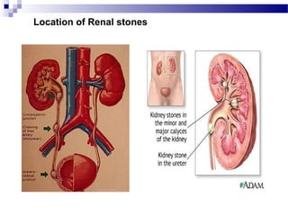
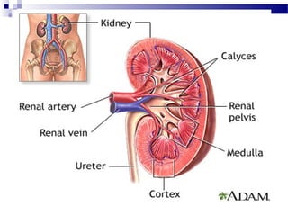
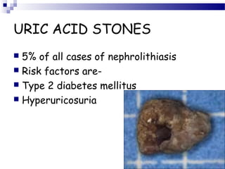
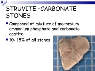
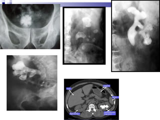
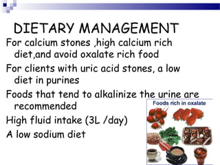
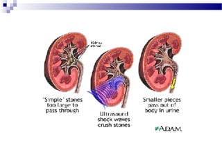
RENAL CALCULI
By
NEETHU.M
MSc (N)
Definition
 Renal calculi are Stones which is small, harden
deposits of mineral and acid salts on the inner
surfaces of the kidneys They originate as
microscopic particles and develop into stones
over time. The medical term for this condition is
nephrolithiasis, or renal stone disease
 Alternative names include:
Renal Lithiasis
Nephrolithiasis (Kidney Stone Disease)
Location of Renal stones
BLOOD
CONSTITUENTS IN
GLOMERULAR
FILTRATE
• WATER
• MINERAL SALTS
• AMINOACIDS
• KETOACIDS
• GLUCOSE,HORMONES
• UREA, CREATININE
• URIC ACID
• TOXINS
• SOME DRUGS
BLOOD
CONSTITUENTS
REMAINING IN
GLOMERULUS
• LEUKOCYTES
• ERYTHROCYTES
• PLATELETS
• PLASMA PROTEINS
• SOME DRUGS
ETIOLOGY
 Immobility and sedentary life style
 Dehydration /decreased fluid intake
 Previous history of urinary calculi
 A diet rich in prurines ,oxalates
 Genetic predisposition
 Warm climate
 Large intake of dietary protein
 Living in mountains ,deserts/tropical
areas
PATHOGENESIS
 SUPERSATURATION
 NUCLEATION
 INHIBITORS OF CRYSTAL
FORMATION
SUPERSATURATION
 Increased solute concentration
 Super saturation
 Crystallization
 Stone formation
Stone Formation
 Kidney stones form when there is a high level
of mineral (s) ; i.e. calcium (hypercalciuria),
oxalate (hyperoxaluria), or uric acid
(hyperuricosuria) in the urine;
 Urine normally contains chemicals—citrate,
magnesium, pyrophosphate—that prevent the
formation of crystals.
 Low levels of these inhibitors can contribute to
the formation of kidney stones.
 Citrate is thought to be the most important
 The chemical composition of stones depends
on the chemical imbalance in the urine.
 The four most common types of stones are
comprised of calcium, uric acid, struvite, and
cystine.
Cont.
TYPES OF STONES
 CALCIUM CONTAINING STONES
 URIC ACID STONES
 STRUVITE –CARBONATE STONES
 CYSTINE STONES
 DRUG RELATED STONES
CALCIUM CONTAINING
STONES
 Approximately 85% of stones are composed
predominantly of calcium compounds.
 The most common cause of calcium stone
production is excess calcium in the urine
(hypercalciuria).
 In hypercalciuria, excess calcium builds up in
the kidneys and urine, where it combines
with other waste products to form stones.
 Low levels of citrate, high levels of oxalate
and uric acid, and inadequate urinary volume
may also cause calcium stone formation.
 Calcium stones are composed of oxalate (calcium
oxalate) or phosphate (calcium phosphate).
 Calcium phosphate stones typically occur in
patients with metabolic or hormonal disorders such
as hyperparathyroidism and renal tubular acidosis.
 These stones come in 2 different types -
monohydrate and dihydrate.
 Calcium oxalate dihydrate stones usually break
easily with lithotripsy.
 Monohydrate stones are among the most difficult
stones to fragment.
Cont.
Cause of hypercalciuria.
 Increased intestinal absorption of calcium
(absorptive hypercalciuria),
 excessive hormone levels
(hyperparathyroidism),
 and renal calcium leak (kidney defect that
causes excessive calcium to enter the
urine)
 Prolonged inactivity also increases urinary
calcium and may cause stones.
 Renal tubular acidosis (inherited condition
in which the kidneys are unable to excrete
acid) significantly reduces urinary citrate
and total acid levels and can lead to stone
formation.
Calcium oxalate monohydrates
Calcium oxalate dihydrates
URIC ACID STONES
 5% of all cases of nephrolithiasis
 Risk factors are-
 Type 2 diabetes mellitus
 Hyperuricosuria
STRUVITE –CARBONATE
STONES
 Composed of mixture of magnesium
ammonium phosphate and carbonate
apatite
 10- 15% of all stones
Struvite Stones
 Also called an infection stone, develops when a
urinary tract infection (e.g., bladder infection)
affects the chemical balance of the urine.
 Bacteria in the urinary tract release chemicals
that neutralize acid in the urine, which enables
bacteria to grow more quickly and promotes
struvite stone development.
 They are capable of splitting urea into
ammonia, decreasing the acidity of the
urine and resulting in favorable conditions
for the formation of struvite stones.
Cont.
 Organisms which alkalinize the urine
can cause struvite stones to form.
 Struvite stones are more common in
women.
 The stones usually develop as jagged
structures called "staghorns" and can
grow to be quite large.
STRUVITE –CARBONATE
STONES
 Urease producing bacteria include-
Proteus , Morganella, Providencia ,
Pseudomonas
 Risk factors –women with recurrent UTI
 Patients with spinal cord injury
 Men with indwelling bladder catheter and
complete spinal cord transection have
higher risk
STRUVITE –CARBONATE
STONES
 Signs and symptoms
 Fever ,hematuria, flank pain, recurrent UTI
 Septicemia
CYSTINE STONES
 Cystinuria is the result of an autosomal
recessive defect in proximal tubular and
jejunum reabsorption of the dibasic
amino acids cysteine ,ornithine lysine,
arginine
 It constitutes of <1% of all stones
 Characteristic hexagonal crystals
present in morning urine
Cystine
DRUG RELATED STONES
 Sulfonamides , triamterene , acyclovir,
antiretroviral agent-indanavir,
 Topiramide
Signs and Symptoms
 Severe flank pain
 Abdominal pain
 Nausea and vomiting
 Fatigue
 Elevated temperature, BP,
and grunting respirations
 Objective Data:
perspiration, clutching of
the abdomen, doubled-over.
 Steady Pain
 Left flank tendernes
 Additional S/S:
 Presence of UTI
 Fever or Chills
 Pain in groin, labia or
testicles
 Dysuria
 Persistent urge to void
 Anuria
http://knol.google.com/k/-/-/27ifsyywko3wx/sqc1f9/kidneystonesymptoms.jpg
DIAGNOSIS
 History collection ,physical examination
 Urine analysis ,blood chemistries
 Stone analysis
 Flat radiographic plate of abdomen
 Ultrasonography
 Intravenous pyelogram
 CT scan
 Cystoscopy
MANAGEMENT
 INCREASE FLUIDS
 REDUCE PAIN
 PREVENT STONE RECURRENCE
 IMPLEMENT DIETARY CHANGES
 ADMINISTER MEDICATIONS
DIETARY MANAGEMENT
For calcium stones ,high calcium rich
diet,and avoid oxalate rich food
For clients with uric acid stones, a low
diet in purines
Foods that tend to alkalinize the urine are
recommended
High fluid intake (3L /day)
A low sodium diet
 High doses of vitamin C (i.e., more than 500 mg
per day) can result in high levels of oxalate in
the urine (hyperoxaluria) and increase the risk
for kidney stones. Oxalate is found in berries,
vegetables (e.g., green beans, beets, spinach,
squash, tomatoes), nuts, chocolate, and tea.
 Stone formers should limit their intake of
cranberries, which contain a moderate amount
of oxalate.
NON SURGICAL
MANGEMENT
 EXTRACORPORAL SHOCK WAVE
LITHOTRIPSY
 PERCUTANEOUSULTRASONIC
LITHOTROPSY
 LASER LITHOTRIPSY
 PERCUTANEOUS
NEPHROSTOLITHOTOMY
 PERCUTANEOUS STONE
DISSOLUTION
OPEN SURGICAL
MANGEMENT
 PYELOLITHOTOMY
 COAGULUM PUELOLITHOTOMY
 NEPHROLITHOTOMY
 NEPHRECTOMY
 URETEROLITHOTOMY
 CYSTOLITHOTOMY
OPEN SURGICAL
MANGEMENT
COMPLICATIONS
 OBSTRUCTION
 INFECTION
 IMPAIRED RENAL FUNCTION
 PERIRENAL HEMATOMA
NURSING MANAGEMENT
 PREOPERATIVE CARE
 Assess knowledge and understanding of
the procedure
 Withhold food and fluids and for bowel
preparation prior to surgery
NURSING MANAGEMENTNURSING MANAGEMENT
• Acute pain related to effect of renal
stone and inadequate pain control or
comfort measures
• Impaired urinary elimination related to
trauma or blockage of ureters or
urethra
• Ineffective therapeutic regimen
management related to lack of
knowledge regarding disease process
prevention of recurrence , diet and fluid
requirements
HEALTH EDUCATION
 Encourage fluid to accelerate passing of
stone particles
 Teach about analgesics for colicky pain
and passage of stone debris
 Encourage frequent walking to assist in
passage of stone fragments
• For patient with calcium oxalate stones
• Instruct to maintain dietary pattern
• Teach purpose of drug therapy
• For patients with uric acid stones
• Teach methods to alkalinze urine to
enhance urate solubility
• Teach the purpose of taking
Allopurinol
• Provide information about reduction
of dietary purine intake
 For patients with infection stone
 Encourage to report signs and symptoms
of urinary infections
 For patients with cystine stones
 teach patient about drug therapywith D-
pencillamine(Depen)
 Instruct the patient to test urine pH
Renal calculi

Renal calculi

  • 1.
  • 2.
    Definition  Renal calculiare Stones which is small, harden deposits of mineral and acid salts on the inner surfaces of the kidneys They originate as microscopic particles and develop into stones over time. The medical term for this condition is nephrolithiasis, or renal stone disease  Alternative names include: Renal Lithiasis Nephrolithiasis (Kidney Stone Disease)
  • 3.
  • 11.
    BLOOD CONSTITUENTS IN GLOMERULAR FILTRATE • WATER •MINERAL SALTS • AMINOACIDS • KETOACIDS • GLUCOSE,HORMONES • UREA, CREATININE • URIC ACID • TOXINS • SOME DRUGS BLOOD CONSTITUENTS REMAINING IN GLOMERULUS • LEUKOCYTES • ERYTHROCYTES • PLATELETS • PLASMA PROTEINS • SOME DRUGS
  • 14.
    ETIOLOGY  Immobility andsedentary life style  Dehydration /decreased fluid intake  Previous history of urinary calculi  A diet rich in prurines ,oxalates  Genetic predisposition  Warm climate  Large intake of dietary protein  Living in mountains ,deserts/tropical areas
  • 15.
  • 16.
    SUPERSATURATION  Increased soluteconcentration  Super saturation  Crystallization  Stone formation
  • 17.
    Stone Formation  Kidneystones form when there is a high level of mineral (s) ; i.e. calcium (hypercalciuria), oxalate (hyperoxaluria), or uric acid (hyperuricosuria) in the urine;  Urine normally contains chemicals—citrate, magnesium, pyrophosphate—that prevent the formation of crystals.
  • 18.
     Low levelsof these inhibitors can contribute to the formation of kidney stones.  Citrate is thought to be the most important  The chemical composition of stones depends on the chemical imbalance in the urine.  The four most common types of stones are comprised of calcium, uric acid, struvite, and cystine. Cont.
  • 19.
    TYPES OF STONES CALCIUM CONTAINING STONES  URIC ACID STONES  STRUVITE –CARBONATE STONES  CYSTINE STONES  DRUG RELATED STONES
  • 20.
    CALCIUM CONTAINING STONES  Approximately85% of stones are composed predominantly of calcium compounds.  The most common cause of calcium stone production is excess calcium in the urine (hypercalciuria).  In hypercalciuria, excess calcium builds up in the kidneys and urine, where it combines with other waste products to form stones.  Low levels of citrate, high levels of oxalate and uric acid, and inadequate urinary volume may also cause calcium stone formation.
  • 21.
     Calcium stonesare composed of oxalate (calcium oxalate) or phosphate (calcium phosphate).  Calcium phosphate stones typically occur in patients with metabolic or hormonal disorders such as hyperparathyroidism and renal tubular acidosis.  These stones come in 2 different types - monohydrate and dihydrate.  Calcium oxalate dihydrate stones usually break easily with lithotripsy.  Monohydrate stones are among the most difficult stones to fragment. Cont.
  • 22.
    Cause of hypercalciuria. Increased intestinal absorption of calcium (absorptive hypercalciuria),  excessive hormone levels (hyperparathyroidism),  and renal calcium leak (kidney defect that causes excessive calcium to enter the urine)  Prolonged inactivity also increases urinary calcium and may cause stones.  Renal tubular acidosis (inherited condition in which the kidneys are unable to excrete acid) significantly reduces urinary citrate and total acid levels and can lead to stone formation.
  • 23.
  • 24.
    URIC ACID STONES 5% of all cases of nephrolithiasis  Risk factors are-  Type 2 diabetes mellitus  Hyperuricosuria
  • 25.
    STRUVITE –CARBONATE STONES  Composedof mixture of magnesium ammonium phosphate and carbonate apatite  10- 15% of all stones
  • 26.
    Struvite Stones  Alsocalled an infection stone, develops when a urinary tract infection (e.g., bladder infection) affects the chemical balance of the urine.  Bacteria in the urinary tract release chemicals that neutralize acid in the urine, which enables bacteria to grow more quickly and promotes struvite stone development.  They are capable of splitting urea into ammonia, decreasing the acidity of the urine and resulting in favorable conditions for the formation of struvite stones.
  • 27.
    Cont.  Organisms whichalkalinize the urine can cause struvite stones to form.  Struvite stones are more common in women.  The stones usually develop as jagged structures called "staghorns" and can grow to be quite large.
  • 28.
    STRUVITE –CARBONATE STONES  Ureaseproducing bacteria include- Proteus , Morganella, Providencia , Pseudomonas  Risk factors –women with recurrent UTI  Patients with spinal cord injury  Men with indwelling bladder catheter and complete spinal cord transection have higher risk
  • 29.
    STRUVITE –CARBONATE STONES  Signsand symptoms  Fever ,hematuria, flank pain, recurrent UTI  Septicemia
  • 30.
    CYSTINE STONES  Cystinuriais the result of an autosomal recessive defect in proximal tubular and jejunum reabsorption of the dibasic amino acids cysteine ,ornithine lysine, arginine  It constitutes of <1% of all stones  Characteristic hexagonal crystals present in morning urine
  • 31.
  • 32.
    DRUG RELATED STONES Sulfonamides , triamterene , acyclovir, antiretroviral agent-indanavir,  Topiramide
  • 33.
    Signs and Symptoms Severe flank pain  Abdominal pain  Nausea and vomiting  Fatigue  Elevated temperature, BP, and grunting respirations  Objective Data: perspiration, clutching of the abdomen, doubled-over.  Steady Pain  Left flank tendernes  Additional S/S:  Presence of UTI  Fever or Chills  Pain in groin, labia or testicles  Dysuria  Persistent urge to void  Anuria http://knol.google.com/k/-/-/27ifsyywko3wx/sqc1f9/kidneystonesymptoms.jpg
  • 34.
    DIAGNOSIS  History collection,physical examination  Urine analysis ,blood chemistries  Stone analysis  Flat radiographic plate of abdomen  Ultrasonography  Intravenous pyelogram  CT scan  Cystoscopy
  • 36.
    MANAGEMENT  INCREASE FLUIDS REDUCE PAIN  PREVENT STONE RECURRENCE  IMPLEMENT DIETARY CHANGES  ADMINISTER MEDICATIONS
  • 37.
    DIETARY MANAGEMENT For calciumstones ,high calcium rich diet,and avoid oxalate rich food For clients with uric acid stones, a low diet in purines Foods that tend to alkalinize the urine are recommended High fluid intake (3L /day) A low sodium diet
  • 38.
     High dosesof vitamin C (i.e., more than 500 mg per day) can result in high levels of oxalate in the urine (hyperoxaluria) and increase the risk for kidney stones. Oxalate is found in berries, vegetables (e.g., green beans, beets, spinach, squash, tomatoes), nuts, chocolate, and tea.  Stone formers should limit their intake of cranberries, which contain a moderate amount of oxalate.
  • 39.
    NON SURGICAL MANGEMENT  EXTRACORPORALSHOCK WAVE LITHOTRIPSY  PERCUTANEOUSULTRASONIC LITHOTROPSY  LASER LITHOTRIPSY  PERCUTANEOUS NEPHROSTOLITHOTOMY  PERCUTANEOUS STONE DISSOLUTION
  • 41.
    OPEN SURGICAL MANGEMENT  PYELOLITHOTOMY COAGULUM PUELOLITHOTOMY  NEPHROLITHOTOMY  NEPHRECTOMY  URETEROLITHOTOMY  CYSTOLITHOTOMY
  • 42.
    OPEN SURGICAL MANGEMENT COMPLICATIONS  OBSTRUCTION INFECTION  IMPAIRED RENAL FUNCTION  PERIRENAL HEMATOMA
  • 43.
    NURSING MANAGEMENT  PREOPERATIVECARE  Assess knowledge and understanding of the procedure  Withhold food and fluids and for bowel preparation prior to surgery
  • 44.
    NURSING MANAGEMENTNURSING MANAGEMENT •Acute pain related to effect of renal stone and inadequate pain control or comfort measures • Impaired urinary elimination related to trauma or blockage of ureters or urethra • Ineffective therapeutic regimen management related to lack of knowledge regarding disease process prevention of recurrence , diet and fluid requirements
  • 45.
    HEALTH EDUCATION  Encouragefluid to accelerate passing of stone particles  Teach about analgesics for colicky pain and passage of stone debris  Encourage frequent walking to assist in passage of stone fragments
  • 46.
    • For patientwith calcium oxalate stones • Instruct to maintain dietary pattern • Teach purpose of drug therapy • For patients with uric acid stones • Teach methods to alkalinze urine to enhance urate solubility • Teach the purpose of taking Allopurinol • Provide information about reduction of dietary purine intake
  • 47.
     For patientswith infection stone  Encourage to report signs and symptoms of urinary infections  For patients with cystine stones  teach patient about drug therapywith D- pencillamine(Depen)  Instruct the patient to test urine pH

Editor's Notes

  • #34 pain typically starts at side or back, just below your ribs, and radiates to ones lower abdomen and groin. Often begins when stone reaches ureters