The document discusses the anatomy of fascial spaces in the head and neck region. It describes several layers of deep cervical fascia including the investing layer, middle layer, visceral layer, vertebral layer and alar fascia. It also outlines the boundaries and contents of various fascial spaces such as the buccal space, retropharyngeal space, submandibular space and submental space. Clinical aspects such as drainage of abscesses in these spaces are also mentioned.
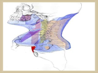
The Indian Dental Academy is the Leader in continuing dental education , training dentists in all aspects of dentistry and
offering a wide range of dental certified courses in different formats.for more details please visit
www.indiandentalacademy.com
the fascial planes of the neck is very important in the spread and containment of infections, as well as being surgical dissection plane during neck surgery.
infections are rare but need to be identified early and treated appropriately to reduce the mortality and morbidity
this is a slightly well illustrated ppt of the previously uploaded one in february 2015
Lung Cancer: Artificial Intelligence, Synergetics, Complex System Analysis, S...Oleg Kshivets
RESULTS: Overall life span (LS) was 2252.1±1742.5 days and cumulative 5-year survival (5YS) reached 73.2%, 10 years – 64.8%, 20 years – 42.5%. 513 LCP lived more than 5 years (LS=3124.6±1525.6 days), 148 LCP – more than 10 years (LS=5054.4±1504.1 days).199 LCP died because of LC (LS=562.7±374.5 days). 5YS of LCP after bi/lobectomies was significantly superior in comparison with LCP after pneumonectomies (78.1% vs.63.7%, P=0.00001 by log-rank test). AT significantly improved 5YS (66.3% vs. 34.8%) (P=0.00000 by log-rank test) only for LCP with N1-2. Cox modeling displayed that 5YS of LCP significantly depended on: phase transition (PT) early-invasive LC in terms of synergetics, PT N0—N12, cell ratio factors (ratio between cancer cells- CC and blood cells subpopulations), G1-3, histology, glucose, AT, blood cell circuit, prothrombin index, heparin tolerance, recalcification time (P=0.000-0.038). Neural networks, genetic algorithm selection and bootstrap simulation revealed relationships between 5YS and PT early-invasive LC (rank=1), PT N0—N12 (rank=2), thrombocytes/CC (3), erythrocytes/CC (4), eosinophils/CC (5), healthy cells/CC (6), lymphocytes/CC (7), segmented neutrophils/CC (8), stick neutrophils/CC (9), monocytes/CC (10); leucocytes/CC (11). Correct prediction of 5YS was 100% by neural networks computing (area under ROC curve=1.0; error=0.0).
CONCLUSIONS: 5YS of LCP after radical procedures significantly depended on: 1) PT early-invasive cancer; 2) PT N0--N12; 3) cell ratio factors; 4) blood cell circuit; 5) biochemical factors; 6) hemostasis system; 7) AT; 8) LC characteristics; 9) LC cell dynamics; 10) surgery type: lobectomy/pneumonectomy; 11) anthropometric data. Optimal diagnosis and treatment strategies for LC are: 1) screening and early detection of LC; 2) availability of experienced thoracic surgeons because of complexity of radical procedures; 3) aggressive en block surgery and adequate lymph node dissection for completeness; 4) precise prediction; 5) adjuvant chemoimmunoradiotherapy for LCP with unfavorable prognosis.
Report Back from SGO 2024: What’s the Latest in Cervical Cancer?bkling
Are you curious about what’s new in cervical cancer research or unsure what the findings mean? Join Dr. Emily Ko, a gynecologic oncologist at Penn Medicine, to learn about the latest updates from the Society of Gynecologic Oncology (SGO) 2024 Annual Meeting on Women’s Cancer. Dr. Ko will discuss what the research presented at the conference means for you and answer your questions about the new developments.
HOT NEW PRODUCT! BIG SALES FAST SHIPPING NOW FROM CHINA!! EU KU DB BK substit...GL Anaacs
Contact us if you are interested:
Email / Skype : kefaya1771@gmail.com
Threema: PXHY5PDH
New BATCH Ku !!! MUCH IN DEMAND FAST SALE EVERY BATCH HAPPY GOOD EFFECT BIG BATCH !
Contact me on Threema or skype to start big business!!
Hot-sale products:
NEW HOT EUTYLONE WHITE CRYSTAL!!
5cl-adba precursor (semi finished )
5cl-adba raw materials
ADBB precursor (semi finished )
ADBB raw materials
APVP powder
5fadb/4f-adb
Jwh018 / Jwh210
Eutylone crystal
Protonitazene (hydrochloride) CAS: 119276-01-6
Flubrotizolam CAS: 57801-95-3
Metonitazene CAS: 14680-51-4
Payment terms: Western Union,MoneyGram,Bitcoin or USDT.
Deliver Time: Usually 7-15days
Shipping method: FedEx, TNT, DHL,UPS etc.Our deliveries are 100% safe, fast, reliable and discreet.
Samples will be sent for your evaluation!If you are interested in, please contact me, let's talk details.
We specializes in exporting high quality Research chemical, medical intermediate, Pharmaceutical chemicals and so on. Products are exported to USA, Canada, France, Korea, Japan,Russia, Southeast Asia and other countries.
Couples presenting to the infertility clinic- Do they really have infertility...Sujoy Dasgupta
Dr Sujoy Dasgupta presented the study on "Couples presenting to the infertility clinic- Do they really have infertility? – The unexplored stories of non-consummation" in the 13th Congress of the Asia Pacific Initiative on Reproduction (ASPIRE 2024) at Manila on 24 May, 2024.
Ethanol (CH3CH2OH), or beverage alcohol, is a two-carbon alcohol
that is rapidly distributed in the body and brain. Ethanol alters many
neurochemical systems and has rewarding and addictive properties. It
is the oldest recreational drug and likely contributes to more morbidity,
mortality, and public health costs than all illicit drugs combined. The
5th edition of the Diagnostic and Statistical Manual of Mental Disorders
(DSM-5) integrates alcohol abuse and alcohol dependence into a single
disorder called alcohol use disorder (AUD), with mild, moderate,
and severe subclassifications (American Psychiatric Association, 2013).
In the DSM-5, all types of substance abuse and dependence have been
combined into a single substance use disorder (SUD) on a continuum
from mild to severe. A diagnosis of AUD requires that at least two of
the 11 DSM-5 behaviors be present within a 12-month period (mild
AUD: 2–3 criteria; moderate AUD: 4–5 criteria; severe AUD: 6–11 criteria).
The four main behavioral effects of AUD are impaired control over
drinking, negative social consequences, risky use, and altered physiological
effects (tolerance, withdrawal). This chapter presents an overview
of the prevalence and harmful consequences of AUD in the U.S.,
the systemic nature of the disease, neurocircuitry and stages of AUD,
comorbidities, fetal alcohol spectrum disorders, genetic risk factors, and
pharmacotherapies for AUD.
Acute scrotum is a general term referring to an emergency condition affecting the contents or the wall of the scrotum.
There are a number of conditions that present acutely, predominantly with pain and/or swelling
A careful and detailed history and examination, and in some cases, investigations allow differentiation between these diagnoses. A prompt diagnosis is essential as the patient may require urgent surgical intervention
Testicular torsion refers to twisting of the spermatic cord, causing ischaemia of the testicle.
Testicular torsion results from inadequate fixation of the testis to the tunica vaginalis producing ischemia from reduced arterial inflow and venous outflow obstruction.
The prevalence of testicular torsion in adult patients hospitalized with acute scrotal pain is approximately 25 to 50 percent
Recomendações da OMS sobre cuidados maternos e neonatais para uma experiência pós-natal positiva.
Em consonância com os ODS – Objetivos do Desenvolvimento Sustentável e a Estratégia Global para a Saúde das Mulheres, Crianças e Adolescentes, e aplicando uma abordagem baseada nos direitos humanos, os esforços de cuidados pós-natais devem expandir-se para além da cobertura e da simples sobrevivência, de modo a incluir cuidados de qualidade.
Estas diretrizes visam melhorar a qualidade dos cuidados pós-natais essenciais e de rotina prestados às mulheres e aos recém-nascidos, com o objetivo final de melhorar a saúde e o bem-estar materno e neonatal.
Uma “experiência pós-natal positiva” é um resultado importante para todas as mulheres que dão à luz e para os seus recém-nascidos, estabelecendo as bases para a melhoria da saúde e do bem-estar a curto e longo prazo. Uma experiência pós-natal positiva é definida como aquela em que as mulheres, pessoas que gestam, os recém-nascidos, os casais, os pais, os cuidadores e as famílias recebem informação consistente, garantia e apoio de profissionais de saúde motivados; e onde um sistema de saúde flexível e com recursos reconheça as necessidades das mulheres e dos bebês e respeite o seu contexto cultural.
Estas diretrizes consolidadas apresentam algumas recomendações novas e já bem fundamentadas sobre cuidados pós-natais de rotina para mulheres e neonatos que recebem cuidados no pós-parto em unidades de saúde ou na comunidade, independentemente dos recursos disponíveis.
É fornecido um conjunto abrangente de recomendações para cuidados durante o período puerperal, com ênfase nos cuidados essenciais que todas as mulheres e recém-nascidos devem receber, e com a devida atenção à qualidade dos cuidados; isto é, a entrega e a experiência do cuidado recebido. Estas diretrizes atualizam e ampliam as recomendações da OMS de 2014 sobre cuidados pós-natais da mãe e do recém-nascido e complementam as atuais diretrizes da OMS sobre a gestão de complicações pós-natais.
O estabelecimento da amamentação e o manejo das principais intercorrências é contemplada.
Recomendamos muito.
Vamos discutir essas recomendações no nosso curso de pós-graduação em Aleitamento no Instituto Ciclos.
Esta publicação só está disponível em inglês até o momento.
Prof. Marcus Renato de Carvalho
www.agostodourado.com
ARTIFICIAL INTELLIGENCE IN HEALTHCARE.pdfAnujkumaranit
Artificial intelligence (AI) refers to the simulation of human intelligence processes by machines, especially computer systems. It encompasses tasks such as learning, reasoning, problem-solving, perception, and language understanding. AI technologies are revolutionizing various fields, from healthcare to finance, by enabling machines to perform tasks that typically require human intelligence.
Knee anatomy and clinical tests 2024.pdfvimalpl1234
This includes all relevant anatomy and clinical tests compiled from standard textbooks, Campbell,netter etc..It is comprehensive and best suited for orthopaedicians and orthopaedic residents.
New Directions in Targeted Therapeutic Approaches for Older Adults With Mantl...i3 Health
i3 Health is pleased to make the speaker slides from this activity available for use as a non-accredited self-study or teaching resource.
This slide deck presented by Dr. Kami Maddocks, Professor-Clinical in the Division of Hematology and
Associate Division Director for Ambulatory Operations
The Ohio State University Comprehensive Cancer Center, will provide insight into new directions in targeted therapeutic approaches for older adults with mantle cell lymphoma.
STATEMENT OF NEED
Mantle cell lymphoma (MCL) is a rare, aggressive B-cell non-Hodgkin lymphoma (NHL) accounting for 5% to 7% of all lymphomas. Its prognosis ranges from indolent disease that does not require treatment for years to very aggressive disease, which is associated with poor survival (Silkenstedt et al, 2021). Typically, MCL is diagnosed at advanced stage and in older patients who cannot tolerate intensive therapy (NCCN, 2022). Although recent advances have slightly increased remission rates, recurrence and relapse remain very common, leading to a median overall survival between 3 and 6 years (LLS, 2021). Though there are several effective options, progress is still needed towards establishing an accepted frontline approach for MCL (Castellino et al, 2022). Treatment selection and management of MCL are complicated by the heterogeneity of prognosis, advanced age and comorbidities of patients, and lack of an established standard approach for treatment, making it vital that clinicians be familiar with the latest research and advances in this area. In this activity chaired by Michael Wang, MD, Professor in the Department of Lymphoma & Myeloma at MD Anderson Cancer Center, expert faculty will discuss prognostic factors informing treatment, the promising results of recent trials in new therapeutic approaches, and the implications of treatment resistance in therapeutic selection for MCL.
Target Audience
Hematology/oncology fellows, attending faculty, and other health care professionals involved in the treatment of patients with mantle cell lymphoma (MCL).
Learning Objectives
1.) Identify clinical and biological prognostic factors that can guide treatment decision making for older adults with MCL
2.) Evaluate emerging data on targeted therapeutic approaches for treatment-naive and relapsed/refractory MCL and their applicability to older adults
3.) Assess mechanisms of resistance to targeted therapies for MCL and their implications for treatment selection
3. •Completely encloses the head and neck
and is generally considerd torepresent a
continuous sheeth of fatty connective
tissue (thin layer of subcutaneous
connective tissue)
extending from the head and neck into
the thorax , shoulders & maxilla.
•In the head , this layers encases the
voluntary layerof face andscalp.
•In the neck , this layer encases the
platysma ,External jugular vein,
and Sup. Lymph nodes.
4. •Lies between the dermis of the skin and
the deepcervical fascia
•Also contains a varying amount of fat -
its distinguishing
characteristic
A potential fascial space within the fatty
tissue superficial to the platysma as well
as between the latter and the deepfascia
termed as “ SpaceI of Grondinisky &
Holyoke”.
5.
6.
7. •the most superficial Deep Fascial Layer
•Surrounds the structures of the neck
•Lies between the superficial cervical fascia
and the muscles
•Splits into superficial and deeplayers to
enclose trapezius, SCM, submandibular gland
and fibrous capsule of parotid gland
•Covers the posterior as well as the anterior
triangle of the neck
8. • Superiorly it attaches to
Superiornuchalline ofoccipital bone(a)
Spinousprocessesofcervical vertebraeandnuchal
ligament(b)
Mastoidprocessesoftemporalbones(c)
Zygomaticarches(d)
Inferiorborderofmandible(e)
Hyoid bone(f)
12. Surrounds infrahyoid (strap) muscles: Sternohyoid,
Sternothyroid,
Omohyoid,
Thyrohyoid
Thickens to form a pulley through which the
intermediate tendon of the digastric musclepasses,
suspending the hyoid bone
MIDDLE LAYER OF DEEP CERVICAL FASCIA
A DERIVATIVE OF INVESTING FASCIA
13. Also tethers the omohyoid muscle, redirecting the
course of the muscle between the two bellies
Fused with the Investing layer of fascia that lies on
the Deep surface ofSCM
MIDDLE LAYER OF DEEP CERVICAL FASCIA
A DERIVATIVE OF INVESTING FASCIA
14. (a)- middle layer of deepcervical fascia
(b)-strap muscles
MIDDLE LAYER OF DEEP CERVICAL FASCIA
A DERIVATIVE OF INVESTING FASCIA
15. Visceral Layer of DeepCervical Fascia
•Liesdeep tothe infrahyoidmuscles, followingthem totheir
origin behindthe sternum,and splits toenclose the
thyroid,trachea,pharynx,andesophagus
•Attachedsuperiorlytothe cricoidcartilage(e), thyroid
cartilage(d), andhyoidbone(f)
•Attachedposteriorlyto the(Pre)Vertebral Fascia
VISCERAL AND PHARYNGEAL LAYER OF
DEEP CERVICAL FASCIA
16. Visceral Layer of DeepCervical Fascia
•Blends laterally with the carotid sheath and
inferiorly with the fibrous pericardium
•Blends posteriorly and superiorly with
pharyngeal fascia of the pharynx
•Continuous with Investing Fascia at lateral
borders of infrahyoid muscles
•Is refered to pretracheal anteriorly(a) and
retrovisceral(c) posteriorly.
VISCERAL AND PHARYNGEAL LAYER OF
DEEP CERVICAL FASCIA
17. Visceral Layer of DeepCervical Fascia
•Thinlayeron thepharynxitself
•OftenbrokendownintoRetropharyngeal(b),Lateral
PharyngealandBuccopharyngealcomponentsasit
posteriorlytoanteriorlyenvelops thepharynx
•Retropharyngealfasciais consideredcontinuousbelow(T2)
withthe visceral fasciaonthe
esophagus(Retrovisceral/RetroesophagealFascia)
•Separatesthemuscularwall ofthe pharynxfromcertain
potentialspaces thatlargely surroundit.
•OtherComponentsof Visceral Fasciadiscussedin detailin
Suprahyoidsection
VISCERAL AND PHARYNGEAL LAYER OF
DEEP CERVICAL FASCIA
18. Vertebral Layer of Deep Cervical Fascia
•Formsatubularsheathforthevertebral columnandthe
musclesassociatedwithit extendingfromthe baseofthe
skull toT3vertebra
•Extendslaterallyas theaxillarysheath– surroundsthe
axillaryvessels, brachialplexus,andsympathetictrunks
•Begins fromcervical spinousprocesses(a) andthe
ligamentum nuchae(b).(Similar tothe Investing Layerof
DeepCervical Fascia)
VERTEBRAL LAYER OF DEEP CERVICAL FASCIA
19. Vertebral Layer of Deep Cervical Fascia
•Initiallylies on theoutersurfaceofthe backmuscles that
extendintothe neck (nuchallayer) andis immediatelydeep
tothe trapeziusmuscle anditssurroundingsuperficiallayer
offascia.
•Coversthe floorof theposteriortriangleofthe neck.
•Isreferedtoasprevertebralanteriorly.
•Theprevertebrallayeroffasciaattachesto thetranverse
processes(c) anddivides intotwolayers/laminaeasit
passesbehind theesophagusandin frontofthe vertebral
column.
VERTEBRAL LAYER OF DEEP CERVICAL FASCIA
20. Alar fascia is the anterior subdivision of
prevertebral fascia that bridges between the
transverse processes(a).
It blends with the (retro)Visceral fascia (posterior
fascia of the esophagus) at the level of T2
vertebral body.
This seals inferiorly the (retro)Pharyngealspace.
It runs from the base of the skull to the superior
mediastinum
ALAR FASCIA – ANTERIOR SUBDIVISION OF
PREVERTEBRAL FASCIA
21. (a)- tongue
(b)- inferior concha
(c)-danger space #4
(d)- alar fascia
(e)- oropharynx
(f)- epiglottis
ALAR FASCIA – ANTERIOR SUBDIVISION OF
PREVERTEBRAL FASCIA
31. BOUNDARIES
Anteroinferiorly : anterior belly of digastric
Posteroinferiorly : posterior belly of
digastric
Superiorly or base : base of the mandible
and the line joining the angle of the
mandible and mastoid process
CONTENT
Submandibular gland; submandibular lymph
nodes; hypoglossal nerve [XII]; mylohyoid
nerve; facial artery and vein
33. BOUNDARIES
Hyoid bone superiorly,
the superior belly of the omohyoid
muscle, and the anterior border of
the sternocleidomastoid muscle
laterally,
and the midline;
CONTENTS
Sternohyoid, omohyoid,
sternothyroid, and thyrohyoid
muscles; thyroid and parathyroid
glands; pharynx
34. BOUNDARIES
superior belly of the omohyoid
muscle anteroinferiorly,
the stylohyoid muscle and posterior
belly of the digastric superiorly, and
the anterior borderof the
sternocleidomastoid muscle
posteriorly.
35. Tributaries to common facial vein
Cervical branch of facial nerve[VII]
Common carotid artery
External and internal carotid arteries
Superiorthyroid; ascending pharyngeal; lingual, facial, and occipital arteries
Internal jugular vein
Vagus [X], accessory [XI],and hypoglossal [XII] nerves
Superiorand inferior roots of ansa cervicalis
Transverse cervical nerve
42. The floor of the posterior triangle is formed by the prevertebal layer
of the deep cervical fasciacoverting the following muscles:
(a) splenius capitus
(b) levator scapule
(c) scalenusmedius andoccasionaly scalenusposterior.
51. Buccal Space Boundaries
•Medial:Buccinator muscle
•Lateral:Zygomaticus (lesser and greater)
and Risoriusmuscles and theirInvesting
Superficiallayerofthe Deep CervicalFascia
(SLDCF)
•Posterior:Parotid,Mandible, Pterygoid
muscles ,Masseter & Masticator space
•Superior:Temporal Fossa
52. Buccal Space contents
- Buccalfat pad
- Space is fierced at right angle by stenson’s duct
-Portion of the buccalpad extends posteriorly beneath
the massetor and the ramusof mandible and become
continuos with adipose tissue of the infratemporal
space.
- Parotid duct
- Accessory parotid gland
53. TheBS is filled primarily with fat, which is
called the buccal fat pad (BFP).
It consists of a central hub of fat with four
projections:
–Superior(Superficialanddeep tothe
temporalismuscle– locatedin thetemporal
fossa)
–Medial ( locatedin theinfratemporalfossa)
–Anterior
–Lateral
54. Superiorly : Levator labii superiorris , Zygomatic
minor & Alar Nasi
Posteriorly : Buccinator muscle
Anteriorly : Orbicularis oris muscle
Source of infection :
Anterior maxillary teeth & pre-molars
Skin of nose
Upper lips
(Drainage is done intraorally and carried through the
periosteum and down to the bone.)
55.
56. This space lies between the superficial fascia and the deep layer of the deep cervical
fascia. (Entire length of neck)
Surrounds platysma
Site of superficial cellulitis of neck usually secondary to supparation of lymph node.
Clinically the signs of infection are obvious as these abscesses point and fluctuant, as
opposed to deep space neck infections.
Treatment : I & D (Treat with incision along Langer’s lines)
and Antibiotics
57. Entirelengthofneck.
Anteriorborder- pharynxandesophagus(buccopharyngealfascia)
Posteriorborder- alar layerofdeepfascia
Superiorborder- skullbase
Inferior border–superiormediastinum
Combines with buccopharyngeal fascia at level of T1-T2( communicate
with the pretracheal space, )
58.
59.
60. Passes downward and is continuous
with the (Retro)Visceral
(retroesophageal) space (which begins
belowthe pharynx) and opens inferiorly
into the posterior mediastinum
Contains retropharyngeal lymph nodes
which drain the adenoids, nasal cavities,
pharynx , nasopharynx, and PNS
(posterior ethmoid sinuses ),Adjacent
muscles , Middle ear.
61. Clinical
Abscess : drained by trans oral drainage.( Horizontal incision is made over the bulging posterior
pharyngeal mucosa to establish the drainage)
If contagious space involvement suggested : External Drainage.
Horizontal incision in the neck and dissecting anterior to the anterior border of the SCM. The
greater vessels are identified and retracted laterally. RPS is then identified and entered with
blunt dissection(between the carotid sheath & and the constrictor muscle) , first laterally and
then posterior to the pharyngeal musculatare. A drain is left in position.
62. Clinical
Commonly regarded as a route through which infections of the mouth and throat reach the
mediastinum. It can break through the posterior wall of the space through the alarfascia, and
can enter Danger Space 4, between the two lamellae of the prevertebral layer of fascia
(extends from the base of the skull to the level of the diaphragm).
A sagitically oriented fibrous septum connects the anterior and the posterior pillar in the midline
and divides it into two compartments called THE SPACES OF GILLETTE. Abscess in this space is
unilateral .
Retropharyngeal edema may occur due to post radiation fluid collection and non infectious
inflammatory processes.
63. Thespacebelow C4 is normallytermed Retrovisceral.
The spacelocatedposteriortotheesophaguscan becalled
retrovisceralas well, however if youwish tobespecific,this
spacecan betermedthe Retroesophageal space.
Inferiorly,like the pretracheal,it extendsintothemediastinum
andendsatT1-T2,wherethespaceisobliteratedthrough
fusionofthe connectivetissueonthe posteriorsurfaceof
theesophagustotheprevertebralAlar layeroffascia.
A prevertebralspaceexistsbelow thislevel -Danger Space4.
64. Clinical
Important pathway by which infections orginating from various locations in
the head andthe upper portion of the neck reach the mediastinum.
Theretrovisceral space may also beinfected directly from posterior
perforations of the esophagus or by infections of the deepcervical nodes
lying adjacentto it.
67. Potential pocket existingbetween the
"prevertebral fasciaand the
vertebral bodies.
Intervertebral discs exist between
vertebrae and are vulnerable to an
infection traveling in this space
Contents : vertebral body , prevertebral
muscles and vertebral vessels.
68. Superiorly : Skullbase
Inferiorly : Coccyx
Anteriorly : RetroPharyngealSpace & Danger Space ,
Anterolaterally : Carotid Space
Posteriorly - vertebral bodies and deep neck muscles
Lateraly – transverse processes
71. Potential Cavity within the carotid sheath
which extends into the mediastinum
3 Deep Cervical fasciallayers:
Investing,
Pretracheal, and
Pre-Vertebral
Condenses to form Carotid Sheath
72. Contains the carotid artery , internal jugular vein , lymphatics , cranial nerves
IX through XII and sympathetic trunk behind.
Extends from the base of skull (where it communicates with the carotid canal or jugular foramen )to
aortic arch at the level of root of neck .
Anterolateral wall is composed of investing layer , deep to sternomastoid , and
pre-tracheal layers.
Posterior and medial wall : Pre-vertebral layer of cervical fascia.
Laterally related with parotid space , anteriorly with parapharyngeal space , and medially with
retropharyngeal space , posteriorly the vertebral bodies of the cervical spine
73. - Common and internal carotid arteries
-Internal Jugular vein
-Vagusnerve(CN X)
-Deepcervical lymph nodes
-Sympathetic fibers
-In the upperpart, the carotid sheath also contains the glossopharyngeal nerve(IX), the
accessory nerve (XI),and the hypoglossal nerve(XII), which pierce the fascia of the carotid
sheath.
-Sheath is extremely strong which prevents easy compression.
(Therefore a problem in the carotid sheath can crush the internal jugular vein and vagus nerve)
74. Clinical :Itcan beinvolved inany neck infectionbecause itis madeofthose three layers:
Investing, Pretrachealand PrevertebralFascia.
Infections (between hyoid and root of the neck) because the sheath is closely adherent to
vessels & also arises from thrombosis of the internal jugular vein (“Picket Fence” type of fever) ,
carotid artery thrombosis , or from infection of those deep cervical lymph nodes that lie within
the sheath .
Drug use (Heroin) usually use carotid route to obtain a fast high. A result can be abscess of the
carotid sheath presenting in a patient who is groggy with a weak pulse (bradycardia) and low
blood pressure due to the compression of the carotid sinus and irritation of the vagus nerve.
75. Vascular Infectious Benign tumors Malignant
tumour
Internal jugular
vein thrombosis
Abscess Paranganglioma Neuroblastoma
Carotid artery
thrombosis
Schwannoma Non-hodgkins
lymphoma
Cartid artery
aneurysm/
pseudo aneurysm
Meningioma from
posterior fossa via
jugular foramen
Extension of
squmous cell ca
and metastases.
76.
77. Is diveded into sub-lingual and sub-maxillary
space.( by the mylohyoid)
Sub-maxillary space is further divided into
subsidiary submental and submaxillary spaces
by attachment of the superficial layer of fascia
to the anterior belly of the digastric muscle.
A cross section reveals that the submental
space represents a median space that
separates the two submaxillary spaces.
78. Inferior : Superfacial layer of the deep cervical fasciaextending from
the hyoidto the mandible.
Laterally and Anteriorly : Body of mandible
Posteriorly : Hyoid bone
Superiorly : Mucosa of floor of the mouth
79. Containing submental lymph nodes
Corresponding to the triangle of the same name, lies medial to the anterior
belly of the digastric
80. Lateral and posterior to sub-mental space
Contains Submandibular Gland with its fascial covering, Facial Artery and
Vein, Hypoglossal Nerve,Vena hypoglossi commitantes
These spaces consist only of an easy line of cleavagebetween the fascia and
the muscles, unless they are abnormally distended.
Theroots of the third, second, and first molars areall below the level of the
mylohyoid.
81. Infection of these teeth pass through the root, directly into the submaxillary
space and then to the lateral pharyngeal space.
Patient canpresent with problems in their airway.
Nevergivea nerveblock if there is an infection of the submandibular space.
Infection can be passed by way of a needle tract infection to a deeperarea ofthe
body.
86. 2.Theparapharyngeal spaceabsess reaches the mediastinum through the
“LINCONS HIGHWAY”(CAROTID SHEATH).
3. EAGELS SYNDROME: it is seenin cases of congenital elongated styloid process oracquired
elongation dueto ossification of the stylohyoid ligament. The elongated styloid process
impinges onthe glossopharyngeal nerveand produces stylalgia.
4.Asuperficial parotid swelling like plemorphic adenoma can pass through the
stylomandibular tunnel and may present in the parappharyngeal space forming a Dumb
Bell tumour.
5. Masses in the surrounding spaces displace the pharyngeal space fat. Assessing the centre od
a deepfascial mass relative to the parapharyngeal space and observing the direction in
which this mass displaces the fat,the site of the lesion can be localized radiologically.
87. The palatinetonsils arepairedlymphoidorgans
foundbetween the palatoglossaland
palatopharyngealfoldsoftheoropharynx.
Theyaresurroundedbya thincapsule thatseperates
the tonsilsfromthesuperior andmiddle
constrictormuscles.
The anteriorandposteriorpliiarsformthefrontand
backlimits ofthe peritonsillarspace.
Superiorly,thispotentialspaceis relatedtothe torus
tubarius,whileinferiorlyiit is boundedbythe
pyriformsinus.
Looseconnectivetissuesis themajorcontent.
88.
89. Applied clinical significance
1.Peritonsillar abscess/quinsy is produced usually following tonsillitis and presents with
odynophagia,hot potato voice,trisums,ipsilateral otalgia. Theinflammation and
suppurative process of the space may extend to involve the soft palate , the lateral wall of
the pharynx and occasionally, the base of the tongue.
2.The tonsillar fossa has a rich network of lymphatic vessels leading to the paraphyngeal
space and the upper cervicallymph nodes.which explains the pattern of adenopathy
Observerdclinically. Ipsilateral upper cervical lymphadenopathy is the result of the spread
ofthe infection to the regional lympahtics. occasionally the severity ofthe suppurative
process may lead to the cervical abscess,especially in veryfulminant orrapidly progressive
cases.
90. 3. Earlyabsesses generally extend superior and anterior. Later
posterior extension allows it to decompress into the parapharyngeal
space. Hence allperitonsillar abscesses are potential parapharyngeal
or retropharyngeal abscesses.
4. The treatment includes Hydration,Analgesics,Antibiotics,Surgical
intervation including I&D with interval tonsillectomy.
94. Clinical
Infections of the zygomatic or temporal bones may pass to the masticator space, and so
may abscesses from the lower molar teeth
Abscesses within this space may apparently point at the anterior aspect of the masseter
muscle, either into the cheek or the mouth, or they may point posteriorly below the
parotid gland.
Drainage : Intraoral approach
External approach : Horizontal incision along the angle of mandible.
95. Congenital / Devolopement Inflammatory/ Infectious Neoplastic
Haemangioma Odontogenic
infections,abscess,
cellulitis
Osteosarcoma
Venolymphatic
malformation
Myositis Rhabdomyosarcoma
Masseteric hypertrophy Non hodgkin’s lymphoma
Deep extension of mucosal
squamous cell carcinoma
Metastic disease
Bening tumors of the
muscle and bone
Nerve sheath tumour
96. Encloses the parotid gland and
its associated lymph nodes and
the facialnerve and great vessels
traversing it.
Attached to its surrounding
fascialikethe submandibular
gland
97. Anteriorly : Masticator space
Anterolaterally : Parapharyngeal Space
Posteromedially : Carotid Space
Laterally : Sub-cutaneous fat
Postero-superiorly : Temporal bone
99. Clinical: Though the deep surface of the parotid gland is strong,
infections (usuallyof the glands or the nodes) may readilypass
deeply and therefore into the important lateralpharyngeal space
lying deep to the parotid gland.
When orally palpating and examining the area, it is important to note
that the deep fasciaaround the parotid gland is weaker medially
than laterally. Therefore an infection in this space can evidence itself
asa bulge that sticksout medially into the oral cavity.
100. Deveopement Infectious Benign tumours Malignancy
Pleomorphic adenoma Mucoepidermoid
carcinoma
Haemangioma Parotitis Warthin’s tumour Adenocystic ca
Venolymphatic
malformation
Parotid abscess Lipoma Acinic cell ca
1st branchial cleft cyst Reactive
lypfadenopathy
Facial nerve
schwannoma
Carcinoma ex
pleomorphic adenoma
Lymphoepithelial cysts
or lesions
Oncocytoma Salivary ductal ca
Squamous cell ca
Nodal and extra nodal
hodgkins lymphoma
and metastases
101. Defined by both the superficial and deep layer of the deep cervical
fascia.
Superior : Skullbase
Inferior : Clavicle
Anterior : Carotid space
Medial : Peri-vertebral Space
Lateral : Sternomastoid & Sub-cutaneous fat.
104. Bounded anteriorly by pretracheal fasciaand posteriorly by
prevertebral fascia
Contents :
Thyroid gland
Parathyroid gland
Larynx
Trachea
Hypopharynx
Osephagus
105. Surroundingthetracheaandlying againstthe anteriorwall ofthe
esophagus
Anteriorly- bytheInvesting Cervical Fascia
Posteriorly- byVisceral Cervical Fascia
Above- bytheattachmentsof theInfra-hyoidMuscles andtheir
FasciaTo TheThyroidCartilage andto theHyoid Bone
Below - continuesintotheanteriorportionofthe Superior
mediastinum
Inferiorly-bytheSternumandScaleneFascia
106. Extends to approximately the arch of the
aorta to about the level of the T4
vertebrae where the posterior surface
of the sternum and the fibrous
pericardium are united by denser
connective tissue
Contents: Infrahyoid Strap muscles
108. Formed superior to the manubrium
wherethe Investing Fasciadivided into
two layers attachedto the anterior and
posterior surfaces of the manubrium.
Encloses the sternal heads of theSCM’s,
the inferior endsof the anterior jugular
veins, the jugularvenous arch, fat anda
few lymphnodes
109. Clinical:
Above this space lies the JACKSON’S TRIANGLE (used for tracheostomy) formed
superiorly by the cricoid cartilage and on eitherside by the anterior borderof the
sternemastoids.
TraumatothisSPACEcancausea bleederandsubsequentlya large
bulgingabovethemanubriumandevenmightdistenddownposteriorto
themanubriumintothesuperiormediastinum.
110. Near the root of the neck the investing layer splits into 2 layers, the
superficial layer attachesto the upper border and the deep layer
attachesto the lower border of the back of the claviclein the
region of the lower part of the post. triangle thereby creating the
omohyoid space
114. IA – Sub-mental nodes
Drains : anterior floor of mouth , lower lip & ventral tongue.
IB – Sub-mandibular nodes
Drains : Other subsites in oral cavity
115. II A – Lies antero-inferior to the spinalaccessory nerve
II B – Lies postero-superior to the spinal accessory nerve
(also known assub-muscular recess)
Drains : oropharynx , larynx , hypopharynx & parotid.
116. LEVEL III is not further sub-divided.
Drains : pharynx & larynx
117. LEVEL IV is not further sub-divided.
Drains : Hypopharynx & larynx
118. VA – Superior To The Inferior Belly Of Omohyoid
Drains : Nasopharynx( contain the chain along the accessory nerve)
VB– Inferior To The Inferior BellyOf Omohyoid
Drains : Thyroid gland ( more related to thryocervical trunk)