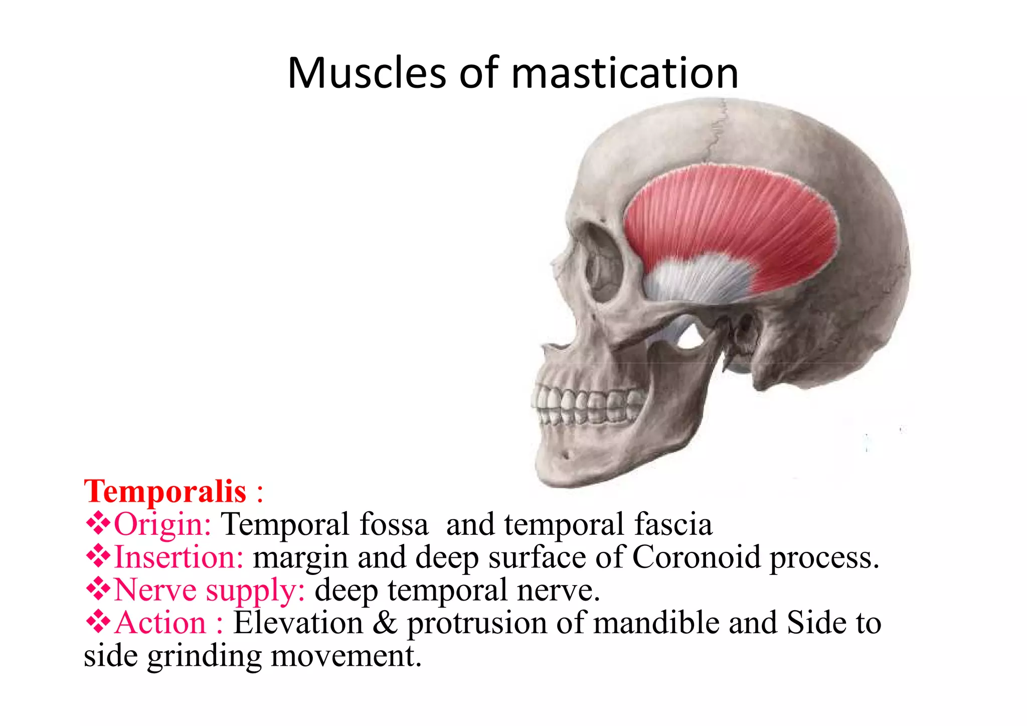
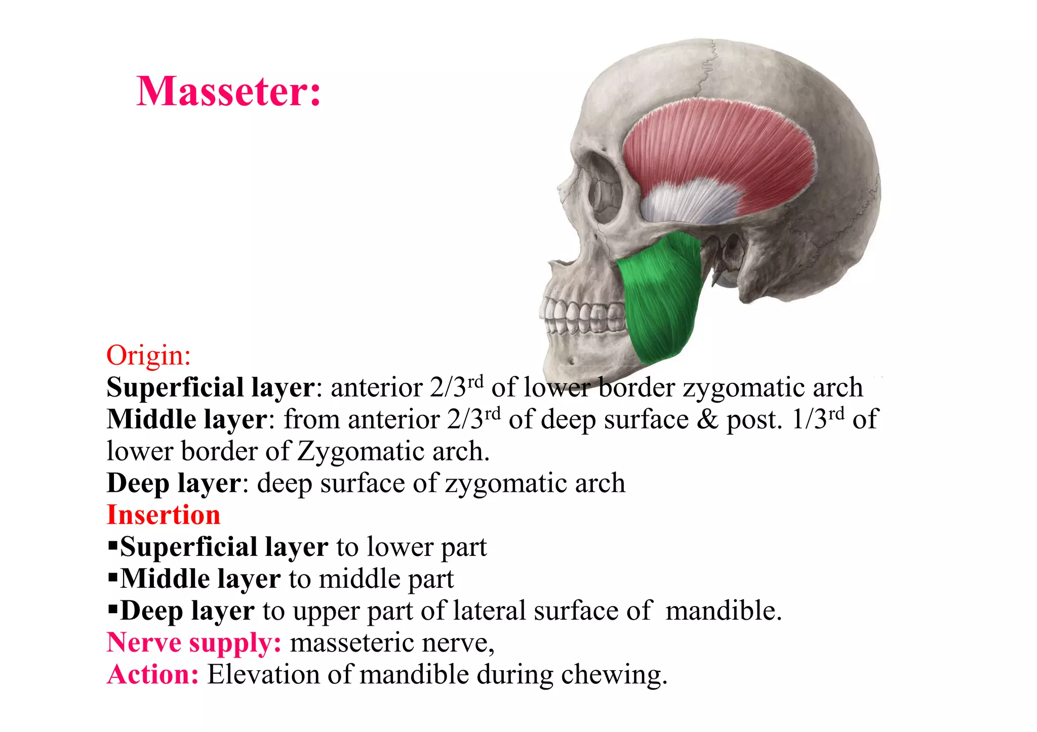
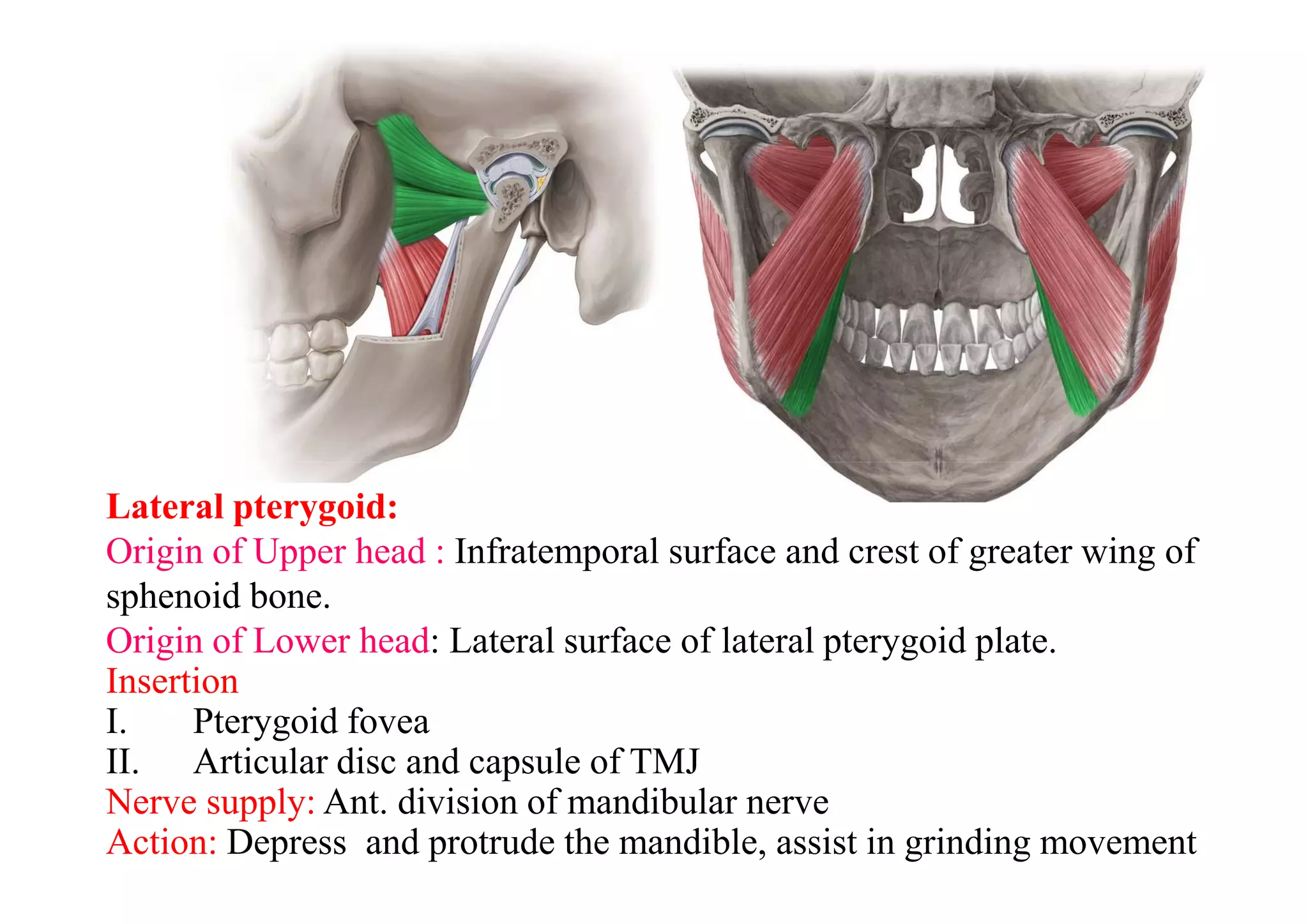
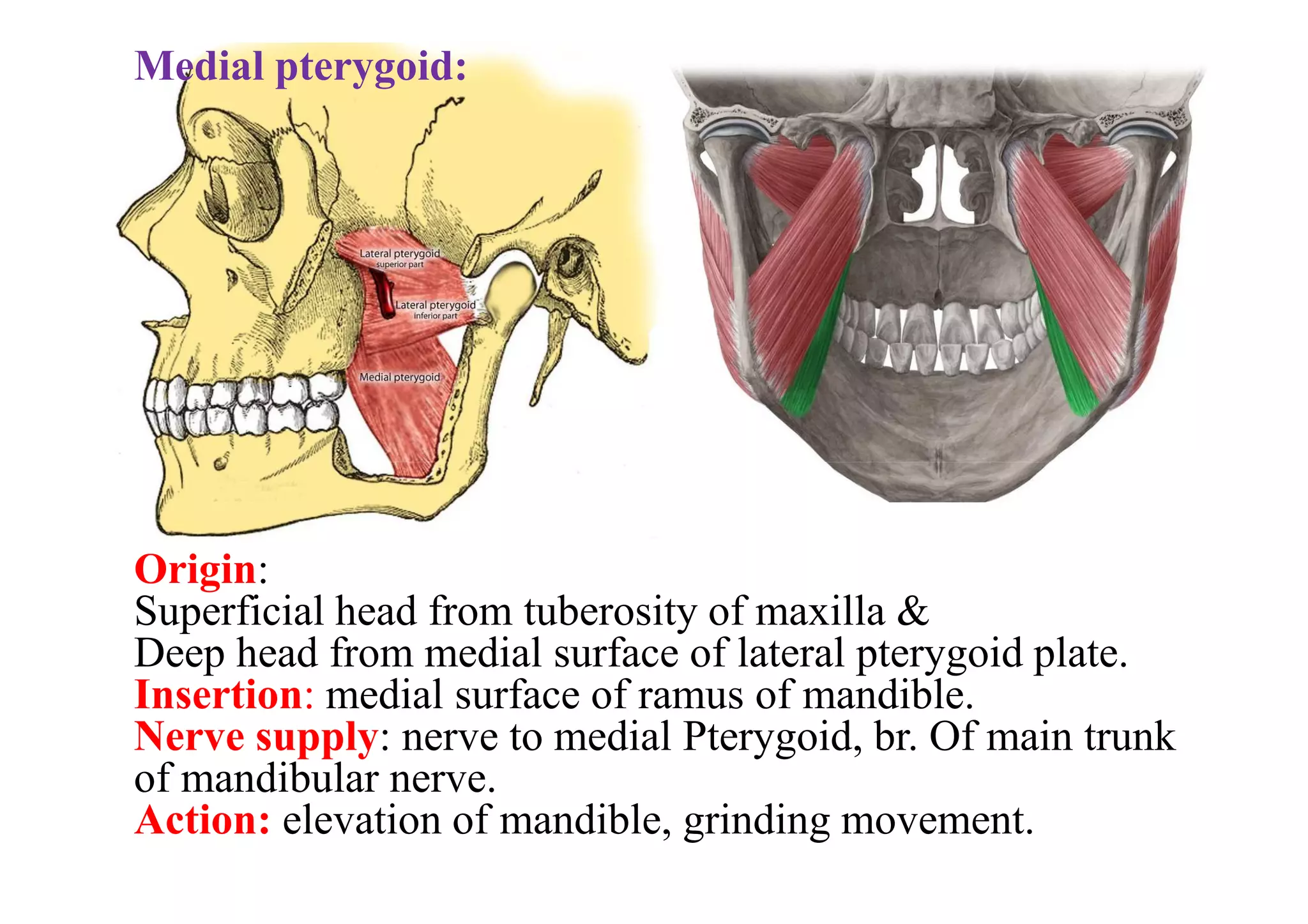
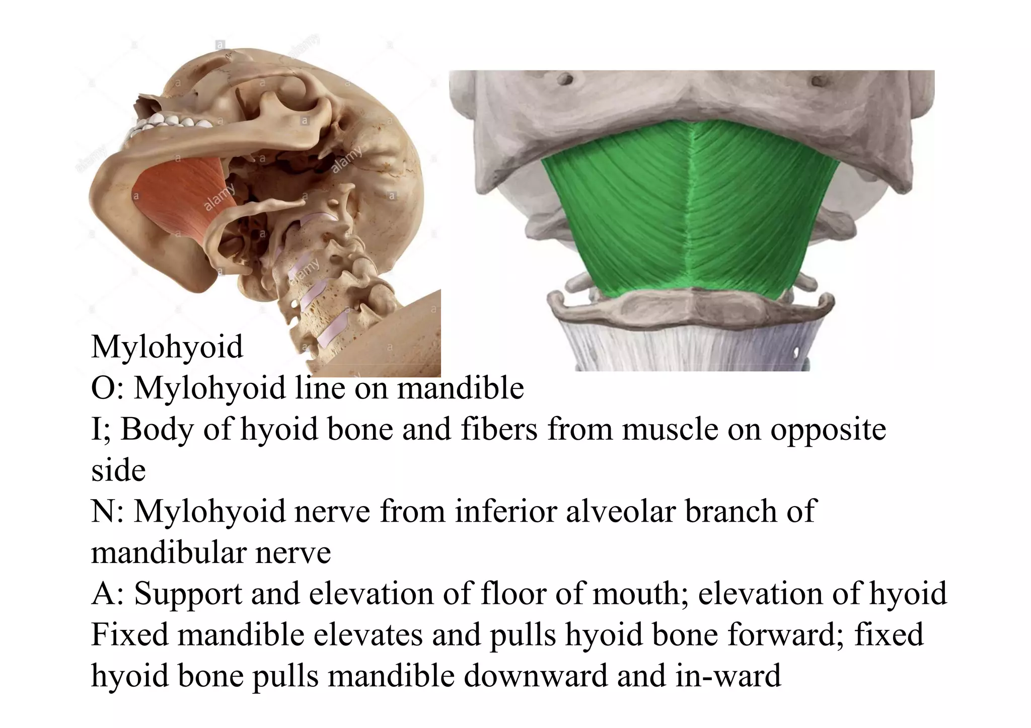
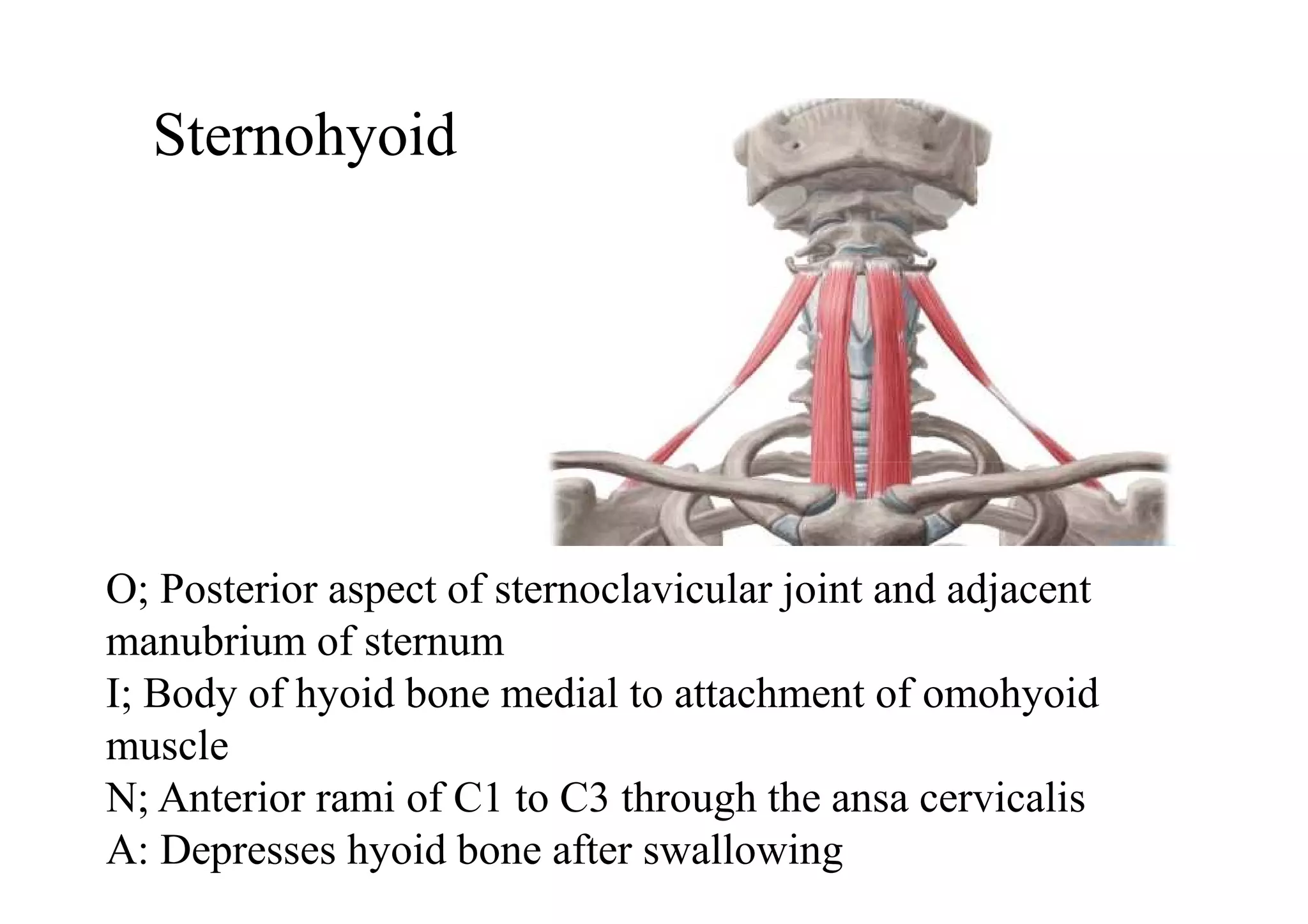
This document summarizes several muscles of the head and neck region. It describes the origin, insertion, nerve supply and action of the temporalis, masseter, lateral pterygoid, medial pterygoid, mylohyoid, geniohyoid, sternohyoid, omohyoid, thyrohyoid, sternothyroid, trapezius, and sternocleidomastoid muscles. It also provides information on torticollis, listing four types: rheumatic, reflex, congenital, and spasmodic.
Chapter 12 Muscular systemMuscles of Head and neck1. Scalene.docxbartholomeocoombs
Chapter 12 Muscular system
Muscles of Head and neck
1. Scalene muscles
The scalene muscles are a group of three pairs of muscles in the lateral neck:
scalenus anterior, scalenus medius, and scalenus posterior
The scalene muscles are related to the thoracic outlet syndrome
i. Scalene anterior:
Origin: transverse processes of C3 – C6
Insertion: scalene tubercle of 1st rib
Nerve supply: anterior rami of C6 – C8
Action: raises the first rib and bends neck to one side
ii. Scalene Medius
Origin: transverse processes of C1 – C6
Insertion: upper surface of 1st and 2nd ribs
Nerve supply: anterior rami of C3 – C8
Action: raises the first rib and bends neck to one side
iii. Scalene Posterior
Origin: transverse processes of C5 – C7
Insertion: upper surface of 2nd rib
Nerve supply: anterior rami of C7 – C8
Action: raises the second rib and bends neck to one side
2. Sternocleidomastoid/ Sternomastoid
It is given the name sternocleidomastoid because it originates from the sternum (sterno) and clavicle (cleido), and inserts into the mastoid process of the temporal bone of the skull. It is also called the sternomastoid muscle.
Origin: manubrium sterni + medial portion of the clavicle
Insertion: mastoid process of the temporal bone and superior nuchal line
Nerve Supply:
motor: accessory nerve
sensory: cervical plexus
Action:
Acting alone it tilts head to its own side and rotates it so the face is turned towards the opposite side.
Acting together, flexes the neck, raises the sternum and assists in forced inspiration.
3. Infrahyoid or “strap” muscles
Strap-like muscles occupy each side of the midline of the neck from the mandible to the manubrium sterni.
Between these 2 points lie the hyoid bone, and below it is the Adam’s apple or thyroid cartilage; to them certain muscles gain attachment.
They include:
Sternohyoid m., Sternothyroid m., Thyro-hyoid m. and Omohyoid muscles.
These four muscles, also known as the "strap" muscles, function to depress the hyoid bone and larynx during swallowing and speaking (except for Thyrohyoid).
i. Sternohyoid
Origin: Posterior surface of manubrium sterni, adjoining parts of clavicle and the posterior sternoclavicular ligament
Insertion: Medial part of lower border of hyoid bone
Nerve supply: Ansa cervicalis
Action: Depresses/ stabilizes hyoid bone
ii. Sternothyroid
Origin: Posterior surface of manubrium sterni and adjoining part of first costal cartilage
Insertion: Oblique line of thyroid cartilage
Nerve supply: Ansa cervicalis
Action: Depresses/ stabilizes thyroid cartilage
iii. Thyrohyoid
Origin: Oblique line of the thyroid cartilage
Insertion: Lower border of the body and the greater cornu of the hyoid bone
Nerve supply: Cervical spinal nerve 1 via the hypoglossal nerve
Action: Elevates larynx + Depresses/ stabilizes hyoid bone
iv. Omohyoid (superior belly)
Origin: Intermediate tendon
Insertion: Hyoid bone
Nerve supply: Ansa cervicalis
Action: Depresses/ stabilizes hyoid bone
v. Omohyoid (inferior belly)
Origin: .
Article titleAuthorsSource (original publication and digest).docxrossskuddershamus
Article title:
Authors:
Source (original publication and digest):
Why did you pick this article:
What was the primary finding and why is this important:
Why was this research done:
What future work or findings might come from this work:
Chapter 12 Muscular system
Muscles of Head and neck
1. Scalene muscles
The scalene muscles are a group of three pairs of muscles in the lateral neck:
scalenus anterior, scalenus medius, and scalenus posterior
The scalene muscles are related to the thoracic outlet syndrome
i. Scalene anterior:
Origin: transverse processes of C3 – C6
Insertion: scalene tubercle of 1st rib
Nerve supply: anterior rami of C6 – C8
Action: raises the first rib and bends neck to one side
ii. Scalene Medius
Origin: transverse processes of C1 – C6
Insertion: upper surface of 1st and 2nd ribs
Nerve supply: anterior rami of C3 – C8
Action: raises the first rib and bends neck to one side
iii. Scalene Posterior
Origin: transverse processes of C5 – C7
Insertion: upper surface of 2nd rib
Nerve supply: anterior rami of C7 – C8
Action: raises the second rib and bends neck to one side
2. Sternocleidomastoid/ Sternomastoid
It is given the name sternocleidomastoid because it originates from the sternum (sterno) and clavicle (cleido), and inserts into the mastoid process of the temporal bone of the skull. It is also called the sternomastoid muscle.
Origin: manubrium sterni + medial portion of the clavicle
Insertion: mastoid process of the temporal bone and superior nuchal line
Nerve Supply:
motor: accessory nerve
sensory: cervical plexus
Action:
Acting alone it tilts head to its own side and rotates it so the face is turned towards the opposite side.
Acting together, flexes the neck, raises the sternum and assists in forced inspiration.
3. Infrahyoid or “strap” muscles
Strap-like muscles occupy each side of the midline of the neck from the mandible to the manubrium sterni.
Between these 2 points lie the hyoid bone, and below it is the Adam’s apple or thyroid cartilage; to them certain muscles gain attachment.
They include:
Sternohyoid m., Sternothyroid m., Thyro-hyoid m. and Omohyoid muscles.
These four muscles, also known as the "strap" muscles, function to depress the hyoid bone and larynx during swallowing and speaking (except for Thyrohyoid).
i. Sternohyoid
Origin: Posterior surface of manubrium sterni, adjoining parts of clavicle and the posterior sternoclavicular ligament
Insertion: Medial part of lower border of hyoid bone
Nerve supply: Ansa cervicalis
Action: Depresses/ stabilizes hyoid bone
ii. Sternothyroid
Origin: Posterior surface of manubrium sterni and adjoining part of first costal cartilage
Insertion: Oblique line of thyroid cartilage
Nerve supply: Ansa cervicalis
Action: Depresses/ stabilizes thyroid cartilage
iii. Thyrohyoid
Origin: Oblique line of the thyroid cartilage
Insertion: Lower border of the body and the greater cornu of the hyoid bone
Nerve supply: Cervical spinal nerve 1 via the hypoglossal nerve
Action.
Temporalis: Located on the side of the head, it helps in closing the jaw.
Masseter: Located at the angle of the jaw, it also aids in closing the jaw.
Frontalis: Covers the forehead and raises the eyebrows.
Orbicularis Oculi: Surrounds the eye and closes the eyelids.
Orbicularis Oris: Encircles the mouth and aids in closing and puckering the lips.
Buccinator: Located in the cheek area, it compresses the cheeks during actions like blowing.
Platysma: Thin, sheet-like muscle covering parts of the neck and jaw, it helps in facial expressions and neck movements.
Sternocleidomastoid: Neck muscle that aids in turning the head and flexing the neck.
Temporalis:
Location: Temporal region of the skull.
Action: Elevates and retracts the mandible (jaw), assisting in closing the jaw during chewing.
Masseter:
Location: Runs from the zygomatic arch to the mandible.
Action: Primary muscle for closing the jaw (elevation), especially during biting and chewing.
Frontalis:
Location: Covers the forehead.
Action: Raises the eyebrows and wrinkles the forehead horizontally.
Orbicularis Oculi:
Location: Surrounds the eye.
Action: Closes the eyelids, protects the eye from light and foreign objects.
Orbicularis Oris:
Location: Encircles the mouth.
Action: Closes and purses the lips, helps in speech and facial expressions, and maintains oral competence (sealing the mouth).
Buccinator:
Location: Located in the cheek area, between the maxilla and mandible.
Action: Compresses the cheeks against the teeth, aids in sucking, blowing, and chewing movements.
Platysma:
Location: Thin, broad muscle covering parts of the neck and jaw.
Action: Depresses the lower jaw and lip, tenses the skin of the neck (e.g., during expressions of tension or fear).
Sternocleidomastoid:
Location: Runs from the sternum and clavicle to the mastoid process of the temporal bone.
Action: Flexes and laterally rotates the head, and flexes the neck. It also acts as an accessory muscle for breathing when the neck is fixed.
We all have good and bad thoughts from time to time and situation to situation. We are bombarded daily with spiraling thoughts(both negative and positive) creating all-consuming feel , making us difficult to manage with associated suffering. Good thoughts are like our Mob Signal (Positive thought) amidst noise(negative thought) in the atmosphere. Negative thoughts like noise outweigh positive thoughts. These thoughts often create unwanted confusion, trouble, stress and frustration in our mind as well as chaos in our physical world. Negative thoughts are also known as “distorted thinking”.
Welcome to TechSoup New Member Orientation and Q&A (May 2024).pdfTechSoup
In this webinar you will learn how your organization can access TechSoup's wide variety of product discount and donation programs. From hardware to software, we'll give you a tour of the tools available to help your nonprofit with productivity, collaboration, financial management, donor tracking, security, and more.
2024.06.01 Introducing a competency framework for languag learning materials ...Sandy Millin
http://sandymillin.wordpress.com/iateflwebinar2024
Published classroom materials form the basis of syllabuses, drive teacher professional development, and have a potentially huge influence on learners, teachers and education systems. All teachers also create their own materials, whether a few sentences on a blackboard, a highly-structured fully-realised online course, or anything in between. Despite this, the knowledge and skills needed to create effective language learning materials are rarely part of teacher training, and are mostly learnt by trial and error.
Knowledge and skills frameworks, generally called competency frameworks, for ELT teachers, trainers and managers have existed for a few years now. However, until I created one for my MA dissertation, there wasn’t one drawing together what we need to know and do to be able to effectively produce language learning materials.
This webinar will introduce you to my framework, highlighting the key competencies I identified from my research. It will also show how anybody involved in language teaching (any language, not just English!), teacher training, managing schools or developing language learning materials can benefit from using the framework.
Students, digital devices and success - Andreas Schleicher - 27 May 2024..pptxEduSkills OECD
Andreas Schleicher presents at the OECD webinar ‘Digital devices in schools: detrimental distraction or secret to success?’ on 27 May 2024. The presentation was based on findings from PISA 2022 results and the webinar helped launch the PISA in Focus ‘Managing screen time: How to protect and equip students against distraction’ https://www.oecd-ilibrary.org/education/managing-screen-time_7c225af4-en and the OECD Education Policy Perspective ‘Students, digital devices and success’ can be found here - https://oe.cd/il/5yV
Palestine last event orientationfvgnh .pptxRaedMohamed3
An EFL lesson about the current events in Palestine. It is intended to be for intermediate students who wish to increase their listening skills through a short lesson in power point.
2. Muscles of mastication
Temporalis :
Origin: Temporal fossa and temporal fascia
Insertion: margin and deep surface of Coronoid process.
Nerve supply: deep temporal nerve.
Action : Elevation & protrusion of mandible and Side to
side grinding movement.
3. Origin:
Masseter:
Origin:
Superficial layer: anterior 2/3rd of lower border zygomatic arch
Middle layer: from anterior 2/3rd of deep surface & post. 1/3rd of
lower border of Zygomatic arch.
Deep layer: deep surface of zygomatic arch
Insertion
Superficial layer to lower part
Middle layer to middle part
Deep layer to upper part of lateral surface of mandible.
Nerve supply: masseteric nerve,
Action: Elevation of mandible during chewing.
4. Lateral pterygoid:
Origin of Upper head : Infratemporal surface and crest of greater wing of
sphenoid bone.
Origin of Lower head: Lateral surface of lateral pterygoid plate.
Insertion
I. Pterygoid fovea
II. Articular disc and capsule of TMJ
Nerve supply: Ant. division of mandibular nerve
Action: Depress and protrude the mandible, assist in grinding movement
5. Medial pterygoid:
Origin:
Superficial head from tuberosity of maxilla &
Deep head from medial surface of lateral pterygoid plate.
Insertion: medial surface of ramus of mandible.
Nerve supply: nerve to medial Pterygoid, br. Of main trunk
of mandibular nerve.
Action: elevation of mandible, grinding movement.
6. MylohyoidMylohyoid
O: Mylohyoid line on mandible
I; Body of hyoid bone and fibers from muscle on opposite
side
N: Mylohyoid nerve from inferior alveolar branch of
mandibular nerve
A: Support and elevation of floor of mouth; elevation of hyoid
Fixed mandible elevates and pulls hyoid bone forward; fixed
hyoid bone pulls mandible downward and in-ward
7. Geniohyoid
O: Inferior mental spine on inner surface of mandible
I; Anterior surface of body of hyoid bone
N: Branch from anterior ramus of C1 (carried along the hypoglossal
nerve)
A; Fixed mandible elevates and pulls hyoid bone forward; fixed hyoid
bone pulls mandible downward and in-ward
8. Sternohyoid
O; Posterior aspect of sternoclavicular joint and adjacent
manubrium of sternum
I; Body of hyoid bone medial to attachment of omohyoid
muscle
N; Anterior rami of C1 to C3 through the ansa cervicalis
A: Depresses hyoid bone after swallowing
9. Omohyoid
O; Superior border of scapula medial to suprascapular notch
I; Lower border of body of hyoid bone just lateral to
attachment of sternohyoid
N; Anterior rami of C1 to C3 through the ansa cervicalis
A; Depresses and fixes hyoid bone
10.
11. Thyrohyoid
O; Oblique line on lamina of thyroid cartilage
I; Greater horn and adjacent aspect of body of hyoid bone
N; Fibers from anterior ramus of C1 carried along hypoglossal nerve
A; Depresses hyoid bone, but when hyoid bone is fixed raises larynx
12. Sternothyroid
O; Posterior surface of manubrium of sternum
I; Oblique line on lamina of thyroid cartilage
N: Anterior rami of C1 to C3 through the ansa cervicalis
A; Draws larynx (thyroid cartilage) downward
13. Trapezius
O ; Superior nuchal line; external occipital protuberance; ligamentum
nuchae; spinous processes of vertebrae CVII to TXII
I: Lateral one-third of clavicle; acromion; spine of scapula
N; Motor-accessory nerve [XI]; proprioception-C3 and C4
A: Assists in rotating the scapula during abduction of humerus above
horizontal; upper fibers-elevate, middle fibers-adduct, lower fibers-
depress scapula
14. Sternocleidomastoid muscle
O; Upper part of anterior surface of manubrium of sternum
I; Lateral one-half of superior nuchal line
O; Superior surface of medial one-third of clavicle
I : Lateral surface of mastoid process
N : Accessory nerve [XI] and branches from anterior rami of C2 to C3 (C4)
A: Individually-will tilt head to-wards shoulder on same side rotating head to turn face
to opposite side; acting to-gether, draw head forwards
15.
16. Torticollis or wry neck- deformity in which the head bent to one
side and the chin points to the other side
1. Rheumatic torticollis- due to exposure to cold or draught
2. Reflex torticollis- due to inflamed or suppurating cervical
lymph nodes which irritate the spinal accessory nerve
3. Congenital torticollis- due to birth injury
4. Spasmodic torticollis- due to central irritation on CN-XI.