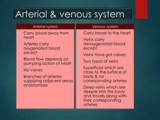
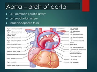
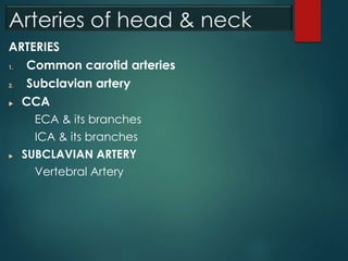
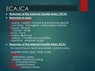
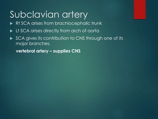
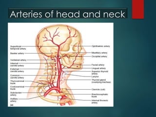
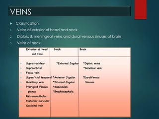
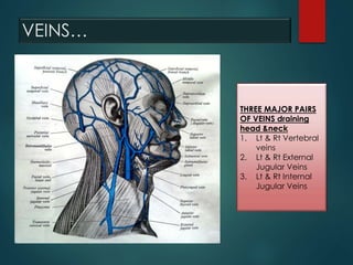
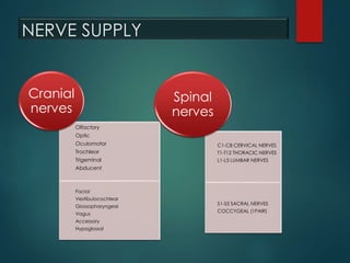
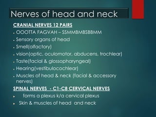
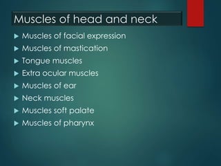
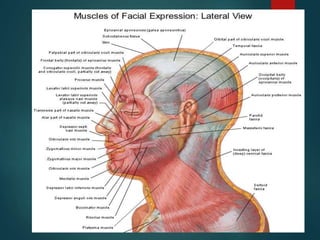
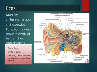
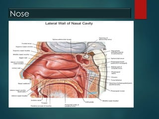
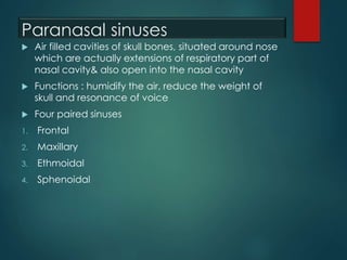
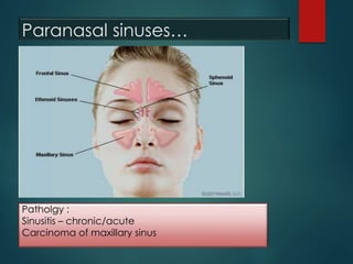
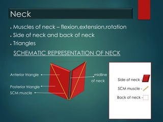
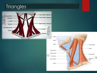
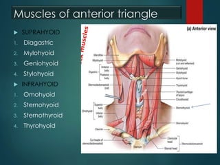
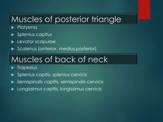
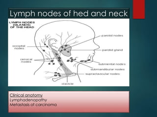
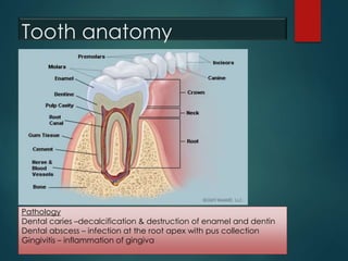
The document provides an overview of human anatomy related to the head and neck, detailing blood vessels, nerves, muscles, and various glands in the area. It describes the arterial and venous systems, nerve supply, muscle functions, and pathology associated with different structures, including the eyes, ears, and mouth. Additionally, it touches on the clinical anatomy of glands and teeth, highlighting common diseases and conditions affecting these regions.
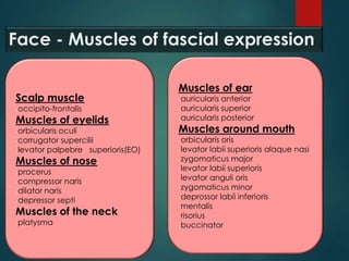


![Muscles of head_&_neck[1]](https://cdn.slidesharecdn.com/ss_thumbnails/musclesofheadneck1-170504175033-thumbnail.jpg?width=640&height=640&fit=bounds)


