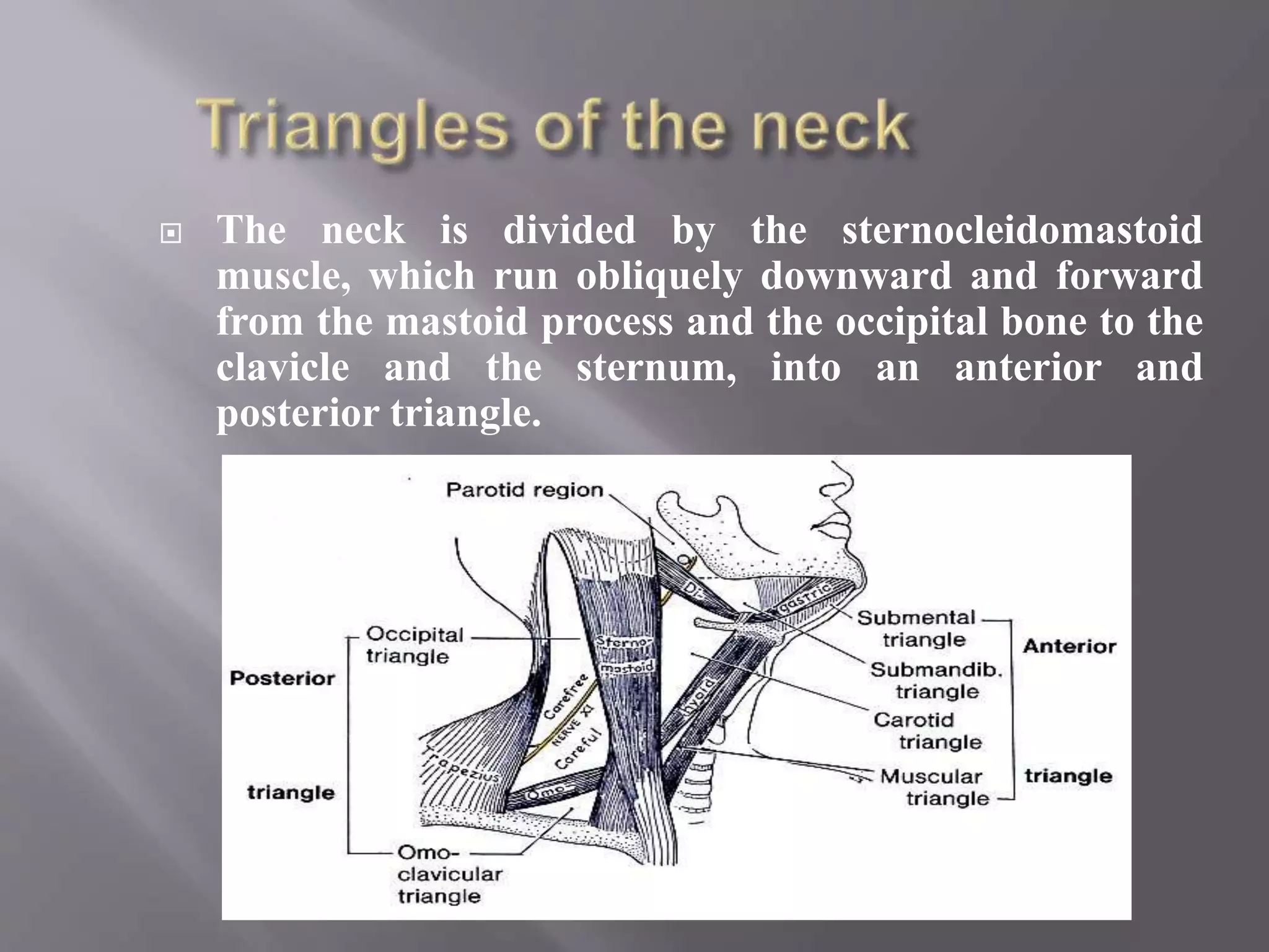
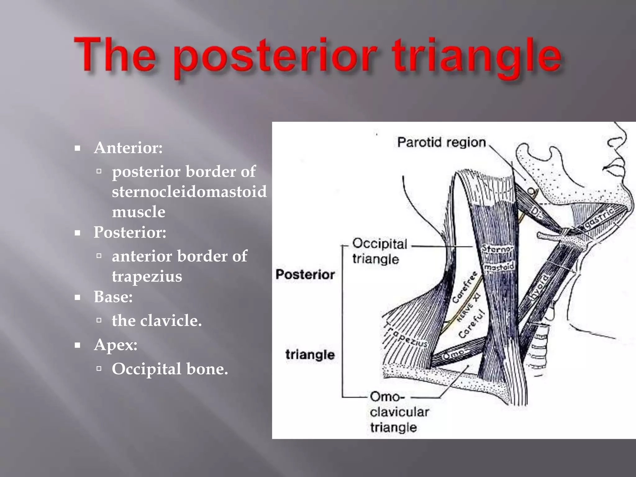
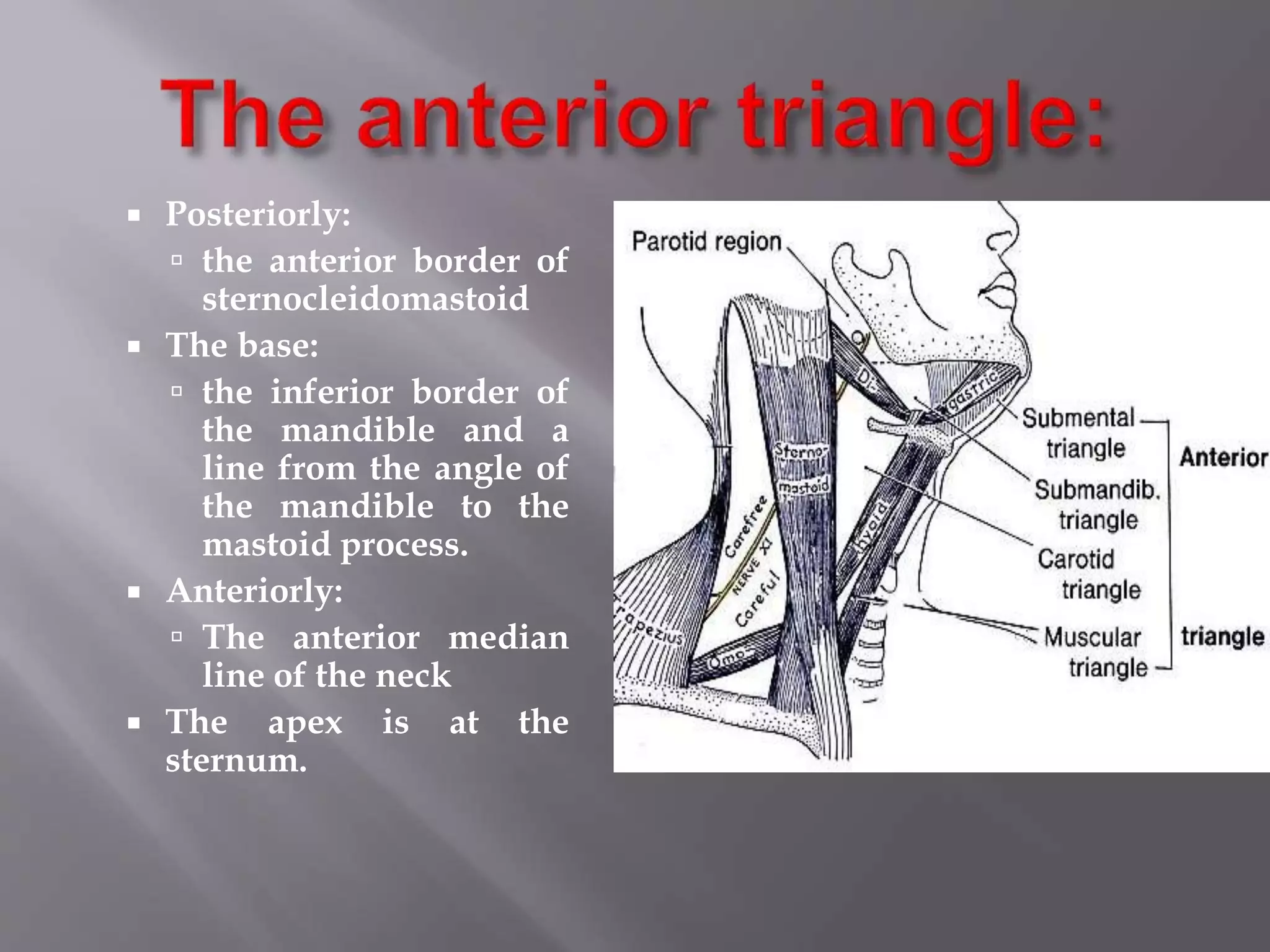
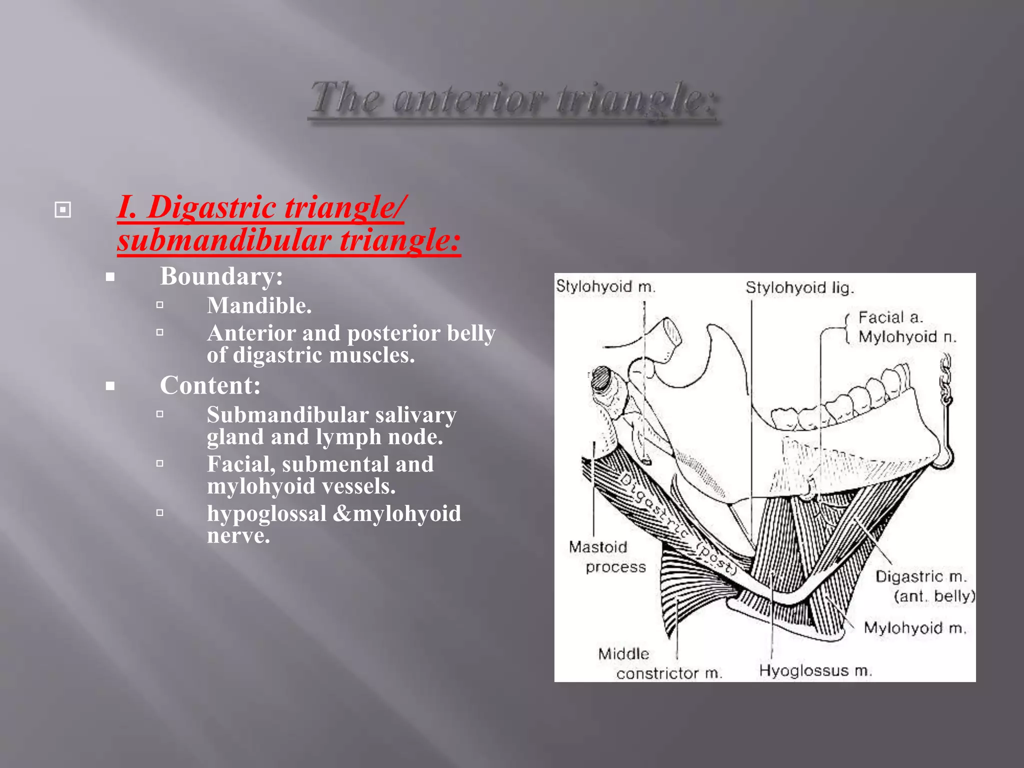
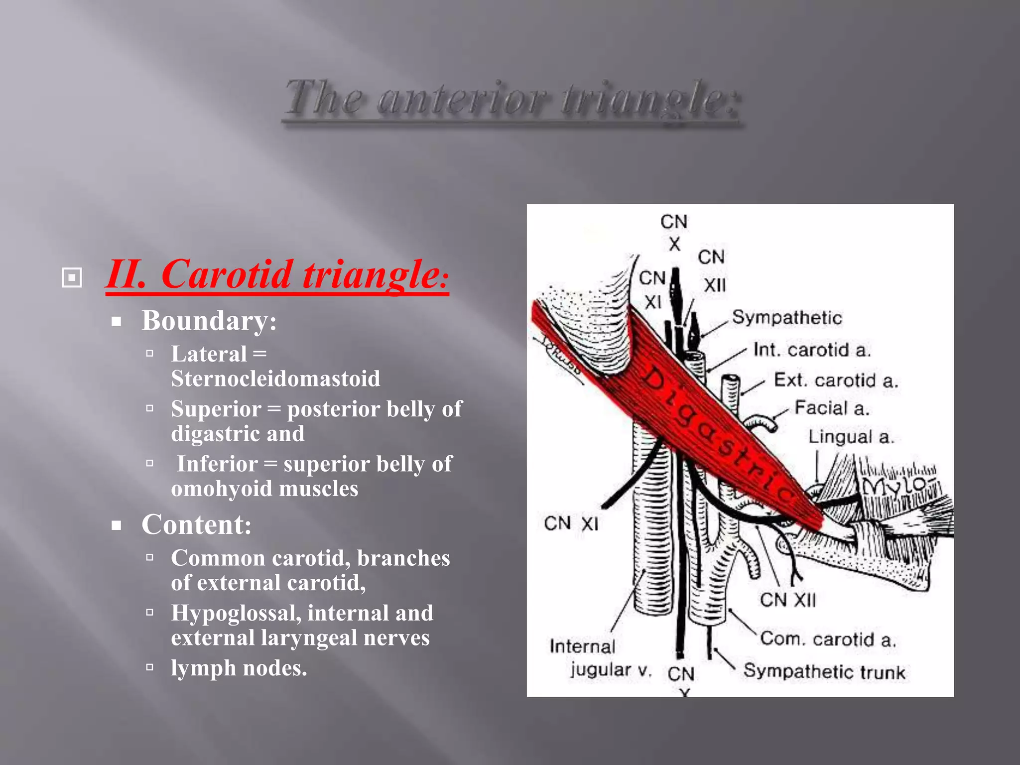
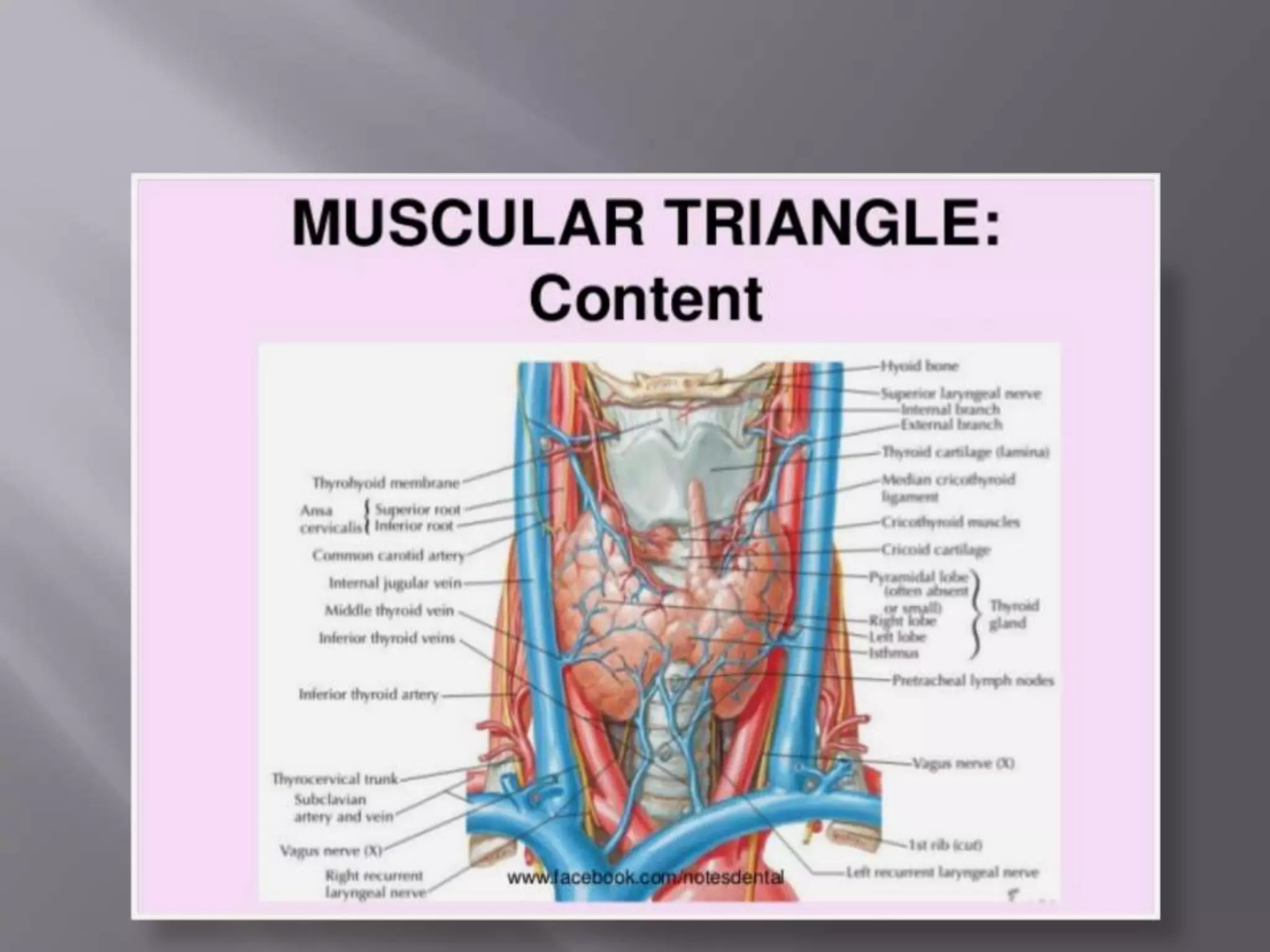
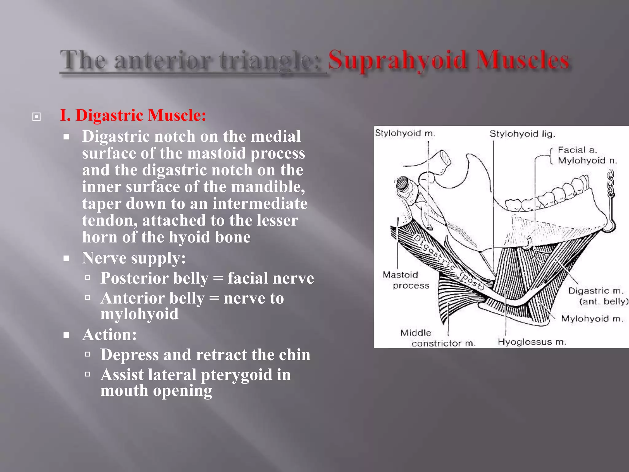
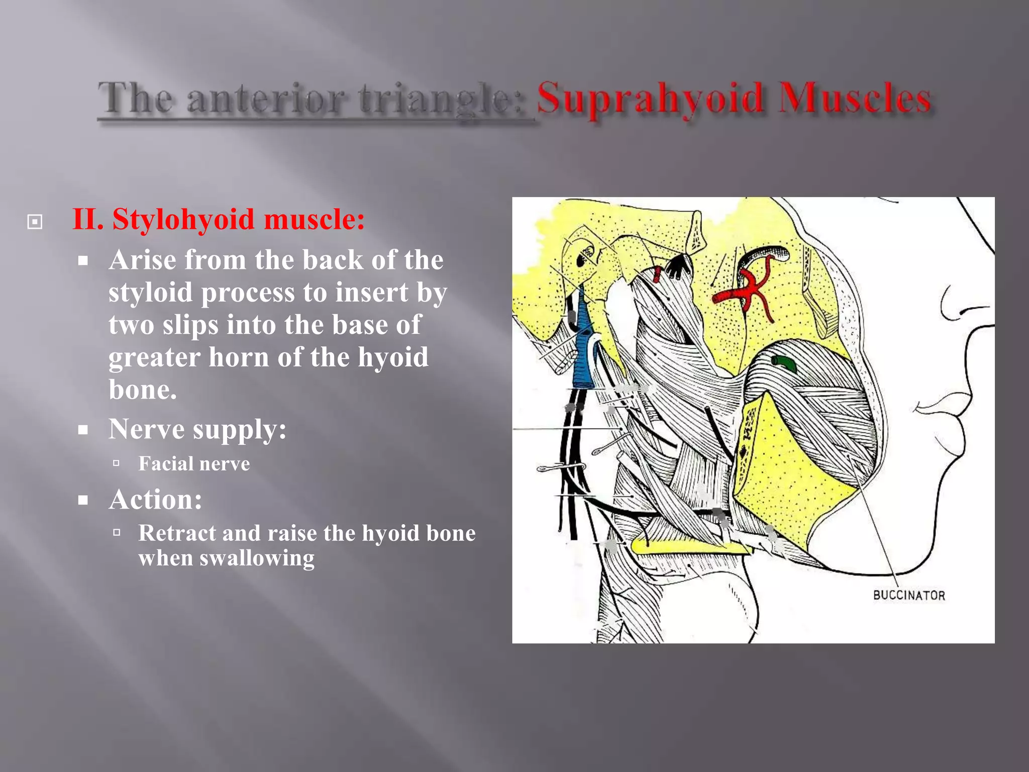
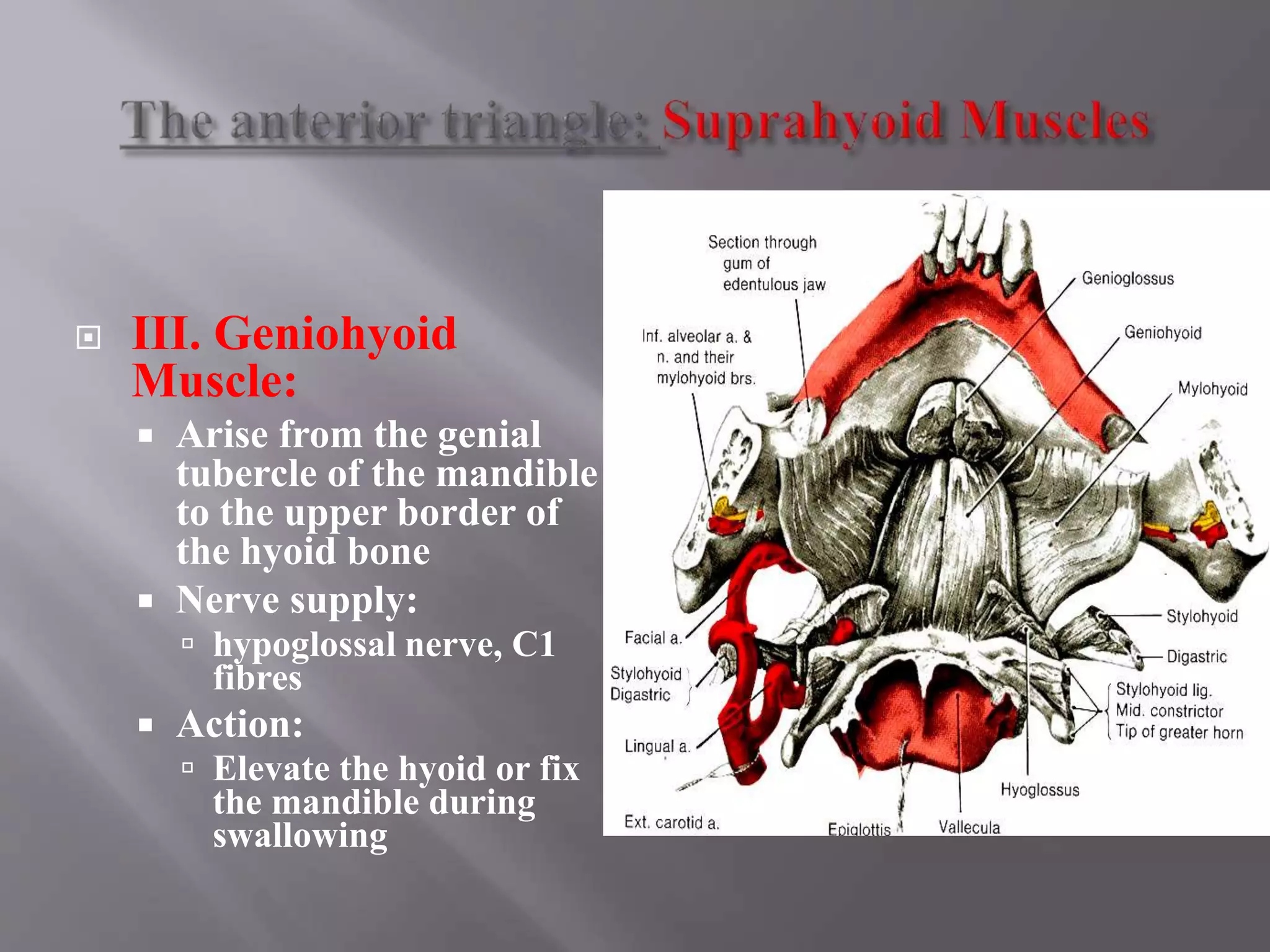
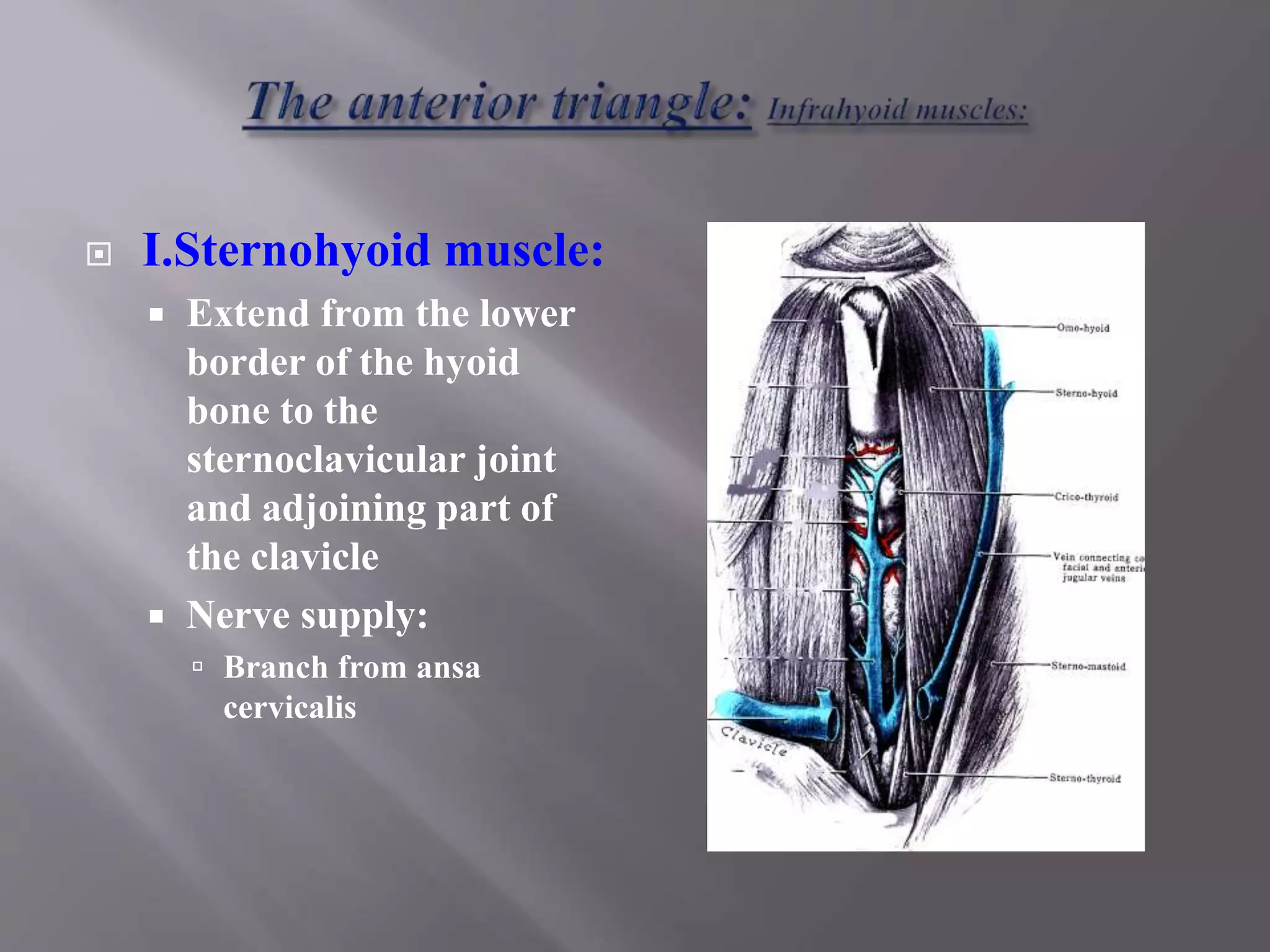
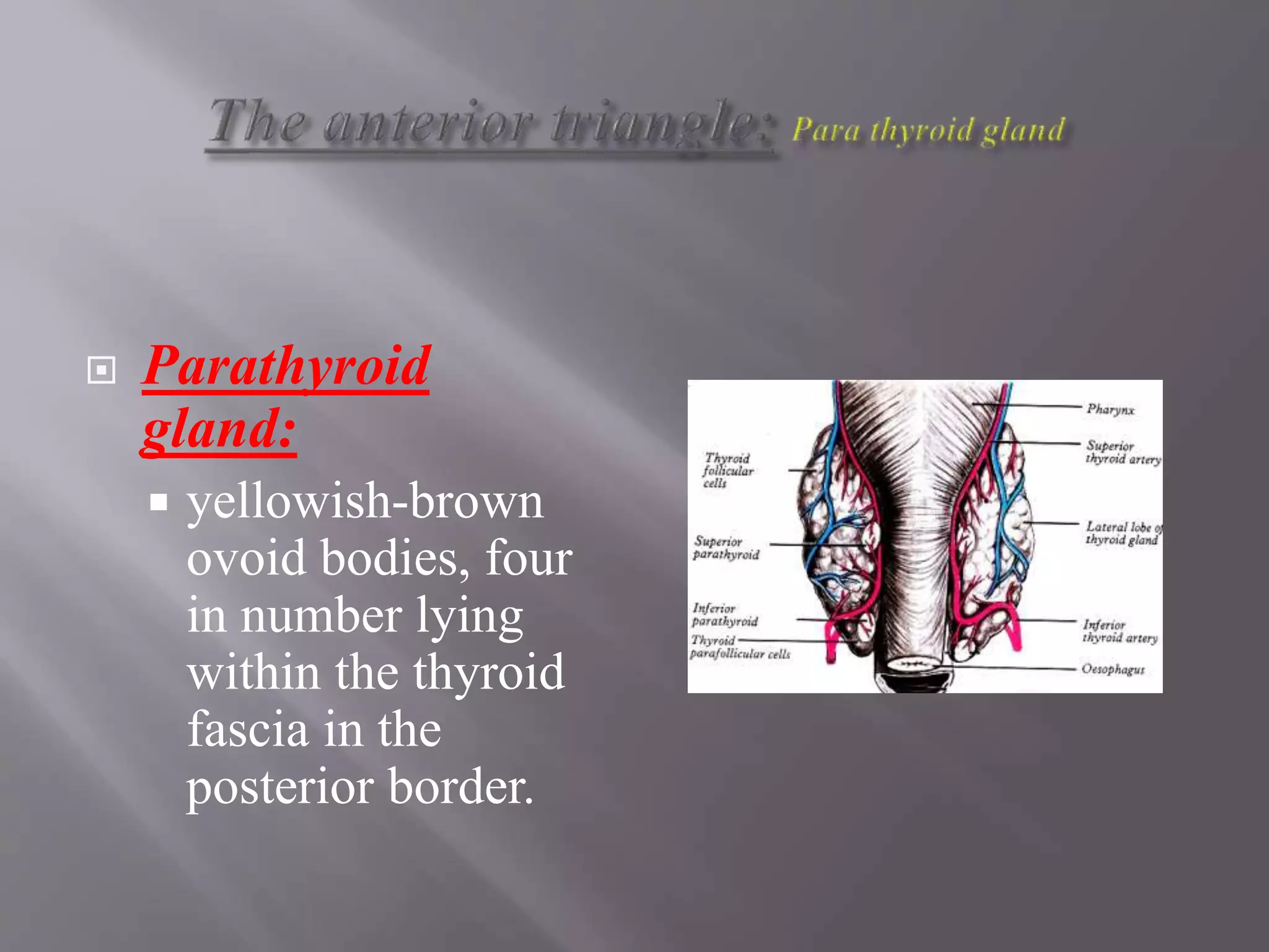
Anatomy of Neck Triangles
Dr. Prabhu Dayal Sinwar
Assistant Professor
 The neck is divided by the sternocleidomastoid
muscle, which run obliquely downward and forward
from the mastoid process and the occipital bone to the
clavicle and the sternum, into an anterior and
posterior triangle.
 Anterior:
 posterior border of
sternocleidomastoid
muscle
 Posterior:
 anterior border of
trapezius
 Base:
 the clavicle.
 Apex:
 Occipital bone.
 The triangle is subdivided by the
inferior belly of the omohyoid
muscle into an I. occipital and II.
supraclavicular triangles.
 The roof:
 the skin and deep cervical
fascia.
 The floor:
 the prevertebral fascia
 The content:
 Occipital lymph nodes,
accessory nerve, greater
auricular nerve and the
cervical plexus.
 Posteriorly:
 the anterior border of
sternocleidomastoid
 The base:
 the inferior border of
the mandible and a
line from the angle of
the mandible to the
mastoid process.
 Anteriorly:
 The anterior median
line of the neck
 The apex is at the
sternum.
 It is covered by the skin, superficial fascia, platysma and investing layer of
deep fascia.
 Under the fascia, two groups of longitudinal muscles(supra and infra hyoid)
extend from the inferior border of the mandible to the sternum.
 The hyoid bone, at the level of C3 vertebra, provide an attachment for the
two groups.
 The muscles are called the strap muscles from their flat shape and they are
in the same plane as the body wall musculature, rectus abdominis
 I. Digastric triangle/
submandibular triangle:
 Boundary:
 Mandible.
 Anterior and posterior belly
of digastric muscles.
 Content:
 Submandibular salivary
gland and lymph node.
 Facial, submental and
mylohyoid vessels.
 hypoglossal &mylohyoid
nerve.
 II. Carotid triangle:
 Boundary:
 Lateral =
Sternocleidomastoid
 Superior = posterior belly of
digastric and
 Inferior = superior belly of
omohyoid muscles
 Content:
 Common carotid, branches
of external carotid,
 Hypoglossal, internal and
external laryngeal nerves
 lymph nodes.
 III. Submental triangle:
 Boundary:
 Posterior = Anterior belly
of digastric
 Anterior = Midline of neck
 Inferior = Body of hyoid
bone
 Floor = Mylohyoid
 Content:
 Anterior jugular vein and
lymph nodes
 IV. Muscular triangle/Inferior carotid triangle:
 Boundary
 Lateral = Sternocleidomastoid,
 Above = superior belly of omohyoid,
 Medial = midline from hyoid bone to sternum
 Content:
 Larynx, thyroid gland and lymph nodes
 The supra hyoid
muscles:
 Above the hyoid bone,
are four in number
 Digastric
 Stylohyoid, superficial
 The mylohyoid
 The geniohyoid deep to
it.
 The infra hyoid
muscles:
 Below the hyoid bone, are
four in number:
 Sternohyoid
 Omohyoid, superficial
 Thyrohyoid
 Sternothyroid deep to them
 I. Digastric Muscle:
 Digastric notch on the medial
surface of the mastoid process
and the digastric notch on the
inner surface of the mandible,
taper down to an intermediate
tendon, attached to the lesser
horn of the hyoid bone
 Nerve supply:
 Posterior belly = facial nerve
 Anterior belly = nerve to
mylohyoid
 Action:
 Depress and retract the chin
 Assist lateral pterygoid in
mouth opening
 II. Stylohyoid muscle:
 Arise from the back of the
styloid process to insert by
two slips into the base of
greater horn of the hyoid
bone.
 Nerve supply:
 Facial nerve
 Action:
 Retract and raise the hyoid bone
when swallowing
 III. Geniohyoid
Muscle:
 Arise from the genial
tubercle of the mandible
to the upper border of
the hyoid bone
 Nerve supply:
 hypoglossal nerve, C1
fibres
 Action:
 Elevate the hyoid or fix
the mandible during
swallowing
 IV. Mylohyoid muscle:
 Arises from the mylohyoid line on the inner surface of the
mandible as far as the posterior surface of last molar
 Slope toward each other, and the posterior quarter is inserted
into the anterior surface of the body of the hyoid bone.
 The anterior two third interdigitate in a midline raphe
extending from the chin to hyoid bone.
 Nerve supply:
 Nerve to mylohyoid
 Action:
 Form a mobile but
stable floor
 Support the weight and
thrust of the tongue
 The two muscles form a
shallow gutter elevating
the tongue and hyoid
bone during swallowing.
 I.Sternohyoid muscle:
 Extend from the lower
border of the hyoid
bone to the
sternoclavicular joint
and adjoining part of
the clavicle
 Nerve supply:
 Branch from ansa
cervicalis
 II.Omohyoid:
 Lie edge to edge with the
sternohyoid on the lateral part
of the inferior border of the
hyoid bone
 It diverge and pass beneath the
sternocleidomastoid over the
carotid sheath, replaced by
fibrous tendon
 Inferior belly pass horizontally
back to be inserted in the
transverse scapular ligament
and upper border of the
scapula.
 Nerve supply:
 Ansa cervicalis
 III.Thyrohyoid:
 Broad muscle, arise from
the greater horn of the
hyoid, undercover of the
other muscles and is
inserted into the oblique
line of the thyroid
cartilage
 Nerve supply:
 Hypoglossal nerve, C1
fibres.
 IV. Sternothyroid:
 Extend from the oblique line
of the thyroid cartilage to
posterior border of the
manubrium and the first
intercostal cartilage
 Nerve supply: Ansa
cervicalis (C2,3)
 Action of infrahyoid muscles
 Depressor of the larynx
 Thyroid gland:
 consist of two
symmetrical lobes
united, in front of the of
the 2nd - 4th tracheal
rings, by an isthmus of
gland tissue.
 Covered by a layer of
pretracheal fascia.
 Pear-shaped with
narrow upper pole and
wide lower pole, with
lateral, medial and
posterior surfaces
 Development:
 Develop as an
endodermal
outgrowth from the
midline of the
pharynx, between the
tuberculum impar,
becoming the thyro-
glossal duct
 The duct elongate
and distal end
become bilobed.
 A solid cord
migrates anterior or
posterior to the hyoid
bone to the larynx
and the trachea
 The solid cord break
up and disappear but
the site of origin
remain as a pit on the
tongue “foramen
cecum” at the junction
of the posterior third
with anterior two-
third.
 Clinically:
 Lingual thyroid:
 Part of the gland remain as a swelling at the
tongue base.
 Thyroglossal cyst:
 Above or below the hyoid bone.
 Mobile with swallowing.
 Thyroid goitre:
 Exert pressure on the structures behind it, trachea
and recurrent laryngeal nerve.
 Parathyroid
gland:
 yellowish-brown
ovoid bodies, four
in number lying
within the thyroid
fascia in the
posterior border.
 Common carotid artery:
 In the right side, the artery arise from the brachio-cephalic
artery and the left, from the arch of the aorta
 It divides into the external and internal carotid artery at
the upper border of the thyroid cartilage
 The carotid sinus is a small dilatation at the bifurcation,
contain numerous nerve ending from glossopharyngeal
nerve and serve as a reflex pressoreceptor
 The carotid body is a small reddish structure,posterior to
the bifurcation and is a chemoreceptor.
Anatomy of neck triangles

Anatomy of neck triangles

  • 1.
    Anatomy of NeckTriangles Dr. Prabhu Dayal Sinwar Assistant Professor
  • 2.
     The neckis divided by the sternocleidomastoid muscle, which run obliquely downward and forward from the mastoid process and the occipital bone to the clavicle and the sternum, into an anterior and posterior triangle.
  • 3.
     Anterior:  posteriorborder of sternocleidomastoid muscle  Posterior:  anterior border of trapezius  Base:  the clavicle.  Apex:  Occipital bone.
  • 4.
     The triangleis subdivided by the inferior belly of the omohyoid muscle into an I. occipital and II. supraclavicular triangles.  The roof:  the skin and deep cervical fascia.  The floor:  the prevertebral fascia  The content:  Occipital lymph nodes, accessory nerve, greater auricular nerve and the cervical plexus.
  • 5.
     Posteriorly:  theanterior border of sternocleidomastoid  The base:  the inferior border of the mandible and a line from the angle of the mandible to the mastoid process.  Anteriorly:  The anterior median line of the neck  The apex is at the sternum.
  • 6.
     It iscovered by the skin, superficial fascia, platysma and investing layer of deep fascia.  Under the fascia, two groups of longitudinal muscles(supra and infra hyoid) extend from the inferior border of the mandible to the sternum.  The hyoid bone, at the level of C3 vertebra, provide an attachment for the two groups.  The muscles are called the strap muscles from their flat shape and they are in the same plane as the body wall musculature, rectus abdominis
  • 9.
     I. Digastrictriangle/ submandibular triangle:  Boundary:  Mandible.  Anterior and posterior belly of digastric muscles.  Content:  Submandibular salivary gland and lymph node.  Facial, submental and mylohyoid vessels.  hypoglossal &mylohyoid nerve.
  • 10.
     II. Carotidtriangle:  Boundary:  Lateral = Sternocleidomastoid  Superior = posterior belly of digastric and  Inferior = superior belly of omohyoid muscles  Content:  Common carotid, branches of external carotid,  Hypoglossal, internal and external laryngeal nerves  lymph nodes.
  • 11.
     III. Submentaltriangle:  Boundary:  Posterior = Anterior belly of digastric  Anterior = Midline of neck  Inferior = Body of hyoid bone  Floor = Mylohyoid  Content:  Anterior jugular vein and lymph nodes
  • 12.
     IV. Musculartriangle/Inferior carotid triangle:  Boundary  Lateral = Sternocleidomastoid,  Above = superior belly of omohyoid,  Medial = midline from hyoid bone to sternum  Content:  Larynx, thyroid gland and lymph nodes
  • 14.
     The suprahyoid muscles:  Above the hyoid bone, are four in number  Digastric  Stylohyoid, superficial  The mylohyoid  The geniohyoid deep to it.
  • 15.
     The infrahyoid muscles:  Below the hyoid bone, are four in number:  Sternohyoid  Omohyoid, superficial  Thyrohyoid  Sternothyroid deep to them
  • 17.
     I. DigastricMuscle:  Digastric notch on the medial surface of the mastoid process and the digastric notch on the inner surface of the mandible, taper down to an intermediate tendon, attached to the lesser horn of the hyoid bone  Nerve supply:  Posterior belly = facial nerve  Anterior belly = nerve to mylohyoid  Action:  Depress and retract the chin  Assist lateral pterygoid in mouth opening
  • 18.
     II. Stylohyoidmuscle:  Arise from the back of the styloid process to insert by two slips into the base of greater horn of the hyoid bone.  Nerve supply:  Facial nerve  Action:  Retract and raise the hyoid bone when swallowing
  • 19.
     III. Geniohyoid Muscle: Arise from the genial tubercle of the mandible to the upper border of the hyoid bone  Nerve supply:  hypoglossal nerve, C1 fibres  Action:  Elevate the hyoid or fix the mandible during swallowing
  • 20.
     IV. Mylohyoidmuscle:  Arises from the mylohyoid line on the inner surface of the mandible as far as the posterior surface of last molar  Slope toward each other, and the posterior quarter is inserted into the anterior surface of the body of the hyoid bone.  The anterior two third interdigitate in a midline raphe extending from the chin to hyoid bone.
  • 21.
     Nerve supply: Nerve to mylohyoid  Action:  Form a mobile but stable floor  Support the weight and thrust of the tongue  The two muscles form a shallow gutter elevating the tongue and hyoid bone during swallowing.
  • 22.
     I.Sternohyoid muscle: Extend from the lower border of the hyoid bone to the sternoclavicular joint and adjoining part of the clavicle  Nerve supply:  Branch from ansa cervicalis
  • 23.
     II.Omohyoid:  Lieedge to edge with the sternohyoid on the lateral part of the inferior border of the hyoid bone  It diverge and pass beneath the sternocleidomastoid over the carotid sheath, replaced by fibrous tendon  Inferior belly pass horizontally back to be inserted in the transverse scapular ligament and upper border of the scapula.  Nerve supply:  Ansa cervicalis
  • 24.
     III.Thyrohyoid:  Broadmuscle, arise from the greater horn of the hyoid, undercover of the other muscles and is inserted into the oblique line of the thyroid cartilage  Nerve supply:  Hypoglossal nerve, C1 fibres.
  • 25.
     IV. Sternothyroid: Extend from the oblique line of the thyroid cartilage to posterior border of the manubrium and the first intercostal cartilage  Nerve supply: Ansa cervicalis (C2,3)  Action of infrahyoid muscles  Depressor of the larynx
  • 26.
     Thyroid gland: consist of two symmetrical lobes united, in front of the of the 2nd - 4th tracheal rings, by an isthmus of gland tissue.  Covered by a layer of pretracheal fascia.  Pear-shaped with narrow upper pole and wide lower pole, with lateral, medial and posterior surfaces
  • 27.
     Development:  Developas an endodermal outgrowth from the midline of the pharynx, between the tuberculum impar, becoming the thyro- glossal duct
  • 28.
     The ductelongate and distal end become bilobed.  A solid cord migrates anterior or posterior to the hyoid bone to the larynx and the trachea
  • 29.
     The solidcord break up and disappear but the site of origin remain as a pit on the tongue “foramen cecum” at the junction of the posterior third with anterior two- third.
  • 30.
     Clinically:  Lingualthyroid:  Part of the gland remain as a swelling at the tongue base.  Thyroglossal cyst:  Above or below the hyoid bone.  Mobile with swallowing.  Thyroid goitre:  Exert pressure on the structures behind it, trachea and recurrent laryngeal nerve.
  • 31.
     Parathyroid gland:  yellowish-brown ovoidbodies, four in number lying within the thyroid fascia in the posterior border.
  • 32.
     Common carotidartery:  In the right side, the artery arise from the brachio-cephalic artery and the left, from the arch of the aorta  It divides into the external and internal carotid artery at the upper border of the thyroid cartilage  The carotid sinus is a small dilatation at the bifurcation, contain numerous nerve ending from glossopharyngeal nerve and serve as a reflex pressoreceptor  The carotid body is a small reddish structure,posterior to the bifurcation and is a chemoreceptor.