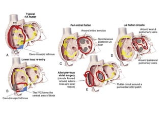
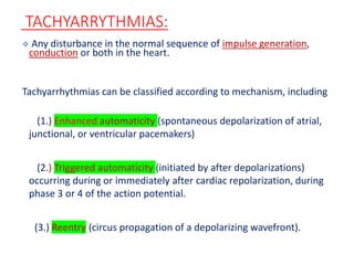
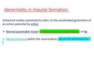
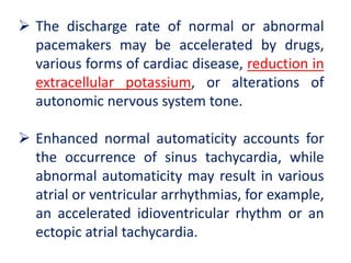
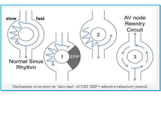
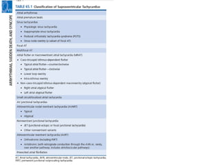
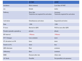
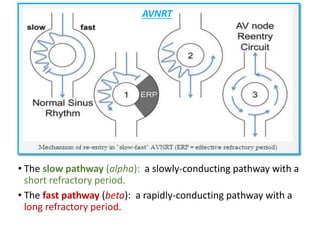
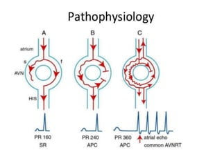
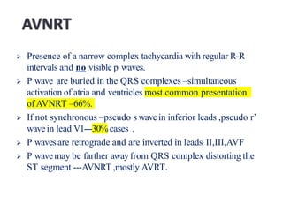
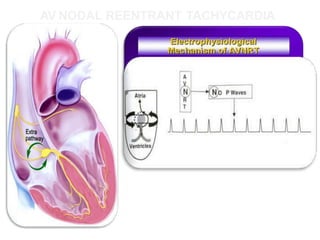
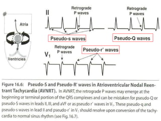
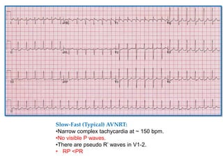
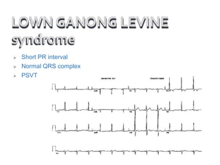
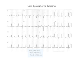
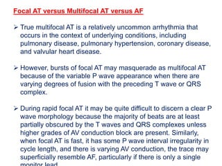
1. This document discusses the approach to evaluating and diagnosing narrow complex tachycardias. It describes the main mechanisms that can cause tachycardias including enhanced automaticity, triggered activity, and reentry.
2. Specific tachycardia types are then discussed in detail including AV nodal reentrant tachycardia (AVNRT), atrioventricular reentrant tachycardia (AVRT), atrial tachycardia (AT), junctional ectopic tachycardia (JET), and inappropriate sinus tachycardia. The diagnostic criteria and distinguishing characteristics of each are provided.
3. A number of other arrhythmias are also briefly covered such
Ozempic: Preoperative Management of Patients on GLP-1 Receptor Agonists Saeid Safari
Preoperative Management of Patients on GLP-1 Receptor Agonists like Ozempic and Semiglutide
ASA GUIDELINE
NYSORA Guideline
2 Case Reports of Gastric Ultrasound
Recomendações da OMS sobre cuidados maternos e neonatais para uma experiência pós-natal positiva.
Em consonância com os ODS – Objetivos do Desenvolvimento Sustentável e a Estratégia Global para a Saúde das Mulheres, Crianças e Adolescentes, e aplicando uma abordagem baseada nos direitos humanos, os esforços de cuidados pós-natais devem expandir-se para além da cobertura e da simples sobrevivência, de modo a incluir cuidados de qualidade.
Estas diretrizes visam melhorar a qualidade dos cuidados pós-natais essenciais e de rotina prestados às mulheres e aos recém-nascidos, com o objetivo final de melhorar a saúde e o bem-estar materno e neonatal.
Uma “experiência pós-natal positiva” é um resultado importante para todas as mulheres que dão à luz e para os seus recém-nascidos, estabelecendo as bases para a melhoria da saúde e do bem-estar a curto e longo prazo. Uma experiência pós-natal positiva é definida como aquela em que as mulheres, pessoas que gestam, os recém-nascidos, os casais, os pais, os cuidadores e as famílias recebem informação consistente, garantia e apoio de profissionais de saúde motivados; e onde um sistema de saúde flexível e com recursos reconheça as necessidades das mulheres e dos bebês e respeite o seu contexto cultural.
Estas diretrizes consolidadas apresentam algumas recomendações novas e já bem fundamentadas sobre cuidados pós-natais de rotina para mulheres e neonatos que recebem cuidados no pós-parto em unidades de saúde ou na comunidade, independentemente dos recursos disponíveis.
É fornecido um conjunto abrangente de recomendações para cuidados durante o período puerperal, com ênfase nos cuidados essenciais que todas as mulheres e recém-nascidos devem receber, e com a devida atenção à qualidade dos cuidados; isto é, a entrega e a experiência do cuidado recebido. Estas diretrizes atualizam e ampliam as recomendações da OMS de 2014 sobre cuidados pós-natais da mãe e do recém-nascido e complementam as atuais diretrizes da OMS sobre a gestão de complicações pós-natais.
O estabelecimento da amamentação e o manejo das principais intercorrências é contemplada.
Recomendamos muito.
Vamos discutir essas recomendações no nosso curso de pós-graduação em Aleitamento no Instituto Ciclos.
Esta publicação só está disponível em inglês até o momento.
Prof. Marcus Renato de Carvalho
www.agostodourado.com
Tom Selleck Health: A Comprehensive Look at the Iconic Actor’s Wellness Journeygreendigital
Tom Selleck, an enduring figure in Hollywood. has captivated audiences for decades with his rugged charm, iconic moustache. and memorable roles in television and film. From his breakout role as Thomas Magnum in Magnum P.I. to his current portrayal of Frank Reagan in Blue Bloods. Selleck's career has spanned over 50 years. But beyond his professional achievements. fans have often been curious about Tom Selleck Health. especially as he has aged in the public eye.
Follow us on: Pinterest
Introduction
Many have been interested in Tom Selleck health. not only because of his enduring presence on screen but also because of the challenges. and lifestyle choices he has faced and made over the years. This article delves into the various aspects of Tom Selleck health. exploring his fitness regimen, diet, mental health. and the challenges he has encountered as he ages. We'll look at how he maintains his well-being. the health issues he has faced, and his approach to ageing .
Early Life and Career
Childhood and Athletic Beginnings
Tom Selleck was born on January 29, 1945, in Detroit, Michigan, and grew up in Sherman Oaks, California. From an early age, he was involved in sports, particularly basketball. which played a significant role in his physical development. His athletic pursuits continued into college. where he attended the University of Southern California (USC) on a basketball scholarship. This early involvement in sports laid a strong foundation for his physical health and disciplined lifestyle.
Transition to Acting
Selleck's transition from an athlete to an actor came with its physical demands. His first significant role in "Magnum P.I." required him to perform various stunts and maintain a fit appearance. This role, which he played from 1980 to 1988. necessitated a rigorous fitness routine to meet the show's demands. setting the stage for his long-term commitment to health and wellness.
Fitness Regimen
Workout Routine
Tom Selleck health and fitness regimen has evolved. adapting to his changing roles and age. During his "Magnum, P.I." days. Selleck's workouts were intense and focused on building and maintaining muscle mass. His routine included weightlifting, cardiovascular exercises. and specific training for the stunts he performed on the show.
Selleck adjusted his fitness routine as he aged to suit his body's needs. Today, his workouts focus on maintaining flexibility, strength, and cardiovascular health. He incorporates low-impact exercises such as swimming, walking, and light weightlifting. This balanced approach helps him stay fit without putting undue strain on his joints and muscles.
Importance of Flexibility and Mobility
In recent years, Selleck has emphasized the importance of flexibility and mobility in his fitness regimen. Understanding the natural decline in muscle mass and joint flexibility with age. he includes stretching and yoga in his routine. These practices help prevent injuries, improve posture, and maintain mobilit
Knee anatomy and clinical tests 2024.pdfvimalpl1234
This includes all relevant anatomy and clinical tests compiled from standard textbooks, Campbell,netter etc..It is comprehensive and best suited for orthopaedicians and orthopaedic residents.
micro teaching on communication m.sc nursing.pdfAnurag Sharma
Microteaching is a unique model of practice teaching. It is a viable instrument for the. desired change in the teaching behavior or the behavior potential which, in specified types of real. classroom situations, tends to facilitate the achievement of specified types of objectives.
New Drug Discovery and Development .....NEHA GUPTA
The "New Drug Discovery and Development" process involves the identification, design, testing, and manufacturing of novel pharmaceutical compounds with the aim of introducing new and improved treatments for various medical conditions. This comprehensive endeavor encompasses various stages, including target identification, preclinical studies, clinical trials, regulatory approval, and post-market surveillance. It involves multidisciplinary collaboration among scientists, researchers, clinicians, regulatory experts, and pharmaceutical companies to bring innovative therapies to market and address unmet medical needs.
Local Advanced Lung Cancer: Artificial Intelligence, Synergetics, Complex Sys...Oleg Kshivets
Overall life span (LS) was 1671.7±1721.6 days and cumulative 5YS reached 62.4%, 10 years – 50.4%, 20 years – 44.6%. 94 LCP lived more than 5 years without cancer (LS=2958.6±1723.6 days), 22 – more than 10 years (LS=5571±1841.8 days). 67 LCP died because of LC (LS=471.9±344 days). AT significantly improved 5YS (68% vs. 53.7%) (P=0.028 by log-rank test). Cox modeling displayed that 5YS of LCP significantly depended on: N0-N12, T3-4, blood cell circuit, cell ratio factors (ratio between cancer cells-CC and blood cells subpopulations), LC cell dynamics, recalcification time, heparin tolerance, prothrombin index, protein, AT, procedure type (P=0.000-0.031). Neural networks, genetic algorithm selection and bootstrap simulation revealed relationships between 5YS and N0-12 (rank=1), thrombocytes/CC (rank=2), segmented neutrophils/CC (3), eosinophils/CC (4), erythrocytes/CC (5), healthy cells/CC (6), lymphocytes/CC (7), stick neutrophils/CC (8), leucocytes/CC (9), monocytes/CC (10). Correct prediction of 5YS was 100% by neural networks computing (error=0.000; area under ROC curve=1.0).
Best Ayurvedic medicine for Gas and IndigestionSwastikAyurveda
Here is the updated list of Top Best Ayurvedic medicine for Gas and Indigestion and those are Gas-O-Go Syp for Dyspepsia | Lavizyme Syrup for Acidity | Yumzyme Hepatoprotective Capsules etc
Muktapishti is a traditional Ayurvedic preparation made from Shoditha Mukta (Purified Pearl), is believed to help regulate thyroid function and reduce symptoms of hyperthyroidism due to its cooling and balancing properties. Clinical evidence on its efficacy remains limited, necessitating further research to validate its therapeutic benefits.
NVBDCP.pptx Nation vector borne disease control programSapna Thakur
NVBDCP was launched in 2003-2004 . Vector-Borne Disease: Disease that results from an infection transmitted to humans and other animals by blood-feeding arthropods, such as mosquitoes, ticks, and fleas. Examples of vector-borne diseases include Dengue fever, West Nile Virus, Lyme disease, and malaria.
Flu Vaccine Alert in Bangalore Karnatakaaddon Scans
As flu season approaches, health officials in Bangalore, Karnataka, are urging residents to get their flu vaccinations. The seasonal flu, while common, can lead to severe health complications, particularly for vulnerable populations such as young children, the elderly, and those with underlying health conditions.
Dr. Vidisha Kumari, a leading epidemiologist in Bangalore, emphasizes the importance of getting vaccinated. "The flu vaccine is our best defense against the influenza virus. It not only protects individuals but also helps prevent the spread of the virus in our communities," he says.
This year, the flu season is expected to coincide with a potential increase in other respiratory illnesses. The Karnataka Health Department has launched an awareness campaign highlighting the significance of flu vaccinations. They have set up multiple vaccination centers across Bangalore, making it convenient for residents to receive their shots.
To encourage widespread vaccination, the government is also collaborating with local schools, workplaces, and community centers to facilitate vaccination drives. Special attention is being given to ensuring that the vaccine is accessible to all, including marginalized communities who may have limited access to healthcare.
Residents are reminded that the flu vaccine is safe and effective. Common side effects are mild and may include soreness at the injection site, mild fever, or muscle aches. These side effects are generally short-lived and far less severe than the flu itself.
Healthcare providers are also stressing the importance of continuing COVID-19 precautions. Wearing masks, practicing good hand hygiene, and maintaining social distancing are still crucial, especially in crowded places.
Protect yourself and your loved ones by getting vaccinated. Together, we can help keep Bangalore healthy and safe this flu season. For more information on vaccination centers and schedules, residents can visit the Karnataka Health Department’s official website or follow their social media pages.
Stay informed, stay safe, and get your flu shot today!
CDSCO and Phamacovigilance {Regulatory body in India}NEHA GUPTA
The Central Drugs Standard Control Organization (CDSCO) is India's national regulatory body for pharmaceuticals and medical devices. Operating under the Directorate General of Health Services, Ministry of Health & Family Welfare, Government of India, the CDSCO is responsible for approving new drugs, conducting clinical trials, setting standards for drugs, controlling the quality of imported drugs, and coordinating the activities of State Drug Control Organizations by providing expert advice.
Pharmacovigilance, on the other hand, is the science and activities related to the detection, assessment, understanding, and prevention of adverse effects or any other drug-related problems. The primary aim of pharmacovigilance is to ensure the safety and efficacy of medicines, thereby protecting public health.
In India, pharmacovigilance activities are monitored by the Pharmacovigilance Programme of India (PvPI), which works closely with CDSCO to collect, analyze, and act upon data regarding adverse drug reactions (ADRs). Together, they play a critical role in ensuring that the benefits of drugs outweigh their risks, maintaining high standards of patient safety, and promoting the rational use of medicines.
Title: Sense of Taste
Presenter: Dr. Faiza, Assistant Professor of Physiology
Qualifications:
MBBS (Best Graduate, AIMC Lahore)
FCPS Physiology
ICMT, CHPE, DHPE (STMU)
MPH (GC University, Faisalabad)
MBA (Virtual University of Pakistan)
Learning Objectives:
Describe the structure and function of taste buds.
Describe the relationship between the taste threshold and taste index of common substances.
Explain the chemical basis and signal transduction of taste perception for each type of primary taste sensation.
Recognize different abnormalities of taste perception and their causes.
Key Topics:
Significance of Taste Sensation:
Differentiation between pleasant and harmful food
Influence on behavior
Selection of food based on metabolic needs
Receptors of Taste:
Taste buds on the tongue
Influence of sense of smell, texture of food, and pain stimulation (e.g., by pepper)
Primary and Secondary Taste Sensations:
Primary taste sensations: Sweet, Sour, Salty, Bitter, Umami
Chemical basis and signal transduction mechanisms for each taste
Taste Threshold and Index:
Taste threshold values for Sweet (sucrose), Salty (NaCl), Sour (HCl), and Bitter (Quinine)
Taste index relationship: Inversely proportional to taste threshold
Taste Blindness:
Inability to taste certain substances, particularly thiourea compounds
Example: Phenylthiocarbamide
Structure and Function of Taste Buds:
Composition: Epithelial cells, Sustentacular/Supporting cells, Taste cells, Basal cells
Features: Taste pores, Taste hairs/microvilli, and Taste nerve fibers
Location of Taste Buds:
Found in papillae of the tongue (Fungiform, Circumvallate, Foliate)
Also present on the palate, tonsillar pillars, epiglottis, and proximal esophagus
Mechanism of Taste Stimulation:
Interaction of taste substances with receptors on microvilli
Signal transduction pathways for Umami, Sweet, Bitter, Sour, and Salty tastes
Taste Sensitivity and Adaptation:
Decrease in sensitivity with age
Rapid adaptation of taste sensation
Role of Saliva in Taste:
Dissolution of tastants to reach receptors
Washing away the stimulus
Taste Preferences and Aversions:
Mechanisms behind taste preference and aversion
Influence of receptors and neural pathways
Impact of Sensory Nerve Damage:
Degeneration of taste buds if the sensory nerve fiber is cut
Abnormalities of Taste Detection:
Conditions: Ageusia, Hypogeusia, Dysgeusia (parageusia)
Causes: Nerve damage, neurological disorders, infections, poor oral hygiene, adverse drug effects, deficiencies, aging, tobacco use, altered neurotransmitter levels
Neurotransmitters and Taste Threshold:
Effects of serotonin (5-HT) and norepinephrine (NE) on taste sensitivity
Supertasters:
25% of the population with heightened sensitivity to taste, especially bitterness
Increased number of fungiform papillae
5. TACHYARRYTHMIAS:
Any disturbance in the normal sequence of impulse generation,
conduction or both in the heart.
Tachyarrhythmias can be classified according to mechanism, including
(1.) Enhanced automaticity (spontaneous depolarization of atrial,
junctional, or ventricular pacemakers)
(2.) Triggered automaticity (initiated by after depolarizations)
occurring during or immediately after cardiac repolarization, during
phase 3 or 4 of the action potential.
(3.) Reentry (circus propagation of a depolarizing wavefront).
6.
7.
8.
9.
10. Enhanced cardiac automaticity refers to the accelerated generation of
an action potential by either
Normal pacemaker tissue (enhanced normal automaticity) or by
Abnormal tissue within the myocardium (abnormal automaticity).
Abnormality in impulse formation:
11. The discharge rate of normal or abnormal
pacemakers may be accelerated by drugs,
various forms of cardiac disease, reduction in
extracellular potassium, or alterations of
autonomic nervous system tone.
Enhanced normal automaticity accounts for
the occurrence of sinus tachycardia, while
abnormal automaticity may result in various
atrial or ventricular arrhythmias, for example,
an accelerated idioventricular rhythm or an
ectopic atrial tachycardia.
12. An increase in automaticity normally causes an
increase in sinus rate and sinus tachycardia.
Abnormal automaticity is due to an increase in the
slope of phase 4 depolarization in myocardium or
reduced threshold for action potential depolarization in
myocardium other than the sinus node.
Abnormal automaticity is thought to be responsible
for most atrial premature complexes (APC) and VPCs.
13. Less commonly, abnormal impulse formation
is due to the development of triggered
activity.
Triggered activity is related to cellular
afterdepolarization that occur at the end of
the action potential, during phase 3, and are
referred to as early afterdepolarization, or
they occur after the action potential, during
phase 4, and are referred to as late/delay
afterdepolarization.
14. ABNORMALITY IN IMPULSE PROPAGATION:
The most common arrhythmia mechanism is reentry.
Reentry is defined as circulation of an activation wave
around an inexcitable obstacle.
Impulse propagation around an inexcitable region such
electrophysiologically dissimilar pathways for impulse
propagation around an inexcitable region such that
unidirectional block occurs in one of the pathways and a
region of excitable tissue exists at the head of the
propagating wavefront .
15.
16. Look for QRS duration.
QRS complex regular/irregular.
Then look for presence of p waves.
P waves morphology
P waves and QRS relationship 1:1
AV block present.
QRS alternation
Termination initiation of tachycardia.
Effect of BBB on tachycardia cycle length.
17.
18.
19.
20.
21. s
AVNRT AVRT
Incidence Most common Less than AVNRT
sex female males
Pathway Slow-fast,
Ventricles not required for activation
Accesory
Ventricles required for activation
Activation Simultaneous activation Sequential activation
Rate <200 >200
P-wave Burried in QRS Will be seen after QRS
Pseudo-r,pseudo-s,pseudo-q present absent
RP-interval <90msec >90msec
ST-T changes Less common more
ST elevation in aVR lesss more
Notch in aVL more less
QRS alternans Rare common
Abberancy Rare common
BBB Doesnot alter rate Alters rate
AV block Possible Not possible in its presence
22. • The slow pathway (alpha): a slowly-conducting pathway with a
short refractory period.
• The fast pathway (beta): a rapidly-conducting pathway with a
long refractory period.
AVNRT
23.
24. Presence of a narrow complex tachycardia with regular R-R
intervals and no visible p waves.
P wave are buried in the QRS complexes –simultaneous
activation of atria and ventricles most common presentation
of AVNRT –66%.
If not synchronous –pseudo s wavein inferior leads ,pseudo r’
wavein lead V1---30%cases .
P waves are retrograde and are inverted in leads II,III,AVF
P wavemay be farther awayfrom QRS complex distorting the
ST segment ---AVNRT ,mostly AVRT.
31. •Slow-Slow AVNRT (Atypical AVNRT)
• 1-5% AVNRT
• Associated with Slow AV nodal pathway for anterograde
conduction and Slow left atrial fibres as the pathway for
retrograde conduction.
• ECG features:
• Tachycardia with a P-wave seen in mid-diastole… effectively
appearing “before” the QRS complex.
• Confusing as a P wave appearing before the QRS complex in the
face of a tachycardia might be read as a sinus tachycardia.
32. What are “Pre-excitation syndromes” ?
• Term coined by Ohnell
• First described in 1930 by Louis Wolff, John Parkinson and Paul Dudley White.
• A group of ECG and Electrophysiological abnormalities in which
• The atrial impulses are conducted partly or completely, PREMATURELY, to
the ventricles via a mechanism other than the normal AV-node.
• Associated with a wide array of tachycardias with both normal QRS and
prolonged QRS durations.
34. WPW
• PR interval <120ms
• Delta wave – slurring slow rise of
initial portion of the QRS
• QRS prolongation >120ms
• ST Segment and T wave discordant
changes – i.e. in the opposite
direction to the major component of
the QRS complex
• Pseudo-infarction pattern can be
seen in up to 70% of patients – due
to negatively deflected delta waves
in the inferior / anterior leads
(“pseudo-Q waves”), or as a
prominent R wave in V1-3
(mimicking posterior infarction).
WPW in sinus rhythm
37. Two types
Orthodromic
Antidromic
Antidromic is wide complex tachycardia
In NSR detected by delta wave.
Can ppt into AF and VF on use of AV nodal blockers
MEMBRANE ACTIVE ANTIARRHTYHMIC DRUGS are safe.
CONCEALED WPW syndrome – no delta wave less risk of AF
38. Typical – RP interval <PR interval
RP interval >90 millisec
Atypical –RP interval >PR interval
Concealed bypass tract – only retrograde conduction
Manifest bypass tract– both anterograde and
retrograde conduction.
Electrical alternans –the amplitude of QRS
complexes varies by 5 mm alternatively.
Rate related BBB occuring and the rate of
tachycardia is decreasing –then the bypass tract is on
the same side of the block.
51. Sinus Node Reentry
Sinus node reentry is clinically defined as a tachycardia that can be
induced and terminated with programmed stimulation and has a P
wave morphology identical or similar to that of the sinus P wave.
It is now well recognized that the sinus node is not a discrete
structure but rather a diffuse pacemaker complex located along the
long axis of the crista terminalis.
AT has been described arising from sites along the length of this
structure, and it may be best to define these simply as crista
terminalis AT.
There has been some debate as to whether tachycardias due to
reentry within the sinus node truly exist.11
52. Inappropriate Sinus Tachycardia
Heart rate > 100 bpm at rest or on minimal exertion.
The mean 24-hour heart rate >90 bpm.
Tachycardia-induced cardiomyopathy are extremely rare.
It is important to rule out secondary causes as a critical
part of the initial evaluation before the diagnosis may be
considered.
A broad range of secondary causes of sinus tachycardia,
including anxiety, anemia volume depletion,
thyrotoxicosis, fever, pulmonary embolus, cardiac failure,
sepsis, stimulants, and drug withdrawal must first be
excluded.
Resting heart rates are frequently in the normal range,
53. Heart rate increases generally occur gradually over 30
seconds to several minutes.
The P wave morphology of IAST reflects an origin in the
region of the superior crista terminalis in the right atrium
with a biphasic positive-negative appearance in V1 and
upright P waves in inferior leads and lead I. This
resembles the morphology of a focal AT originating in
the same anatomic region.
The differential diagnosis also includes both postural
orthostatic tachycardia syndrome (POTS) and physical
deconditioning, which may have many overlapping
features.
54. THE MECHANISM OF IAST:
Is not well understood, and both intrinsic
(sinus node hyperactivity) and extrinsic
(related to dysautonomia or
neurohormonal dysregulation)
mechanisms have been suggested.
A number of recent reports have
described the development of
inappropriate sinus tachycardia with
elevated mean heart rates and reduced
heart rate variability as a manifestation of
55. Atrial Tachycardias
Tachycardias that originate in the atria and do not require the
participation of the AV node for maintenance of the
arrhythmia.9
AT can be: Focal or Macroreentry.
Focal AT has been defined as atrial activation starting
rhythmically at a small area (focus) from which it spreads
out centrifugally.
In Macroreentry, activation occurs around a large central obstacle,
such as an anatomic structure or region of scarring; electrical activity
can be recorded throughout the entire atrial cycle length. These include
typical AFL and other well-characterized macroreentrant circuits in the
right and left atrium, which are also frequently referred to as types of
“atypical AFL.” More recently a third category of AT has been described
although not routinely included in all classifications. These have been
termed “small circuit” or “localized” reentry .
56. Focal Atrial Tachycardia
Focal AT is a form of SVT characterized by regular, organized atrial activity
with discrete P waves and typically an isoelectric segment between P waves.
However, when focal AT rate is particularly rapid, an isoelectric interval may
not be apparent .
Focal AT may display some irregularity particularly at onset (“warm-up”) and
termination (“cool-down”),1 usually occurring over several beats. Atrial
mapping reveals a focal point of origin.
Mechanisms of focal AT include abnormal or enhanced automaticity
(abnormal impulse initiation in an individual or cluster of myocytes), triggered
activity (abnormal impulse initiation due to oscillations of membrane potential,
termed early or delayed after-depolarizations) and reentry (when myocardial
regions activated later in propagation reexcite regions that have already
recovered excitability).
The arrhythmia may also demonstrate cycle length variability.
Finally, focal AT due to either microreentry or triggered activity may be
terminated with adenosine.
57.
58. EPIDEMIOLOGY.
Focal AT is the least common mechanism of PSVT (10% to
20% )
Focal AT : Female>male
AT can occur across the age spectrum but gradually
increases in prevalence with age and peaking between
the age of 40 and 60 years.
Automatic AT tends to be more common in younger
populations.
Focal AT due to microreentry is more common in older
populations.
59. SYMPTOMS
Palpitations,
Dizziness,
Chest pain,
Dyspnea,
Fatigue, and
Presyncope.
Syncope is unusual unless tachycardia rates are extremely
rapid or there is associated underlying structural heart
disease.
Tachycardia-mediated cardiomyopathy (TMC) has been
reported to occur in a small percentage of patients with
incessant ATs.
Embolic events and stroke have rarely been reported in
patients with AT, and treatment with anticoagulation is
generally not indicated.
Spontaneous remission of focal AT has been reported in both
adults and children after cessation of medical therapy. In fact,
60.
61.
62. At least three consequtive p waves with different morphologies
with a rate >100 bpm to be present.
Isoelectric baseline between p waves.
Also called as choatic atrial tachycardia
Mostly seen in COPD ,electrolyte abn,theophylline
Rate usually does not exceed 130-140 bpm.
63.
64.
65. JUNCTIONAL ECTOPIC TACHYCARDIA (NONREENTRANT
JUNCTIONAL TACHYCARDIA).
JET is a rare arrhythmia
It may occur early after surgical repair of CHD with an incidence of 1%
to 5%.
As a congenital arrhythmia presenting in the first 6 months of life, in
which it has been associated with a high morbidity and mortality.51
In this age group the arrhythmia is more likely to be incessant and has
higher rates.
Complete suppression of the arrhythmia is rare; more frequently the
rate and frequency of episodes is decreased.
In up to 20% of cases, antiarrhythmics are completely ineffective.
The agent with highest reported efficacy is AMIODARONE.
Catheter ablation is playing an increasing role using either
radiofrequency or cryoablation approaches.
66.
67. Diagnosis and Differential Diagnosis
Inappropriate Sinus Tachycardia versus AT
P wave in AT usually has a morphology different from that
of the sinus P wave.
When AT arises from the superior crista terminalis these
differences may be subtle.
AT usually demonstrates an abrupt onset and termination
or may warm up and cool down over 3 or 4 beats. In
contrast, IAST gradually increases in rate over
approximately 30 seconds to several minutes.
When onset occurs with a tightly coupled P wave,
particularly located in the preceding T wave, is virtually
diagnostic of AT.
68. AT versus AVNRT/AVRT
The most important differentiating factor is R-P relationship.
Both typical AVNRT and AVRT have a short R-P interval that does
not vary.
AT can occur with either a short R-P interval or a long R-P interval
depending on the tachycardia rate and the speed of AV nodal
conduction. It can therefore mimic either AVNRT or AVRT.
In AT the R-P relationship is incidental and hence possibly variable.
In AVRT and AVNRT this relationship will be constant because it is
integral to the tachycardia mechanism.
The presence of an inferior P wave axis excludes AVRT or AVNRT.
Automatic AT may also manifest with recurrent self-limiting bursts of
tachycardia that can exhibit warm-up and cool-down phases.
69. Focal AT versus Macroreentrant AT
Focal AT it is possible to observe a discrete P wave with an intervening
isoelectric interval.
However, when the atrial rate is very rapid and, if atrial conduction
slowing is present, there may be no isoelectric baseline and the
appearance may mimic that of macroreentrant AT .
Macroreentrant AT (including typical AFL) frequently demonstrates a
continuous undulation without an isoelectric period on the ECG.
Variation in the tachycardia rate with spontaneous termination and then
reinitiation is diagnostic of a focal AT, usually the result of enhanced
automaticity or triggered activity.
Spontaneous bursts of tachycardia do not occur with macroreentrant
tachycardias.
70. Focal AT versus Multifocal AT versus AF
True multifocal AT is a relatively uncommon arrhythmia that
occurs in the context of underlying conditions, including
pulmonary disease, pulmonary hypertension, coronary disease,
and valvular heart disease.
However, bursts of focal AT may masquerade as multifocal AT
because of the variable P wave appearance when there are
varying degrees of fusion with the preceding T wave or QRS
complex.
During rapid focal AT it may be quite difficult to discern a clear P
wave morphology because the majority of beats are at least
partially obscured by the T waves and QRS complexes unless
higher grades of AV conduction block are present. Similarly,
when focal AT is fast, it has some P wave interval irregularity in
cycle length, and there is varying AV conduction, the trace may
superficially resemble AF, particularly if there is only a single
71. APPROACH TO A
PATIENT WITH
NARROW COMPLEX
TACHYCARDIA
CONTI………
Speaker-Dr Ramdhan kumar
kamat
Moderator-Dr T.Mukherjee
72. PERMANENT JUNCTIONAL RECIPROCATING TACHYCARDIA
Persistent or permanent junctional reciprocating tachycardia
(PJRT) is an uncommon arrhythmia characterised by an
incessant orthodromic tachycardia.
Anterograde conduction over the atrioventricular node and by
retrograde conduction via an accessory pathway usually located
in the posteroseptal region with slow and decremental
conduction.
The arrhythmia is commonly incessant from birth or infancy.
Its persistence over a long period of time may lead to a
tachycardia induced cardiomyopathy that is reversible with rate
control.
73.
74. THE EKG CRITERIA FOR THE DIAGNOSIS OF PJRT
RP interval is longer than PR interval (because of the
location and decremental conduction properties of the
AP)
Inverted P wave are often visible in the inferior leads, II,
III, and AVF
The rates are typically slightly slower than typical SVT
(can be confused with sinus tachycardia)
75. Response to vagal maneuvers with gradual slowing
of the tachycardia because of prolongation of both
RP and PR intervals and eventual termination but
with recurrence shortly afterwards.
The AV conduction is usually sensitive to adenosine
with tachycardia terminating with AV or VA block
but again recurring shortly afterwards.
PR interval is never prolonged.
QRS is normal and narrow during sinus rhythm and
in tachycardia.
76.
77. TREATMENT
MEDICAL MANAGEMENT
PJRT can be difficult to treat and refractory to medical
management.
The similarity of conduction between the AV node and the AP are
such that both antegrade and retrograde limbs are similarly
affected by antiarrhythmic agents, making it difficult to achieve
sustained block in just one limb of the reentrant circuit.
Many infants and children require treatment with more than a
single antiarrhythmic agent.
78. Earlier studies reported amiodarone, digoxin, and verapamil
in combinations as the most effective regimen with
reasonable success.
The latter two medications are now rarely used and currently,
combination therapy with flecainide and amiodarone has
been shown to be slightly more successful in controlling
tachycardia and reversing cardiomyopathy, if detected and
treated early.
INTERVENTIONAL MANAGEMENT
Ablation is generally indicated when there is difficulty in
medical management and some feel that it needs to be
attempted early in PJRT after appropriate electrophysiologic
mapping.
82. CAVO-TRICUSPID ISTHMUS DEPENDENT FLUTTER.
Typical flutter is as a macroreentrant circuit within the
right atrium. It can be considered to be a broad activation
wavefront rotating between the tricuspid annulus (TA)
anteriorly and the crista terminalis-eustachian
ridge/inferior vena cava (IVC) posteriorly .
In the most common form (approximately 90%) the circuit
rotates in a counterclockwise direction when viewed in the
frontal plane.
In 10%, rotation is clockwise.
83.
84. The ECG of typical counterclockwise AFL is
characterized by the classic inferior lead flutter wave
appearance (“saw-tooth” pattern) demonstrating an initial
gradual downsloping segment followed by a deeply
inverted component with a terminal positive component.
In the precordial leads, V1 classically demonstrates an
initial isoelectric component followed by an upright
component.
With progression across the precordial leads, the initial
component becomes inverted and the second
component isoelectric such that V5 and V6 demonstrate
an inverted flutter wave.
Lead I is low amplitude/isoelectric and aVL usually
upright.
85.
86. In clockwise AFL, although the anatomic
boundaries are identical to those of
counterclockwise AFL, the wavefront is reversed.
87. Non Cavo-Tricuspid Isthmus–Dependent Flutter
(Atypical Atrial Flutter).
Atypical flutter may occur primarily in the right or the left
atrium.
When the V1 flutter wave is deeply inverted, this is highly
likely to represent a right atrial circuit. Conversely, when
the V1 flutter wave is upright, this generally indicates an
LA circuit.
However, many variations exist and these
findings lack sensitivity and specificity.
The atrial rate in atypical flutter has wide limits (120 to
300 bpm) depending on the underlying circuit and
pathology).
88. The pathology underlying atypical flutter is highly variable, and
these circuits may occur in the context of
(1) Prior corrective atrial surgery (congenital heart disease [CHD],
valvular heart disease, after a Maze procedure or cardiac
transplantation),
(2) Previous AF ablation,
(3) Advanced atrial disease associated with atrial enlargement
(these patients frequently have underlying pathologies such as
heart failure [systolic or diastolic] or unoperated valvular heart
disease such as severe mitral regurgitation),
(4) In patients with normal atrial size and without an obvious
underlying pathologic condition. In these patients, spontaneous
scarring of unknown cause may be found at the time of atrial
mapping. These circuits have particularly been described in the RA
free wall.
89. The circuits involved in atypical (non–isthmus dependent)
AFL are highly variable and involve a range of anatomic
boundaries.
These might be anatomic structures, surgical scars, or
regions of low voltage and slowed conduction.
Stereotypical anatomic locations associated with certain
underlying pathologic conditions or procedures have been
defined.
These include surgical repair of complex CHD such as
Mustard or Senning repair, Fontan repair, or simpler atrial
surgeries such as atrial septal defect (ASD) repair ,Mitral
valve surgery (repair or replacement).
90. Dual-loop or figure-of-8 reentry has been described
when there are two simultaneous circuits. This may occur
in either the left atrium (31 In patients with prior atrial
surgery, circuits particularly involve atriotomy scars. In
patients with prior LA ablation, circuits may be around the
pulmonary veins (roof dependent) or around the mitral
annulus (mitral isthmus dependent).
91. SMALL CIRCUIT REENTRY.
A third category of AT termed small circuit reentry has
been increasing recognized in the era of high-density
three-dimensional mapping.
These reentrant circuits occur in a localized region with a
diameter of 1 to 2 cm;
These circuits generally occur in the context of advanced
atrial remodeling, and although they have been
recognized across a range of pathologies they are most
commonly observed in patients with a history of persistent
AF, atrial enlargement, and prior AF ablation.
92.
93. RELATIONSHIP BETWEEN ATRIAL FLUTTER AND ATRIAL
FIBRILLATION.
AFL and AF have been described as two sides of the same
coin.
The two arrhythmias frequently coexist clinically with
documented AF in up to 75% of AFL patients.
In both animal and human studies, the onset of AFL is
usually preceded by a transitional period of AF. When AFL
terminates it is also generally via transitional AF (Fig. 65.9).
94.
95.
96. Due to the frequent coexistence of AF and
AFL, anticoagulant management of patients
with AFL generally follows the same
recommendations as for AF both at the time
of reversion and during chronic
management, in which decisions should be
dictated by the CHADS2-VA2Sc score rather
than apparent rhythm control.
Editor's Notes
ATRIAL TACHYCARDIA – FOCAL, MACRO RE ENTRANT, SINOATRIAL REENTRY
ATRIAL FLUTTER- RT- CLOCKWISE , COUNTER CLOCKWISE LT- MITRAL RE ENTRY, SCAR MEDIATED,PULMONARY VEIN
ALMOST ALL IRREGULAR TACHYS ARE AV NOT INDEPENDENT