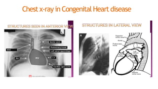
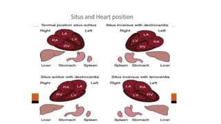
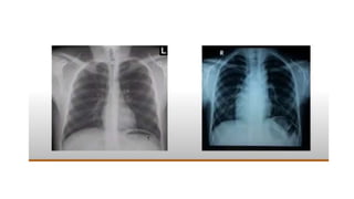
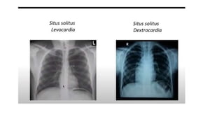
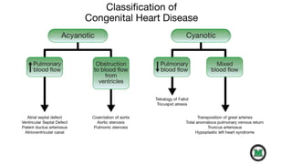
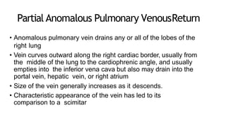
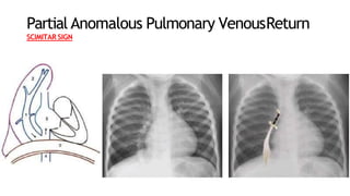
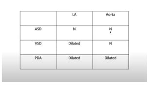
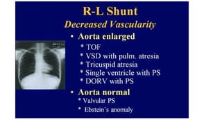
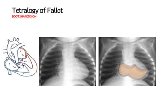
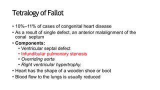
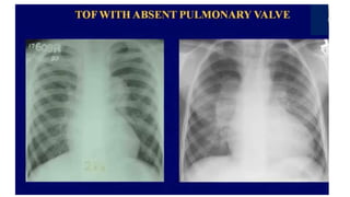
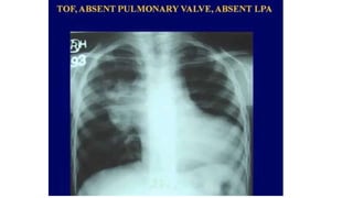
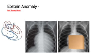
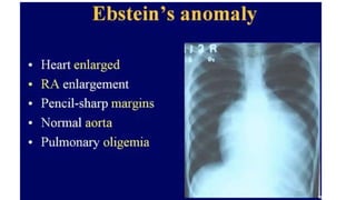
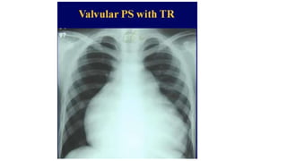
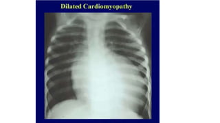
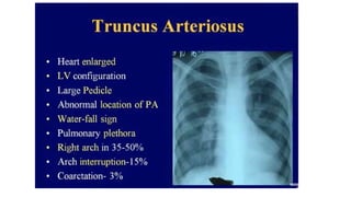
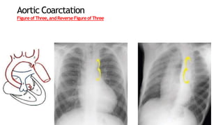
Chest x-ray in Congenital Heart disease
75
Partial Anomalous Pulmonary VenousReturn
• Anomalous pulmonary vein drains any or all of the lobes of the
right lung
• Vein curves outward along the right cardiac border, usually from
the middle of the lung to the cardiophrenic angle, and usually
empties into the inferior vena cava but also may drain into the
portal vein, hepatic vein, or right atrium
• Size of the vein generally increases as it descends.
• Characteristic appearance of the vein has led to its
comparison to a scimitar
Partial Anomalous Pulmonary VenousReturn
SCIMITAR SIGN
Tetralogy of Fallot
BOOT SHAPED SIGN
Tetralogy ofFallot
• 10%–11% of cases of congenital heart disease
• As a result of single defect, an anterior malalignment of the
conal septum
• Components:
• Ventricular septal defect
• Infundibular pulmonary stenosis
• Overriding aorta
• Right ventricular hypertrophy.
• Heart has the shape of a wooden shoe or boot
• Blood flow to the lungs is usually reduced
EbsteinAnomaly
• 0.5%–0.7% of cases of congenital heart disease.
• Characterized by the downward displacement of the septal leaflets and
posterior leaflets
of the tricuspid valve into the inflow portion of the right ventricle.
• Results in the formation of a common right ventriculoatrial chamber and causes
tricuspid regurgitation.
• Insufficiency of the tricuspid valve leads to dilatation of the right ventricular outflow
tract
and all proximal right heart structures,
• Most consistent imaging feature is right atrial enlargement
Ebstein Anomaly–
Box ShapedHeart
TRANSPOSITION OF THE GREAT
VESSELS
• D-Transposition of the Great Vessels
• 5%–7% of congenital cardiac malformations
• isolated in 90%
• Produced by a ventriculoarterial discordance in which the aorta arises
from the morphologic right ventricle and the pulmonary artery arises
from the morphologic left ventricle
• Pulmonary artery is situated to the right of its normal location
• Results in the apparent narrowing of the superior mediastinum on
radiographs
• Patent ASD, VSD, Foramen ovale, systemic collaterals to sustain
life
• Atrial border is abnormally convex, and the left atrium commonly is
enlarged because of increased pulmonary blood flow.
D-Transposition of greatvessels
EGG ON STRINGSIGN
TotalAnomalous Pulmonary VenousReturn
SNOWMAN SIGN
Endocardial CushionDefects
• Interruption of the normal development of the endocardial tissues during
gestation
• Endocardial cushion forms the lower portion of the atrial septum, the upper
portion of the interventricular septum, and the septal leaflets of the mitral valve
and the tricuspid valve
• 4% of all cases of congenital heart disease
• Gooseneck-shaped deformity
• Caused by a deficiency of both the conus and sinus portions of the
interventricular septum, with narrowing of the left ventricular outflow tract.
• Characteristic shape by concavity of the interventricular septum below the
mitral
valve, along with the elongation and narrowing of the left ventricular outflow
tract
Endocardial cushion defect-
GOOSENECK SIGN
AorticCoarctation
• 5%–10% of congenital cardiac lesions
• Produced by a deformity of the aortic media and intima, which causes a
prominent
posterior infolding of the aortic lumen
• Occurs at or near the junction of the aortic arch and the descending thoracic
aorta
• Infolding cause eccentric narrowing of the lumen at the level where the
ductus or ligamentum arteriosus inserts anteromedially
• Resultant luminal narrowing in turn obstructs the flow of blood from
the left ventricle
• Classic radiologic signs
• Figure-of-three sign
• Reverse figure-of-three sign
Aortic Coarctation
Figureof Three,and Reverse Figureof Three
Thank you
References
• Michael Y. M. Chen, Thomas L. Pope, David J. Ott. Basic
Radiology. 2nd ed. Mc. Grow hill. P-86-9.
• Cochard, Larry R.,Netter, Frank H. Netter's Introduction to
Imaging. Elseiver. P-54-9.
CHD CHEST X RAY.pptx

CHD CHEST X RAY.pptx

  • 1.
    Chest x-ray inCongenital Heart disease
  • 17.
  • 36.
    Partial Anomalous PulmonaryVenousReturn • Anomalous pulmonary vein drains any or all of the lobes of the right lung • Vein curves outward along the right cardiac border, usually from the middle of the lung to the cardiophrenic angle, and usually empties into the inferior vena cava but also may drain into the portal vein, hepatic vein, or right atrium • Size of the vein generally increases as it descends. • Characteristic appearance of the vein has led to its comparison to a scimitar
  • 37.
    Partial Anomalous PulmonaryVenousReturn SCIMITAR SIGN
  • 44.
  • 45.
    Tetralogy ofFallot • 10%–11%of cases of congenital heart disease • As a result of single defect, an anterior malalignment of the conal septum • Components: • Ventricular septal defect • Infundibular pulmonary stenosis • Overriding aorta • Right ventricular hypertrophy. • Heart has the shape of a wooden shoe or boot • Blood flow to the lungs is usually reduced
  • 51.
    EbsteinAnomaly • 0.5%–0.7% ofcases of congenital heart disease. • Characterized by the downward displacement of the septal leaflets and posterior leaflets of the tricuspid valve into the inflow portion of the right ventricle. • Results in the formation of a common right ventriculoatrial chamber and causes tricuspid regurgitation. • Insufficiency of the tricuspid valve leads to dilatation of the right ventricular outflow tract and all proximal right heart structures, • Most consistent imaging feature is right atrial enlargement
  • 52.
  • 60.
    TRANSPOSITION OF THEGREAT VESSELS
  • 61.
    • D-Transposition ofthe Great Vessels • 5%–7% of congenital cardiac malformations • isolated in 90% • Produced by a ventriculoarterial discordance in which the aorta arises from the morphologic right ventricle and the pulmonary artery arises from the morphologic left ventricle • Pulmonary artery is situated to the right of its normal location • Results in the apparent narrowing of the superior mediastinum on radiographs • Patent ASD, VSD, Foramen ovale, systemic collaterals to sustain life • Atrial border is abnormally convex, and the left atrium commonly is enlarged because of increased pulmonary blood flow.
  • 62.
  • 66.
  • 68.
    Endocardial CushionDefects • Interruptionof the normal development of the endocardial tissues during gestation • Endocardial cushion forms the lower portion of the atrial septum, the upper portion of the interventricular septum, and the septal leaflets of the mitral valve and the tricuspid valve • 4% of all cases of congenital heart disease • Gooseneck-shaped deformity • Caused by a deficiency of both the conus and sinus portions of the interventricular septum, with narrowing of the left ventricular outflow tract. • Characteristic shape by concavity of the interventricular septum below the mitral valve, along with the elongation and narrowing of the left ventricular outflow tract
  • 69.
  • 70.
    AorticCoarctation • 5%–10% ofcongenital cardiac lesions • Produced by a deformity of the aortic media and intima, which causes a prominent posterior infolding of the aortic lumen • Occurs at or near the junction of the aortic arch and the descending thoracic aorta • Infolding cause eccentric narrowing of the lumen at the level where the ductus or ligamentum arteriosus inserts anteromedially • Resultant luminal narrowing in turn obstructs the flow of blood from the left ventricle • Classic radiologic signs • Figure-of-three sign • Reverse figure-of-three sign
  • 72.
  • 73.
  • 74.
    References • Michael Y.M. Chen, Thomas L. Pope, David J. Ott. Basic Radiology. 2nd ed. Mc. Grow hill. P-86-9. • Cochard, Larry R.,Netter, Frank H. Netter's Introduction to Imaging. Elseiver. P-54-9.