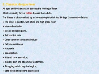
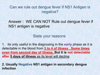
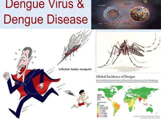
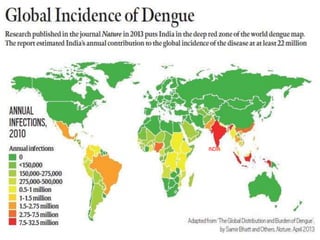
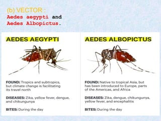
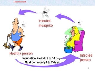
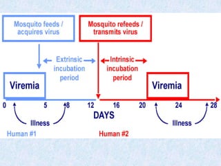
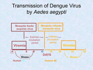
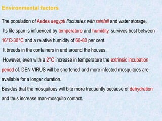
Infected Aedes mosquito
THE DENGUE SYNDROME
PROBLEM STATEMENT
Dengue and DHF is endemic in more than 100 countries in the WHO region. South-East Asia and
Western pacific regions are most seriously affected.
South-East Asia region are divided into 3 categories
Category A (Bangladesh, India, Indonesia, Maldives, Myanmar, Sri Lanka, Thailand and Timor-Leste)
a. Major public health problem;
b. Leading cause of hospitalization and death among children;
c. Hyperendemicity with all 4 serotypes circulating in
urban areas; and
d. Spreading to rural areas
.
Category B (Bhutan, Nepal)
a. Endemicity uncertain;
b. Bhutan reported first outbreak in 2004; and
c. Nepal reported first indigenous case in 2004.
Category C (DPR Korea)
No evidence of endemicity.
INDIA
In India, the risk of dengue has shown an increase in
recent years due to
 Rapid urbanization,
Lifestyle changes and
Deficient water management.
Dengue is endemic in 31 states/UTs. During 2013, about 74,168 cases were reported
with 168 deaths. The case fatality rate was 0.22 per cent., the highest number of
cases were reported from Punjab followed by Tamil Nadu, Gujarat, Kerala and
Andhra
All the four serotypes i.e. dengue 1, 2, 3 and 4 have been isolated in India but at
present DENV-1 and DENV-2 serotypes are widespread.
4
INDIA
Agent factors
(a) AGENT : The dengue virus is flavivirus . There are four virus serotypes
which are designated as DENV-1, DENV-2, DENV-3 and DENV-4.
Infection with any one serotype confers lifelong immunity to
that virus serotype.Secondary infection with dengue serotype 2 or
multiple infection with different serotypes lead to severe
form dengue DHF/DSS.
The first infection probably sensitizes the patient, while the second infection
with
different serotype appears to produce immunological catastrophy.
(b) VECTOR :
Aedes aegypti and
Aedes Albopictus.
Transmission
Transmission of disease
The Aedes mosquito becomes infective by feeding on a patient
from the day before onset to the 5th day (viraemia stage) of illness.
After an extrinsic incubation period of 8 to 10 days, the mosquito
becomes infective, and is able to transmit the infection. Once the
mosquito becomes infective, it remains so for life. The genital tract
of the mosquito gets infected and transovarian transmission of
dengue virus occur.
Transmission of Dengue Virus
by Aedes aegypti
Viremia Viremia
Extrinsic
incubation
period
DAYS
0 5 8 12 16 20 24 28
Human #1 Human #2
Illness
Mosquito feeds/ /
acquires virus
Mosquito refeeds/ /
transmits virus
Intrinsic
incubation
period
Illness
Environmental factors
The population of Aedes aegypti fluctuates with rainfall and water storage.
Its life span is influenced by temperature and humidity, survives best between
16°C-30°C and a relative humidity of 60-80 per cent.
It breeds in the containers in and around the houses.
However, even with a 2°C increase in temperature the extrinsic incubation
period of. DEN VIRUS will be shortened and more infected mosquitoes are
available for a longer duration.
Besides that the mosquitoes will bite more frequently because of dehydration
and thus increase man-mosquito contact.
Few common and favoured
breeding places/sites of
Ae. aegypti
Severity of the disease
All the four serotypes are able to produce DHF cases. However, during
sequential infections,only 2% to 4% of individuals develop severe disease.
Studies in Thailand have revealed that the DENV-1/DENV-2 sequence of
infection was associated with a
500 fold risk of DHF compared with primary infection.
For the DENV-3/DENV-2 sequence the risk was
150-fold,
and a
DENV-4/DENV-2 sequence had a
50-fold risk of DHF.
High risk patients
The following host factors contributeto more severe
disease and its complications:
1. infants and elderly;
2. obesity;
3. pregnancy;
4. peptic ulcer disease;
5. women whoare in menstruation or have abnormal
bleeding;
6. haemolytic disease suchas G-6PD, thalassemiaand
other haemoglobinopathies;
7. congenital heart disease;
8. chronic diseases such as diabetes mellitus, hypertension,
asthma, ischaemicheart disease,chronic renalfailure,
liver cirrhosis; and
9. patients on steroid or NSAIDtreatment.
Clinical mainfestations
CLASSIFICATION
Dengue
virus
infection
Asymtomatic
“ICE BERRG”
Symptomatic
Undifferentiated
fever{ viral
syndrome}
Dengue fever
Without
haemorrhage
With unusual
haemorrhage
Dengue
haemorrhagic
fever
Unusual
manifestation/
expanded dengue
syndrome
DHF 1
DHF 2
DHF3
DHF4
DSS
D
S
S
1.Undifferentiated fever
Infants, children and adults who have been infected
with denuge virus, especially for the first time (i.e.
Primary dengue infection}, may develop a simple fever
indistinguishable from other viral infection.
2. Classical dengue fever
All ages and both sexes are susceptible to dengue fever.
Children usually have a milder disease than adults.
The illness is characterized by an incubation period of 3 to 14 days (commonly 4-7days).
The onset is sudden, with chills and high grade fever,
Intense headache,
Muscle and joint pains,
Retroorbital pain,
Other common symptoms include
Extreme weakness,
 Anorexia,
Constipation,.
 Altered taste sensation,
 Colicky pain and abdominal tenderness,
 Dragging pain in inguinal region,
Sore throat and general depression.
Fever is usually between 39°C and 40°C.
Fever lasts for about 5 days, rarely more than 7 days.
The case fatality is low.
The skin eruptions appear in 80 per cent of cases during the remission or
during second febrile phase, which lasts for 1-2 days.
It starts on the chest and trunk and may spread to the extremities and
rarely to the face.
3. Dengue haemorrhagic fever
Dengue haemorrhagic fever (DHF) is a severe
form of dengue fever. The course of dengue
illness can be divided into three phases-
febrile phase,
critical phase and
recovery phase,
27
1.Febrile phase (Day 1 to 4)
 High grade fever
Facial flushing
Headache.
Anorexia
Vomiting
Epigastric discomfort
 Tenderness at the right costal margin and
 Generalized abdominal pain are common.
 Febrile convulsions may occur particul in infants
A positive tournicate test
Symptoms – Dengue Fever
Positive tourniquet test
Goal of the test :-
 To asses fragility of capillary walls
 To identify thrombocytopenia
 In DHF grade 1, a positive
tourniquet test serves as the only
indicator of haemorrhagic tendency
• 20 or more
petechiae per 1
square inch.
How to do ?
1. Take the patient's blood pressure and record it, for
example, 100/70.
2. Inflate the cuff to a point midway between SBP and
DBP and maintain for 5 minutes. (100 + 70) ÷ 2 = 85
mm Hg
3. Reduce the pressure and wait 2 minutes.
4. Count petechiae below antecubital fossa.
5. A positive test is 20 or more petechiae per 1 square
inch.
Rash
http://www.itg.be/itg/DistanceLearning/LectureNotesVandenEndenE/imagehtml/ppages/CD_1038_061c.htm. Used with permission
33
CRITICAL
2. Critical phase (4th-7th day)
Around the time of defervescence, when the temperature drops
to 37.5-38°C or less, and remains below this level, usually on
days 4-7 of illness, an increase in capillary permeability in
parallel with increasing haematocrit levels may occur. This
marks the beginning of the critical phase. The period of clinically
significant plasma leakage usually lasts 24-48 hours.
Progressive leukopenia followed by a rapid decrease in platelet count usually
precedes plasma leakage. At this point patients without an increase in capillary
permeability will improve, while those with increased capillary permeability may
become worse as a result of lost plasma volume. The degree of plasma leakage
varies.
Pleural effusion mostly on right side and
Ascites may be clinically detectable depending on the degree of plasma leakage
and the volume of fluid therapy.
Gall bladder oedema has been found to precede plasma leakage.
Hence chest X-ray and abdominal ultrasound can be useful tools for diagnosis.
The degree of increase above the baseline haematocrit often reflects the severity of
plasma leakage.
Shock occurs when a critical volume of plasma is lost through leakage. It is often
preceded by warning signs of
Abdominal pain or tenderness,
 Persistent vomiting,
 Clinical fluid accumulation,
Mucosal bleeding,
lethargy,
 restlessness,
liver enlargement more than 2 cm. and
oliguria.
With prolonged shock, the consequent organ hypoperfusion results in progressive
organ impairment,
metabolic acidosis and
disseminated intravascular coagulation.
This in turn leads to severe haemorrhage causing the haematocrit to decrease in
severe shock. Instead of the leukopenia usually seen during this phase of dengue,
the total white cell count may increase in patients with severe bleeding.
In addition, severe organ impairment such as
 severe hepatits,
encephalitis or
myocarditis and/or
 severe bleeding may also
develop without obvious plasma leakage or shock.
38
3. Recovery phase(7th-10th)
If the patient survives the 24-48 hour critical phase, a gradual reabsorption
of extravascular compartment fluid takes place in the following 48-72
hours. General well-being improves, appetite returns, gastrointestinal
symptoms abate, haemodynamic status stabilizes. Some may experience
generalized pruritus. Bradycardia and electrocardiographic changes are
common during this stage.
Recovery Phase
 After critical phase, 48-72 hours of reabsorption of
extravascular fluid
 Well-being, appetite improves
 Bradycardia common
 Hemodynamic status improves
 GI symptoms abate
 Blood counts normalize (RBC>WBC>Plt)
 Diuresis occurs
 Prolonged convalescence
Complications
1-Febrile phase -Dehydration
2-Critical phase -Shock from plasma
leakage: severe haemorrhage; organ
impairment
= Dengue Shock Syndrome
3-Recovery phase -Hypervolaemia
D
S
S
CRITERIA FOR CLINICAL DIAGNOSIS
A summary of clinical diagnosis of DF and DHF is as follows:
Dengue fever
Probable diagnosis
Acute febrile illness with two or more of the following;
headache,
retro-orbital pain,
myalgia,
 arthralgia/bone pain,
 rash,
 haemorrhagic manifestations,
 leucopenia (wbc ≤ 5000 cells/mm3),
 thrombocytopenia (platelet count <150,000 cells/mm3),
 rising haematocrit (5-10%);
and at least one of following :
supportive serology on single serum sample: titre ≥1280 with haemagglutination
inhibition test, comparable IgG titre with enzyme-linked immunosorbent assay, or
testing positive in lgM antibody test, and
-occurrence at the same location and time as confirmed cases of dengue fever.
Confirmed diagnosis
Probable case with at least one of the following :
- isolation of dengue virus from serum, CSF or autopsy samples.
- fourfold or greater increase in serum lgG (by haemagglutination inhibition test) or
increase in lgM antibody specific to dengue virus.
- detection of dengue virus or antigen in tissue, serum or cerebrospinal fluid by
immunohistochemistry, immunofluorescence or enzyme-linked immunosorbent
assay.
- detection of dengue virus genomic sequences by reverse transcription polymerase
chain reaction.
Dengue haemorrhagic fever
All of following :
-acute onset of fever of two to seven days duration.
-haemorrhagic manifestations, shown by any of the following;
1. positive tourniquet test,
2. petechiae, ecchymoses or purpura, or
3. bleeding from mucosa, gastrointestinal tract, injection sites, or other locations
platelet count ≤ 100,000 cells/mm3
-objective evidence of plasma leakage due to increased vascular permeability
shown by any of the following :
-Rising haematocrit/haemoconcentration ≥ 20% from baseline or evidence of
plasma leakage such as
 pleural effusion,
ascites or
 hypoproteinaemia/albuminaemia.
Dengue shock syndrome
Criteria for dengue haemorrhagic fever as above with
signs of shock including :
-tachycardia,
cool extremities,
delayed capillary refill,
weak pulse,
lethargy or restlessness, which may be a sign of reduced brain perfusion.
-pulse pressure ≤ 20 mmHg with increased diastolic pressure, e.g. 100/80
mmHg.
-hypotension by age, defined as systolic pressure <80 mmHg for those
aged <5 years, or ‹ 90 mmHg for adults.
Diagnosis
History Clinical Lab
History tells us the endemic area, previous dengue infection and etc
Clinical diagnosis are all the symptoms. We can make only provisional diagnosis
Lab Diagnosis is the confirmatory
Laboratory Tests in Dengue Fever
• Routine Clinical laboratory tests
CBC;
WBC→Leukopenia, Lymphocytosis.
 platelets→Thrombocytopenia
Hematocrit→Haemoconcentration(Hct. inc. >20%)*
(Plasma leakge)
Albumin→Hypoalbumenia(Plasma leakge)
Liver function tests→Elevated SGOT &SGPT
 Urine--check for microscopic hematuria
56
Dengue-specific tests
57
59
1. Non Structural Protein (NS1 antigen) Test- to
detect NS1 antigen
2. Serological Test using ELISA – To Detect
Antibody( Ig M and Ig G )
• Antigen Detection
– NS1, E/M antigens
– Antigen capture ELISA,
– lateral flow antigen detection,
– NS1 IgM, IgG responses
Most widely used Diagnostic Test
1. NS1 antigen
(Non Structural Protein) Test
 Latest diagnostic tool for diagnosing dengue
 Useful in the diagnosing in the early phase
 Sensitivity in first 5 days – febrile phase(3 to 4 of
illness) Some times even from second day of illness
 But It is not useful after 5 days of illness .
 Criteria for primary infection
 Postive NS1 antigen( >90% for primary infection)
 Criteria for secondary infection(60-80% for secondary
infection.)
 Usually Negative NS1 antigen rarely Can be Positive
as well.
63
NS- 1 Ag
 Structural protein secreted by all flaviviridae
 Detectable upto 10 days after onset of illness
 Disappear once seroconversion has occurred
 ELISA/rapid test lat flow serology NS-1 specific IgM, IgG
 Many commercial rapid test :15mn
 Not FDA approved
 Cross reactivity due to other flaviviridae.
 Lateral flow test : IgG, IgM, NS-1 Ag
2. Serological Test by ELISA –
To Detect Antibody (IGm and IgG)
Criteria for primary infection
Positive IgM after 5 to 7 days of illness
Ig G present after 7 days
Criteria for secondary infection
Positive Ig G after 5 to 7 days onwards
 Usually Absence or slight increase in IgM after 5 to 7 days
onwards
65
 Most of the studies haveshown
that a combination rapidtest
comprising
immunochromatographicassay
for detection of boththe NS1
Antigenand the anti-dengue Igm
together yields satisfactory
clinical results, insteadof sole
NS1 antigen detection.
 The IgM Capture or the MAC-ELISA is the most widely
used serological test
 Sensitivity 61.5-99%
 FDA approved MAC -ELISA in april 2011
 Serum, Saliva, dried blood sample collected in Filter paper
and CSF can be used as sample
 Can even detect a rise in dengue-specific IgM in acute
phase at 1-day to 2-day interval
 Specimens collected at an interval of 2-3 days spanning
the day of defervescence are usually diagnostic
Serology: IgM capture ELISA
Lab (Elisa method)
–ELISA (serology)
• MAC(IgM-Antibody-Capture)-ELISA, IgG ELISA
• Cross reactive with malaria, lepto, old dengue infections
• IgM/IgG (Primary infection >1.2 (1/100 dilution), >1.4 (1/20
dilution; secondary infection if ratio is less) not standardized
• IgA (peaks at Day 8 after fever; undetectable by Day 40;
cant distinguish primary vs. secondary infections)
Rapid Diagnostic Tests (RDT’s)
Important for:
 Quick diagnosis (lab results take time and require
labs)
 In resource-limited settings
 Alerts a unit to ID threats
 Helpful for triage during outbreaks
 Curtail geographic spread of infectious diseases
 Stability operations and infrastructure building
 Worldwide demand for better diagnostics to manage
treatment and prevention
Standard Diagnostics Dengue Duo (NS-1) RDT
NS1 Ag
3 drops (110 μl) of plasma or serum
for early acute phase samples (day 1 ~5)
IgG/IgM Ab
10 μl of plasma or serum for early convalescence
phase samples (after day 5 ~ 14)
Ag/Ablevel
Day
NS1 Ag
IgM
IgG
Ag/Ablevel
Day
NS1 Ag
IgM
IgG
1 2 3 5 7 10 12 1 2 3 5 7 10 12
Slide courtesy of Dr. Subhamoy Pal
Interpretation
SecondaryPrimaryNegative
71
72WHO: Guidelines for diagnosis, treatment, prevention and control Geneva 2009
Can we rule out dengue fever if NS1 Antigen is
negative?
Answer : WE CAN NOT Rule out dengue fever if
NS1 antigen is negative
State your reasons
1. Its only useful in the diagnosing in the early phase as it is
detectable in the blood from 3 to 4 of illness . Some times
even from second day of illness. But It is not detectable
after 5 days of illness as its level will decline
2. Usually Negative NS1 antigen in secondary dengue
infection
74
Day of illness = 4
Ig M - Positive
Ig G - Negative
NS1 - Antigen Positive
Diagnosis – ?
SCENARIO 1
75
Dengue Fever–Primary infection
76
Day of illness = 4
Ig M Negative
Ig G Positive
NS1 Negative
Diagnosis - ?
SCENARIO 2
77
Dengue Fever–Secondary infection
78
Day of illness = 3
Ig M Negative
Ig G Negative
NS1 Positive
Diagnosis - ?
SCENARIO 3
79
Dengue Fever–Primary infection
THANK YOU

Dengue

  • 1.
  • 2.
    THE DENGUE SYNDROME PROBLEMSTATEMENT Dengue and DHF is endemic in more than 100 countries in the WHO region. South-East Asia and Western pacific regions are most seriously affected. South-East Asia region are divided into 3 categories Category A (Bangladesh, India, Indonesia, Maldives, Myanmar, Sri Lanka, Thailand and Timor-Leste) a. Major public health problem; b. Leading cause of hospitalization and death among children; c. Hyperendemicity with all 4 serotypes circulating in urban areas; and d. Spreading to rural areas . Category B (Bhutan, Nepal) a. Endemicity uncertain; b. Bhutan reported first outbreak in 2004; and c. Nepal reported first indigenous case in 2004. Category C (DPR Korea) No evidence of endemicity.
  • 3.
    INDIA In India, therisk of dengue has shown an increase in recent years due to  Rapid urbanization, Lifestyle changes and Deficient water management. Dengue is endemic in 31 states/UTs. During 2013, about 74,168 cases were reported with 168 deaths. The case fatality rate was 0.22 per cent., the highest number of cases were reported from Punjab followed by Tamil Nadu, Gujarat, Kerala and Andhra All the four serotypes i.e. dengue 1, 2, 3 and 4 have been isolated in India but at present DENV-1 and DENV-2 serotypes are widespread.
  • 4.
  • 5.
    Agent factors (a) AGENT: The dengue virus is flavivirus . There are four virus serotypes which are designated as DENV-1, DENV-2, DENV-3 and DENV-4. Infection with any one serotype confers lifelong immunity to that virus serotype.Secondary infection with dengue serotype 2 or multiple infection with different serotypes lead to severe form dengue DHF/DSS. The first infection probably sensitizes the patient, while the second infection with different serotype appears to produce immunological catastrophy.
  • 6.
    (b) VECTOR : Aedesaegypti and Aedes Albopictus.
  • 7.
  • 8.
    Transmission of disease TheAedes mosquito becomes infective by feeding on a patient from the day before onset to the 5th day (viraemia stage) of illness. After an extrinsic incubation period of 8 to 10 days, the mosquito becomes infective, and is able to transmit the infection. Once the mosquito becomes infective, it remains so for life. The genital tract of the mosquito gets infected and transovarian transmission of dengue virus occur.
  • 10.
    Transmission of DengueVirus by Aedes aegypti Viremia Viremia Extrinsic incubation period DAYS 0 5 8 12 16 20 24 28 Human #1 Human #2 Illness Mosquito feeds/ / acquires virus Mosquito refeeds/ / transmits virus Intrinsic incubation period Illness
  • 11.
    Environmental factors The populationof Aedes aegypti fluctuates with rainfall and water storage. Its life span is influenced by temperature and humidity, survives best between 16°C-30°C and a relative humidity of 60-80 per cent. It breeds in the containers in and around the houses. However, even with a 2°C increase in temperature the extrinsic incubation period of. DEN VIRUS will be shortened and more infected mosquitoes are available for a longer duration. Besides that the mosquitoes will bite more frequently because of dehydration and thus increase man-mosquito contact.
  • 12.
    Few common andfavoured breeding places/sites of Ae. aegypti
  • 13.
    Severity of thedisease All the four serotypes are able to produce DHF cases. However, during sequential infections,only 2% to 4% of individuals develop severe disease. Studies in Thailand have revealed that the DENV-1/DENV-2 sequence of infection was associated with a 500 fold risk of DHF compared with primary infection. For the DENV-3/DENV-2 sequence the risk was 150-fold, and a DENV-4/DENV-2 sequence had a 50-fold risk of DHF.
  • 14.
    High risk patients Thefollowing host factors contributeto more severe disease and its complications: 1. infants and elderly; 2. obesity; 3. pregnancy; 4. peptic ulcer disease; 5. women whoare in menstruation or have abnormal bleeding; 6. haemolytic disease suchas G-6PD, thalassemiaand other haemoglobinopathies; 7. congenital heart disease; 8. chronic diseases such as diabetes mellitus, hypertension, asthma, ischaemicheart disease,chronic renalfailure, liver cirrhosis; and 9. patients on steroid or NSAIDtreatment.
  • 15.
  • 16.
    CLASSIFICATION Dengue virus infection Asymtomatic “ICE BERRG” Symptomatic Undifferentiated fever{ viral syndrome} Denguefever Without haemorrhage With unusual haemorrhage Dengue haemorrhagic fever Unusual manifestation/ expanded dengue syndrome DHF 1 DHF 2 DHF3 DHF4 DSS
  • 17.
  • 18.
    1.Undifferentiated fever Infants, childrenand adults who have been infected with denuge virus, especially for the first time (i.e. Primary dengue infection}, may develop a simple fever indistinguishable from other viral infection.
  • 19.
    2. Classical denguefever All ages and both sexes are susceptible to dengue fever. Children usually have a milder disease than adults. The illness is characterized by an incubation period of 3 to 14 days (commonly 4-7days). The onset is sudden, with chills and high grade fever, Intense headache, Muscle and joint pains, Retroorbital pain, Other common symptoms include Extreme weakness,  Anorexia, Constipation,.  Altered taste sensation,  Colicky pain and abdominal tenderness,  Dragging pain in inguinal region, Sore throat and general depression.
  • 21.
    Fever is usuallybetween 39°C and 40°C. Fever lasts for about 5 days, rarely more than 7 days. The case fatality is low. The skin eruptions appear in 80 per cent of cases during the remission or during second febrile phase, which lasts for 1-2 days. It starts on the chest and trunk and may spread to the extremities and rarely to the face.
  • 22.
    3. Dengue haemorrhagicfever Dengue haemorrhagic fever (DHF) is a severe form of dengue fever. The course of dengue illness can be divided into three phases- febrile phase, critical phase and recovery phase,
  • 26.
  • 27.
    1.Febrile phase (Day1 to 4)  High grade fever Facial flushing Headache. Anorexia Vomiting Epigastric discomfort  Tenderness at the right costal margin and  Generalized abdominal pain are common.  Febrile convulsions may occur particul in infants A positive tournicate test
  • 28.
    Symptoms – DengueFever Positive tourniquet test Goal of the test :-  To asses fragility of capillary walls  To identify thrombocytopenia  In DHF grade 1, a positive tourniquet test serves as the only indicator of haemorrhagic tendency • 20 or more petechiae per 1 square inch.
  • 29.
    How to do? 1. Take the patient's blood pressure and record it, for example, 100/70. 2. Inflate the cuff to a point midway between SBP and DBP and maintain for 5 minutes. (100 + 70) ÷ 2 = 85 mm Hg 3. Reduce the pressure and wait 2 minutes. 4. Count petechiae below antecubital fossa. 5. A positive test is 20 or more petechiae per 1 square inch.
  • 30.
  • 31.
  • 32.
    2. Critical phase(4th-7th day) Around the time of defervescence, when the temperature drops to 37.5-38°C or less, and remains below this level, usually on days 4-7 of illness, an increase in capillary permeability in parallel with increasing haematocrit levels may occur. This marks the beginning of the critical phase. The period of clinically significant plasma leakage usually lasts 24-48 hours.
  • 33.
    Progressive leukopenia followedby a rapid decrease in platelet count usually precedes plasma leakage. At this point patients without an increase in capillary permeability will improve, while those with increased capillary permeability may become worse as a result of lost plasma volume. The degree of plasma leakage varies. Pleural effusion mostly on right side and Ascites may be clinically detectable depending on the degree of plasma leakage and the volume of fluid therapy. Gall bladder oedema has been found to precede plasma leakage. Hence chest X-ray and abdominal ultrasound can be useful tools for diagnosis. The degree of increase above the baseline haematocrit often reflects the severity of plasma leakage.
  • 34.
    Shock occurs whena critical volume of plasma is lost through leakage. It is often preceded by warning signs of Abdominal pain or tenderness,  Persistent vomiting,  Clinical fluid accumulation, Mucosal bleeding, lethargy,  restlessness, liver enlargement more than 2 cm. and oliguria.
  • 35.
    With prolonged shock,the consequent organ hypoperfusion results in progressive organ impairment, metabolic acidosis and disseminated intravascular coagulation. This in turn leads to severe haemorrhage causing the haematocrit to decrease in severe shock. Instead of the leukopenia usually seen during this phase of dengue, the total white cell count may increase in patients with severe bleeding. In addition, severe organ impairment such as  severe hepatits, encephalitis or myocarditis and/or  severe bleeding may also develop without obvious plasma leakage or shock.
  • 36.
  • 37.
    3. Recovery phase(7th-10th) Ifthe patient survives the 24-48 hour critical phase, a gradual reabsorption of extravascular compartment fluid takes place in the following 48-72 hours. General well-being improves, appetite returns, gastrointestinal symptoms abate, haemodynamic status stabilizes. Some may experience generalized pruritus. Bradycardia and electrocardiographic changes are common during this stage.
  • 38.
    Recovery Phase  Aftercritical phase, 48-72 hours of reabsorption of extravascular fluid  Well-being, appetite improves  Bradycardia common  Hemodynamic status improves  GI symptoms abate  Blood counts normalize (RBC>WBC>Plt)  Diuresis occurs  Prolonged convalescence
  • 39.
    Complications 1-Febrile phase -Dehydration 2-Criticalphase -Shock from plasma leakage: severe haemorrhage; organ impairment = Dengue Shock Syndrome 3-Recovery phase -Hypervolaemia
  • 41.
  • 42.
    CRITERIA FOR CLINICALDIAGNOSIS A summary of clinical diagnosis of DF and DHF is as follows: Dengue fever Probable diagnosis Acute febrile illness with two or more of the following; headache, retro-orbital pain, myalgia,  arthralgia/bone pain,  rash,  haemorrhagic manifestations,  leucopenia (wbc ≤ 5000 cells/mm3),  thrombocytopenia (platelet count <150,000 cells/mm3),  rising haematocrit (5-10%); and at least one of following : supportive serology on single serum sample: titre ≥1280 with haemagglutination inhibition test, comparable IgG titre with enzyme-linked immunosorbent assay, or testing positive in lgM antibody test, and -occurrence at the same location and time as confirmed cases of dengue fever.
  • 43.
    Confirmed diagnosis Probable casewith at least one of the following : - isolation of dengue virus from serum, CSF or autopsy samples. - fourfold or greater increase in serum lgG (by haemagglutination inhibition test) or increase in lgM antibody specific to dengue virus. - detection of dengue virus or antigen in tissue, serum or cerebrospinal fluid by immunohistochemistry, immunofluorescence or enzyme-linked immunosorbent assay. - detection of dengue virus genomic sequences by reverse transcription polymerase chain reaction.
  • 44.
    Dengue haemorrhagic fever Allof following : -acute onset of fever of two to seven days duration. -haemorrhagic manifestations, shown by any of the following; 1. positive tourniquet test, 2. petechiae, ecchymoses or purpura, or 3. bleeding from mucosa, gastrointestinal tract, injection sites, or other locations platelet count ≤ 100,000 cells/mm3 -objective evidence of plasma leakage due to increased vascular permeability shown by any of the following : -Rising haematocrit/haemoconcentration ≥ 20% from baseline or evidence of plasma leakage such as  pleural effusion, ascites or  hypoproteinaemia/albuminaemia.
  • 45.
    Dengue shock syndrome Criteriafor dengue haemorrhagic fever as above with signs of shock including : -tachycardia, cool extremities, delayed capillary refill, weak pulse, lethargy or restlessness, which may be a sign of reduced brain perfusion. -pulse pressure ≤ 20 mmHg with increased diastolic pressure, e.g. 100/80 mmHg. -hypotension by age, defined as systolic pressure <80 mmHg for those aged <5 years, or ‹ 90 mmHg for adults.
  • 46.
    Diagnosis History Clinical Lab Historytells us the endemic area, previous dengue infection and etc Clinical diagnosis are all the symptoms. We can make only provisional diagnosis Lab Diagnosis is the confirmatory
  • 47.
    Laboratory Tests inDengue Fever • Routine Clinical laboratory tests CBC; WBC→Leukopenia, Lymphocytosis.  platelets→Thrombocytopenia Hematocrit→Haemoconcentration(Hct. inc. >20%)* (Plasma leakge) Albumin→Hypoalbumenia(Plasma leakge) Liver function tests→Elevated SGOT &SGPT  Urine--check for microscopic hematuria
  • 48.
  • 49.
  • 51.
  • 53.
    1. Non StructuralProtein (NS1 antigen) Test- to detect NS1 antigen 2. Serological Test using ELISA – To Detect Antibody( Ig M and Ig G ) • Antigen Detection – NS1, E/M antigens – Antigen capture ELISA, – lateral flow antigen detection, – NS1 IgM, IgG responses Most widely used Diagnostic Test
  • 54.
    1. NS1 antigen (NonStructural Protein) Test  Latest diagnostic tool for diagnosing dengue  Useful in the diagnosing in the early phase  Sensitivity in first 5 days – febrile phase(3 to 4 of illness) Some times even from second day of illness  But It is not useful after 5 days of illness .  Criteria for primary infection  Postive NS1 antigen( >90% for primary infection)  Criteria for secondary infection(60-80% for secondary infection.)  Usually Negative NS1 antigen rarely Can be Positive as well.
  • 55.
    63 NS- 1 Ag Structural protein secreted by all flaviviridae  Detectable upto 10 days after onset of illness  Disappear once seroconversion has occurred  ELISA/rapid test lat flow serology NS-1 specific IgM, IgG  Many commercial rapid test :15mn  Not FDA approved  Cross reactivity due to other flaviviridae.  Lateral flow test : IgG, IgM, NS-1 Ag
  • 56.
    2. Serological Testby ELISA – To Detect Antibody (IGm and IgG) Criteria for primary infection Positive IgM after 5 to 7 days of illness Ig G present after 7 days Criteria for secondary infection Positive Ig G after 5 to 7 days onwards  Usually Absence or slight increase in IgM after 5 to 7 days onwards
  • 57.
    65  Most ofthe studies haveshown that a combination rapidtest comprising immunochromatographicassay for detection of boththe NS1 Antigenand the anti-dengue Igm together yields satisfactory clinical results, insteadof sole NS1 antigen detection.
  • 58.
     The IgMCapture or the MAC-ELISA is the most widely used serological test  Sensitivity 61.5-99%  FDA approved MAC -ELISA in april 2011  Serum, Saliva, dried blood sample collected in Filter paper and CSF can be used as sample  Can even detect a rise in dengue-specific IgM in acute phase at 1-day to 2-day interval  Specimens collected at an interval of 2-3 days spanning the day of defervescence are usually diagnostic Serology: IgM capture ELISA
  • 59.
    Lab (Elisa method) –ELISA(serology) • MAC(IgM-Antibody-Capture)-ELISA, IgG ELISA • Cross reactive with malaria, lepto, old dengue infections • IgM/IgG (Primary infection >1.2 (1/100 dilution), >1.4 (1/20 dilution; secondary infection if ratio is less) not standardized • IgA (peaks at Day 8 after fever; undetectable by Day 40; cant distinguish primary vs. secondary infections)
  • 60.
    Rapid Diagnostic Tests(RDT’s) Important for:  Quick diagnosis (lab results take time and require labs)  In resource-limited settings  Alerts a unit to ID threats  Helpful for triage during outbreaks  Curtail geographic spread of infectious diseases  Stability operations and infrastructure building  Worldwide demand for better diagnostics to manage treatment and prevention
  • 61.
    Standard Diagnostics DengueDuo (NS-1) RDT NS1 Ag 3 drops (110 μl) of plasma or serum for early acute phase samples (day 1 ~5) IgG/IgM Ab 10 μl of plasma or serum for early convalescence phase samples (after day 5 ~ 14) Ag/Ablevel Day NS1 Ag IgM IgG Ag/Ablevel Day NS1 Ag IgM IgG 1 2 3 5 7 10 12 1 2 3 5 7 10 12 Slide courtesy of Dr. Subhamoy Pal
  • 62.
  • 63.
  • 64.
    72WHO: Guidelines fordiagnosis, treatment, prevention and control Geneva 2009
  • 65.
    Can we ruleout dengue fever if NS1 Antigen is negative? Answer : WE CAN NOT Rule out dengue fever if NS1 antigen is negative State your reasons 1. Its only useful in the diagnosing in the early phase as it is detectable in the blood from 3 to 4 of illness . Some times even from second day of illness. But It is not detectable after 5 days of illness as its level will decline 2. Usually Negative NS1 antigen in secondary dengue infection
  • 66.
    74 Day of illness= 4 Ig M - Positive Ig G - Negative NS1 - Antigen Positive Diagnosis – ? SCENARIO 1
  • 67.
  • 68.
    76 Day of illness= 4 Ig M Negative Ig G Positive NS1 Negative Diagnosis - ? SCENARIO 2
  • 69.
  • 70.
    78 Day of illness= 3 Ig M Negative Ig G Negative NS1 Positive Diagnosis - ? SCENARIO 3
  • 71.
  • 73.