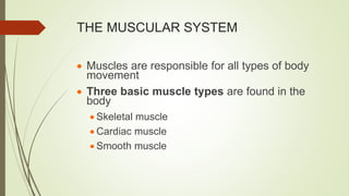
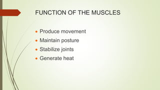
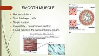
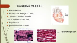
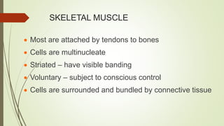
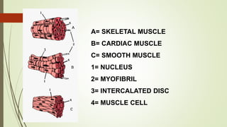
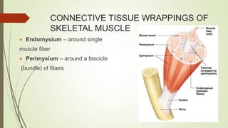
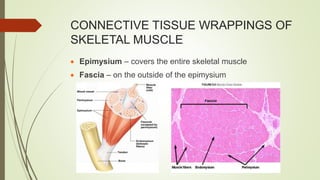
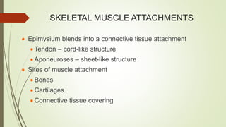
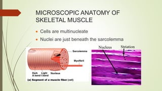
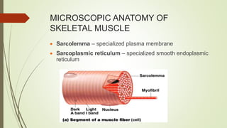
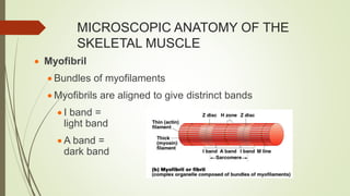
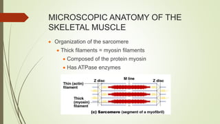
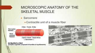
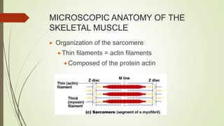
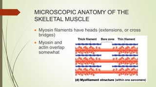
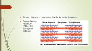
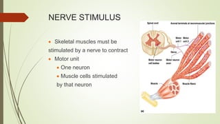
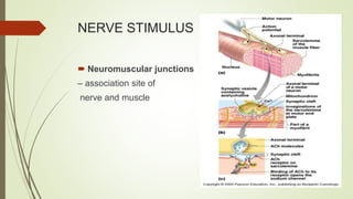
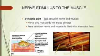
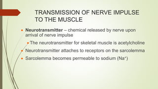
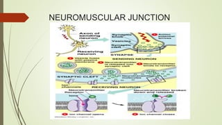
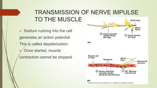
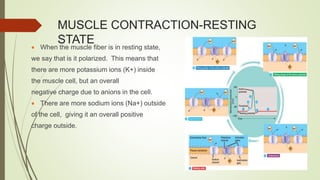
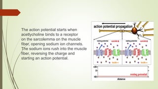
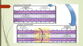
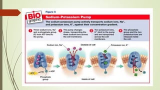
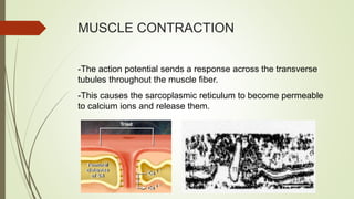
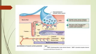
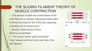
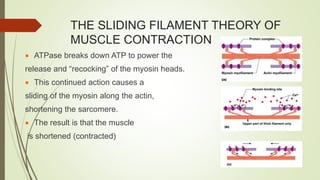
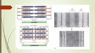
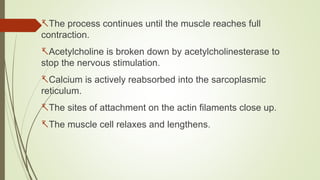
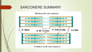
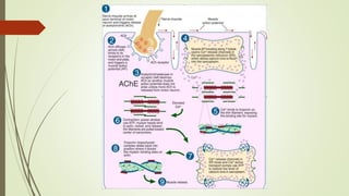
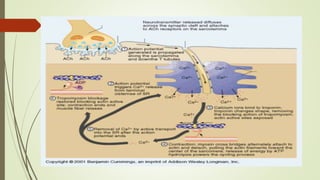
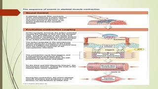
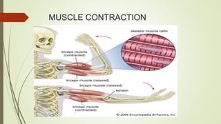
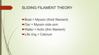
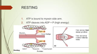
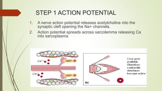
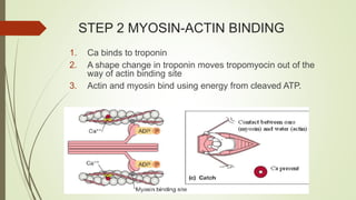
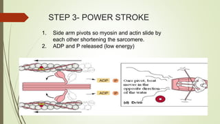
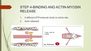
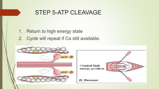
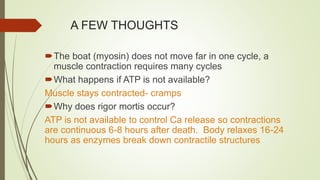
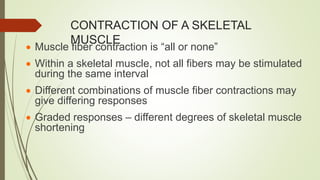
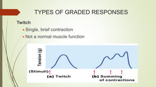
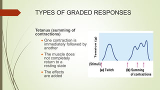
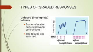
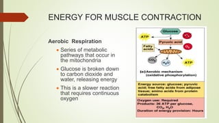
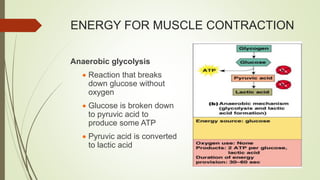
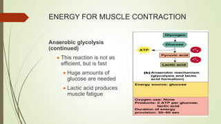
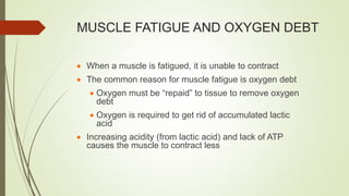
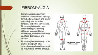
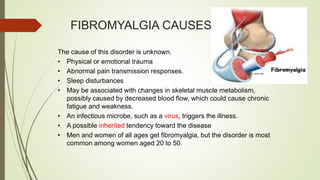
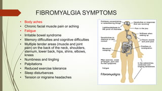
MUSCULAR SYSTEM
RIO D. DOMALAON,RN,MAN,JD
THE MUSCULAR SYSTEM
 Muscles are responsible for all types of body
movement
 Three basic muscle types are found in the
body
 Skeletal muscle
 Cardiac muscle
 Smooth muscle
FUNCTION OF THE MUSCLES
 Produce movement
 Maintain posture
 Stabilize joints
 Generate heat
CHARACTERISTICS OF MUSCLES
 Muscle cells are elongated
(muscle cell = muscle fiber)
 Contraction of muscles is due to the movement of
microfilaments
 All muscles share some terminology
Prefix myo refers to muscle
Prefix mys refers to muscle
Prefix sarco refers to flesh
SMOOTH MUSCLE
 Has no striations
 Spindle-shaped cells
 Single nucleus
 Involuntary – no conscious control
 Found mainly in the walls of hollow organs
CARDIAC MUSCLE
 Has striations
 Usually has a single nucleus
 Joined to another muscle
cell at an intercalated disc
 Involuntary
 Found only in the heart
Branching Fiber
SKELETAL MUSCLE
 Most are attached by tendons to bones
 Cells are multinucleate
 Striated – have visible banding
 Voluntary – subject to conscious control
 Cells are surrounded and bundled by connective tissue
A= SKELETAL MUSCLE
B= CARDIAC MUSCLE
C= SMOOTH MUSCLE
1= NUCLEUS
2= MYOFIBRIL
3= INTERCALATED DISC
4= MUSCLE CELL
CONNECTIVE TISSUE WRAPPINGS OF
SKELETAL MUSCLE
 Endomysium – around single
muscle fiber
 Perimysium – around a fascicle
(bundle) of fibers
CONNECTIVE TISSUE WRAPPINGS OF
SKELETAL MUSCLE
 Epimysium – covers the entire skeletal muscle
 Fascia – on the outside of the epimysium
SKELETAL MUSCLE ATTACHMENTS
 Epimysium blends into a connective tissue attachment
 Tendon – cord-like structure
 Aponeuroses – sheet-like structure
 Sites of muscle attachment
 Bones
 Cartilages
 Connective tissue covering
MICROSCOPIC ANATOMY OF
SKELETAL MUSCLE
 Cells are multinucleate
 Nuclei are just beneath the sarcolemma
MICROSCOPIC ANATOMY OF
SKELETAL MUSCLE
 Sarcolemma – specialized plasma membrane
 Sarcoplasmic reticulum – specialized smooth endoplasmic
reticulum
MICROSCOPIC ANATOMY OF THE
SKELETAL MUSCLE
 Myofibril
 Bundles of myofilaments
 Myofibrils are aligned to give distrinct bands
 I band =
light band
 A band =
dark band
MICROSCOPIC ANATOMY OF THE
SKELETAL MUSCLE
 Organization of the sarcomere
 Thick filaments = myosin filaments
 Composed of the protein myosin
 Has ATPase enzymes
MICROSCOPIC ANATOMY OF THE
SKELETAL MUSCLE
 Sarcomere
 Contractile unit of a muscle fiber
MICROSCOPIC ANATOMY OF THE
SKELETAL MUSCLE
 Organization of the sarcomere
 Thin filaments = actin filaments
 Composed of the protein actin
MICROSCOPIC ANATOMY OF THE
SKELETAL MUSCLE
 Myosin filaments have heads (extensions, or cross
bridges)
 Myosin and
actin overlap
somewhat
 At rest, there is a bare zone that lacks actin filaments
 Sarcoplasmic
reticulum
(SR) – for
storage of
calcium
PROPERTIES OF SKELETAL ACTIVITY
 Irritability – ability to receive and respond to a
stimulus
 Contractility – ability to shorten when an
adequate stimulus is received
NERVE STIMULUS
 Skeletal muscles must be
stimulated by a nerve to contract
 Motor unit
 One neuron
 Muscle cells stimulated
by that neuron
NERVE STIMULUS
 Neuromuscular junctions
– association site of
nerve and muscle
NERVE STIMULUS TO THE MUSCLE
 Synaptic cleft – gap between nerve and muscle
 Nerve and muscle do not make contact
 Area between nerve and muscle is filled with interstitial fluid
TRANSMISSION OF NERVE IMPULSE
TO THE MUSCLE
 Neurotransmitter – chemical released by nerve upon
arrival of nerve impulse
 The neurotransmitter for skeletal muscle is acetylcholine
 Neurotransmitter attaches to receptors on the sarcolemma
 Sarcolemma becomes permeable to sodium (Na+)
NEUROMUSCULAR JUNCTION
TRANSMISSION OF NERVE IMPULSE
TO THE MUSCLE
 Sodium rushing into the cell
generates an action potential.
This is called depolarization.
 Once started, muscle
contraction cannot be stopped
MUSCLE CONTRACTION-RESTING
STATE
 When the muscle fiber is in resting state,
we say that is it polarized. This means that
there are more potassium ions (K+) inside
the muscle cell, but an overall
negative charge due to anions in the cell.
 There are more sodium ions (Na+) outside
of the cell, giving it an overall positive
charge outside.
The action potential starts when
acetlycholine binds to a receptor
on the sarcolemma on the muscle
fiber, opening sodium ion channels.
The sodium ions rush into the muscle
fiber, reversing the charge and
starting an action potential.
MUSCLE CONTRACTION
-The action potential sends a response across the transverse
tubules throughout the muscle fiber.
-This causes the sarcoplasmic reticulum to become permeable
to calcium ions and release them.
THE SLIDING FILAMENT THEORY OF
MUSCLE CONTRACTION
 The calcium causes the conformation of the
actin filament to change, exposing binding sites.
It moves the troponin out of the way, exposing
a binding site on tropomyosin.
 Myosin heads bond to these,
forming crossbridges
 The myosin heads “grab and swivel”
then release and bind to the next site of the
thin filament
THE SLIDING FILAMENT THEORY OF
MUSCLE CONTRACTION
 ATPase breaks down ATP to power the
release and “recocking” of the myosin heads.
 This continued action causes a
sliding of the myosin along the actin,
shortening the sarcomere.
 The result is that the muscle
is shortened (contracted)
The process continues until the muscle reaches full
contraction.
Acetylcholine is broken down by acetylcholinesterase to
stop the nervous stimulation.
Calcium is actively reabsorbed into the sarcoplasmic
reticulum.
The sites of attachment on the actin filaments close up.
The muscle cell relaxes and lengthens.
SARCOMERE SUMMARY
MUSCLE CONTRACTION
SLIDING FILAMENT THEORY
Boat = Myosin (thick filament)
Oar = Myosin side arm
Water = Actin (thin filament)
Life ring = Calcium
RESTING
1. ATP is bound to myosin side arm.
2. ATP cleaves into ADP + P (high energy)
STEP 1 ACTION POTENTIAL
1. A nerve action potential releases acetylcholine into the
synaptic cleft opening the Na+ channels.
2. Action potential spreads across sarcolemma releasing Ca
into sarcoplasma
STEP 2 MYOSIN-ACTIN BINDING
1. Ca binds to troponin
2. A shape change in troponin moves tropomyocin out of the
way of actin binding site
3. Actin and myosin bind using energy from cleaved ATP.
STEP 3- POWER STROKE
1. Side arm pivots so myosin and actin slide by
each other shortening the sarcomere.
2. ADP and P released (low energy)
STEP 4-BINDING AND ACTIN-MYOSIN
RELEASE
1. A different ATPmolecule binds to active site.
2. Actin released
STEP 5-ATP CLEAVAGE
1. Return to high energy state
2. Cycle will repeat if Ca still available.
A FEW THOUGHTS
The boat (myosin) does not move far in one cycle, a
muscle contraction requires many cycles
What happens if ATP is not available?
Muscle stays contracted- cramps
Why does rigor mortis occur?
ATP is not available to control Ca release so contractions
are continuous 6-8 hours after death. Body relaxes 16-24
hours as enzymes break down contractile structures
CONTRACTION OF A SKELETAL
MUSCLE
 Muscle fiber contraction is “all or none”
 Within a skeletal muscle, not all fibers may be stimulated
during the same interval
 Different combinations of muscle fiber contractions may
give differing responses
 Graded responses – different degrees of skeletal muscle
shortening
TYPES OF GRADED RESPONSES
Twitch
 Single, brief contraction
 Not a normal muscle function
TYPES OF GRADED RESPONSES
Tetanus (summing of
contractions)
 One contraction is
immediately followed by
another
 The muscle does
not completely
return to a
resting state
 The effects
are added
TYPES OF GRADED RESPONSES
Unfused (incomplete)
tetanus
 Some relaxation
occurs between
contractions
 The results are
summed
TYPES OF GRADED RESPONSES
Fused (complete)
tetanus
 No evidence of
relaxation before the
following contractions
 The result is a
sustained muscle
contraction
MUSCLE RESPONSE TO STRONG
STIMULI
 Muscle force depends upon the
number of fibers stimulated
 More fibers contracting results in
greater muscle tension
 Muscles can continue to contract
unless they run out of energy
ENERGY FOR MUSCLE CONTRACTION
 Initially, muscles used stored ATP for energy
 Bonds of ATP are broken to release energy
 Only 4-6 seconds worth of ATP is stored by muscles
 After this initial time, other pathways must be utilized to
produce ATP
ENERGY FOR MUSCLE CONTRACTION
Direct phosphorylation
 Muscle cells contain
creatine phosphate (CP)
 CP is a high-energy
molecule
 After ATP is depleted,
ADP is left
 CP transfers energy to
ADP, to regenerate ATP
 CP supplies are
exhausted in about 20
seconds
ENERGY FOR MUSCLE CONTRACTION
Aerobic Respiration
 Series of metabolic
pathways that occur in
the mitochondria
 Glucose is broken down
to carbon dioxide and
water, releasing energy
 This is a slower reaction
that requires continuous
oxygen
ENERGY FOR MUSCLE CONTRACTION
Anaerobic glycolysis
 Reaction that breaks
down glucose without
oxygen
 Glucose is broken down
to pyruvic acid to
produce some ATP
 Pyruvic acid is converted
to lactic acid
ENERGY FOR MUSCLE CONTRACTION
Anaerobic glycolysis
(continued)
 This reaction is not as
efficient, but is fast
 Huge amounts of
glucose are needed
 Lactic acid produces
muscle fatigue
MUSCLE FATIGUE AND OXYGEN DEBT
 When a muscle is fatigued, it is unable to contract
 The common reason for muscle fatigue is oxygen debt
 Oxygen must be “repaid” to tissue to remove oxygen
debt
 Oxygen is required to get rid of accumulated lactic
acid
 Increasing acidity (from lactic acid) and lack of ATP
causes the muscle to contract less
TYPES OF MUSCLE CONTRACTION
Isotonic contractions
 Myofilaments are able to slide past each other during
contractions
 The muscle shortens and movement results
 Ex.- Lifting a light weight, turning your head
Isometric contractions
 Tension in the muscles increases
 The muscle is unable to shorten
 Ex.- Trying to lift something too heavy, legs pressing on the
floor while standing
MUSCLE TONE
 Some fibers are contracted even in a relaxed muscle
 Different fibers contract at different times to provide
muscle tone
 The process of stimulating various fibers is under
involuntary control
MUSCLES AND BODY MOVEMENT
Movement is
attained due to a
muscle moving an
attached bone
MUSCLES AND BODY MOVEMENT
 Muscles are attached to at
least two points
 Origin – attachment to a
immoveable bone
 Insertion – attachment to
an movable bone
EFFECTS OF EXERCISE ON MUSCLE
Results of increased muscle use
 Increase in muscle size (more protein fibers
added to existing muscle fibers)
 Increase in muscle strength
 Increase in muscle efficiency
 Muscle becomes more fatigue resistant
(increase in number of mitochondria)
TYPES OF ORDINARY BODY
MOVEMENTS
 Flexion- decreasing the angle of the joint
 Ex.- Bending your arm or fingers, bringing your
chin towards your chest
 Extension- increasing the angle of the joint
 Ex.- Straightening a bent arm or fingers
 Rotation- turning on an axis
 Ex.- Turning your head side to side, “Parade
wave”
TYPES OF ORDINARY BODY
MOVEMENTS
 Abduction- move away from the midline
 Ex.- Moving clasped hands away from one another
 Adduction- move toward the midline
 Ex.- Bringing your hands together to clap
 Circumduction- cone-like movement in space
 Ex.- Moving your arm, phalanges! or leg around in a circle
BODY MOVEMENTS
SPECIAL MOVEMENTS
 Dorsiflexion- pointing toe upward
 Plantar flexion- pointing toe downward
 Inversion- turning sole of foot medially
 Eversion- turning sole of foot laterally
 Supination- downward to upward motion of the hand
 Pronation- upward to downward motion of the hand
TYPES OF MUSCLES
 Prime mover – muscle with the major responsibility for a
certain movement
 Antagonist – muscle that opposes or reverses a prime mover
 Synergist – muscle that aids a prime mover in a movement
and helps prevent rotation
 Fixator – stabilizes the origin of a prime mover
NAMING OF SKELETAL MUSCLES
Direction of muscle fibers
 Example: rectus
(straight)
Relative size of the muscle
 Example: maximus
(largest)
NAMING OF SKELETAL MUSCLES
Location of the muscle
 Example: many
muscles are named
for bones (e.g.,
temporalis)
Number of origins
 Example: triceps
(three heads)
NAMING OF SKELETAL MUSCLES
Location of the muscles
origin and insertion
 Example: sterno (on
the sternum)
NAMING OF SKELETAL MUSCLES
Shape of the muscle
 Example: deltoid
(triangular)
Action of the muscle
 Example: flexor and
extensor (flexes or
extends a bone)
DEVELOPMENTAL ASPECTS
A pregnant mother can feel the muscular contractions of her baby often
by the 16th week of development. (quickening)
A newborn’s movements are very gross at first, and then develop in a
cephalo-caudal and proximal-distal fashion.
Muscle disuse causes atrophy (decrease in mass) of the tissue.
Over time, the connective tissue and muscle mass decreases,
lowering the strength.
MUSCULAR DISORDERS
MYOSITIS
• Myositis is inflammation of your skeletal muscles, which
are also called the voluntary muscles. These are the
muscles you consciously control that help you move
your body. An injury, infection or autoimmune disease
can cause myositis.
• The diseases dermatomyositis and polymyositis both
involve myositis. Polymyositis causes muscle weakness,
usually in the muscles closest to the trunk of your body.
Dermatomyositis causes muscle weakness, plus a skin
rash. Both diseases are usually treated with prednisone,
a steroid medicine, and sometimes other medicines.
FIBROMYALGIA
• Fibromyalgia is a common
condition characterized by long-
term, body-wide pain and tender
points in joints, muscles,
tendons, and other soft tissues.
Fibromyalgia has also been
linked to fatigue, morning
stiffness, sleep problems,
headaches, numbness in hands
and feet, depression, and
anxiety.
• Fibromyalgia can develop on its
own or along with other
musculoskeletal conditions such
as rheumatoid arthritis or lupus.
FIBROMYALGIA CAUSES
The cause of this disorder is unknown.
• Physical or emotional trauma
• Abnormal pain transmission responses.
• Sleep disturbances
• May be associated with changes in skeletal muscle metabolism,
possibly caused by decreased blood flow, which could cause chronic
fatigue and weakness.
• An infectious microbe, such as a virus, triggers the illness.
• A possible inherited tendency toward the disease
• Men and women of all ages get fibromyalgia, but the disorder is most
common among women aged 20 to 50.
FIBROMYALGIA SYMPTOMS
• Body aches
• Chronic facial muscle pain or aching
• Fatigue
• Irritable bowel syndrome
• Memory difficulties and cognitive difficulties
• Multiple tender areas (muscle and joint
pain) on the back of the neck, shoulders,
sternum, lower back, hips, shins, elbows,
knees
• Numbness and tingling
• Palpitations
• Reduced exercise tolerance
• Sleep disturbances
• Tension or migraine headaches
FIBROMYALGIA TREATMENTS
• In mild cases, symptoms may go away when stress is decreased or lifestyle
changes are implemented.
• Physical therapy and counseling are usually recommended.
• Pregabalin (Lyrica) ,Cymbalta
• Anti-inflammatory pain medications and medications that work on pain
transmission pathways.
• Eating a well-balanced diet and avoiding caffeine may help with problems
sleeping, and may help reduce the severity of the symptoms. Lifestyle
measures to improve the quality of sleep can be effective for fibromyalgia.
• Some reports indicate that fish oil, magnesium/malic acid combinations, or
vitamins may be effective.
• Reducing stress and improving coping skills may also help reduce painful
symptoms.
MUSCULAR DYSTROPHY
• Muscular dystrophy is a group of disorders that involve muscle
weakness and loss of muscle tissue that get worse over time.
There are many kinds of Muscular Dystrophy
• Becker's muscular dystrophy
• Duchenne muscular dystrophy
• Emery-Dreifuss muscular dystrophy
• Facioscapulohumeral muscular dystrophy
• Limb-girdle muscular dystrophy
• Myotonia congenita
• Myotonic dystrophy
MUSCULAR DYSTROPHY CAUSE
• Duchenne muscular dystrophy is a rapidly-worsening form of muscular dystrophy.
Other muscular dystrophies (including Becker's muscular dystrophy) get worse
much more slowly.
• Duchenne muscular dystrophy is caused by a defective gene for dystrophin (a
protein in the muscles). However, it often occurs in people without a known family
history of the condition.
• Because of the way the disease is inherited, males are more likely to develop
symptoms than are women. The sons of females who are carriers of the disease
(women with a defective gene but no symptoms themselves) each have a 50%
chance of having the disease. The daughters each have a 50% chance of being
carriers.
• Duchenne muscular dystrophy occurs in approximately 1 out of every 3,600 male
infants. Because this is an inherited disorder, risks include a family history of
Duchenne muscular dystrophy.
MUSCULAR DYSTROPHY SYMPTOMS
• Symptoms vary with the different types of muscular dystrophy.
• All of the muscles may be affected. Or, only specific groups of muscles may be
affected, such as those around the pelvis, shoulder, or face. Muscular dystrophy can
affect adults, but the more severe forms tend to occur in early childhood.
• Symptoms include:
• Mental retardation(only present in some types of the condition)
• Muscle weakness that slowly gets worse
– Delayed development of muscle motor skills
– Difficulty using one or more muscle groups
– Drooling
– Eyelid drooping (ptosis)
– Frequent falls
– Problems walking (delayed walking)
MUSCULAR DYSTROPHY TREATMENT
• There are no known cures for the various muscular dystrophies. The goal
of treatment is to control symptoms.
• Physical therapy may help patients maintain muscle strength and function.
Orthopedic appliances such as braces and wheelchairs can improve
mobility and self-care abilities. In some cases, surgery on the spine or
legs may help improve function.
• Corticosteroids taken by mouth are sometimes prescribed to children to
keep them walking for as along as possible (Reduce inflammation).
• The person should be as active as possible. Complete inactivity (such as
bedrest) can make the disease worse.
MYASTHENIA GRAVIS
 Myasthenia gravis is a
neuromuscular disorder
characterized by variable
weakness of voluntary
muscles, which often
improves with rest and
worsens with activity. The
condition is caused by an
abnormal immune
response.
MYASTHENIA GRAVIS CAUSES
• In myasthenia gravis, weakness occurs when the nerve impulse to initiate or
sustain movement does not adequately reach the muscle cells. This is caused
when immune cells target and attack the body's own cells (an autoimmune
response). This immune response produces antibodies that attach to affected
areas, preventing muscle cells from receiving chemical messages
(neurotransmitters) from the nerve cell.
• The cause of autoimmune disorders such as myasthenia gravis is unknown.
In some cases, myasthenia gravis may be associated with tumors of the
thymus (an organ of the immune system). Patients with myasthenia gravis
have a higher risk of having other autoimmune disorders like thyrotoxicosis,
rheumatoid arthritis, and systemic lupus erythematosus.
MYASTHENIA GRAVIS SYMPTOMS
• Muscle weakness, including:
– Swallowing difficulty, frequent gagging, or choking
– Paralysis
– Muscles that function best after rest
– Drooping head
– Difficulty climbing stairs
– Difficulty lifting objects
– Need to use hands to rise from sitting positions
– Difficulty talking
– Difficulty chewing
MYASTHENIA GRAVIS SYMPTOMS
• Vision problems:
– Double vision
– Difficulty maintaining steady gaze
– Eyelid drooping
• Additional symptoms that may be associated with this disease:
• Hoarseness or changing voice
• Fatigue
• Facial paralysis
• Drooling
• Breathing difficulty
MYASTHENIA GRAVIS TREATMENT
• There is no known cure for myasthenia gravis. However, treatment may result in prolonged
periods of remission.
• Lifestyle adjustments may enable continuation of many activities. Activity should be
planned to allow scheduled rest periods. An eye patch may be recommended if double
vision is bothersome. Stress and excessive heat exposure should be avoided because
they can worsen symptoms.
• Some medications, such as neostigmine or pyridostigmine, improve the communication
between the nerve and the muscle. Prednisone and other medications that suppress the
immune response (such as azathioprine, cyclosporine, or mycophenolate mofetil) may be
used if symptoms are severe and there is inadequate response to other medications.
• Plasmapheresis, a technique in which blood plasma containing antibodies against the
body is removed from the body and replaced with fluids (donated antibody-free plasma or
other intravenous fluids), may reduce symptoms for up to 4 - 6 weeks and is often used to
optimize conditions before surgery.
SPRAINS AND STRAINS
• Sprains and strains are common injuries that share similar signs and symptoms, but
involve different parts of your body.
• A sprain is a stretching or tearing of ligaments — the tough bands of fibrous tissue that
connect one bone to another in your joints. The most common location for a sprain is in
your ankle.
• A strain is a stretching or tearing of muscle or tendon. A tendon is a fibrous cord of tissue
that connects muscles to bones. Strains often occur in the lower back and in the
hamstring muscle in the back of your thigh.
• These cause pain and swelling around the affected site.
SPRAINS AND STRAINS TREATMENT
 Medications. For mild sprains and strains, your doctor likely will recommend basic self-
care measures and an over-the-counter pain reliever such as ibuprofen (Advil, Motrin,
others) or acetaminophen (Tylenol, others).
 Therapy. In cases of a mild or moderate sprain or strain, apply ice to the area as soon
as possible to minimize swelling. In cases of severe sprain or strain, your doctor may
immobilize the area with a brace or splint.
 Surgery. If you have a torn ligament or ruptured muscle, surgery may be an option.
REFERENCES
Copyright © 2003 Pearson Education, Inc.
publishing as Benjamin Cummings
Marieb,E.N. (2019).Essentials of Human
Anatomy and
Physiology.Jurong,Singapore:Pearson
Education South Asia Pte Ltd.
www.youtube.com

MUSCULAR SYSTEM 7.pptx

  • 1.
    MUSCULAR SYSTEM RIO D.DOMALAON,RN,MAN,JD
  • 3.
    THE MUSCULAR SYSTEM Muscles are responsible for all types of body movement  Three basic muscle types are found in the body  Skeletal muscle  Cardiac muscle  Smooth muscle
  • 4.
    FUNCTION OF THEMUSCLES  Produce movement  Maintain posture  Stabilize joints  Generate heat
  • 5.
    CHARACTERISTICS OF MUSCLES Muscle cells are elongated (muscle cell = muscle fiber)  Contraction of muscles is due to the movement of microfilaments  All muscles share some terminology Prefix myo refers to muscle Prefix mys refers to muscle Prefix sarco refers to flesh
  • 6.
    SMOOTH MUSCLE  Hasno striations  Spindle-shaped cells  Single nucleus  Involuntary – no conscious control  Found mainly in the walls of hollow organs
  • 7.
    CARDIAC MUSCLE  Hasstriations  Usually has a single nucleus  Joined to another muscle cell at an intercalated disc  Involuntary  Found only in the heart Branching Fiber
  • 8.
    SKELETAL MUSCLE  Mostare attached by tendons to bones  Cells are multinucleate  Striated – have visible banding  Voluntary – subject to conscious control  Cells are surrounded and bundled by connective tissue
  • 9.
    A= SKELETAL MUSCLE B=CARDIAC MUSCLE C= SMOOTH MUSCLE 1= NUCLEUS 2= MYOFIBRIL 3= INTERCALATED DISC 4= MUSCLE CELL
  • 10.
    CONNECTIVE TISSUE WRAPPINGSOF SKELETAL MUSCLE  Endomysium – around single muscle fiber  Perimysium – around a fascicle (bundle) of fibers
  • 11.
    CONNECTIVE TISSUE WRAPPINGSOF SKELETAL MUSCLE  Epimysium – covers the entire skeletal muscle  Fascia – on the outside of the epimysium
  • 12.
    SKELETAL MUSCLE ATTACHMENTS Epimysium blends into a connective tissue attachment  Tendon – cord-like structure  Aponeuroses – sheet-like structure  Sites of muscle attachment  Bones  Cartilages  Connective tissue covering
  • 13.
    MICROSCOPIC ANATOMY OF SKELETALMUSCLE  Cells are multinucleate  Nuclei are just beneath the sarcolemma
  • 14.
    MICROSCOPIC ANATOMY OF SKELETALMUSCLE  Sarcolemma – specialized plasma membrane  Sarcoplasmic reticulum – specialized smooth endoplasmic reticulum
  • 15.
    MICROSCOPIC ANATOMY OFTHE SKELETAL MUSCLE  Myofibril  Bundles of myofilaments  Myofibrils are aligned to give distrinct bands  I band = light band  A band = dark band
  • 16.
    MICROSCOPIC ANATOMY OFTHE SKELETAL MUSCLE  Organization of the sarcomere  Thick filaments = myosin filaments  Composed of the protein myosin  Has ATPase enzymes
  • 17.
    MICROSCOPIC ANATOMY OFTHE SKELETAL MUSCLE  Sarcomere  Contractile unit of a muscle fiber
  • 18.
    MICROSCOPIC ANATOMY OFTHE SKELETAL MUSCLE  Organization of the sarcomere  Thin filaments = actin filaments  Composed of the protein actin
  • 19.
    MICROSCOPIC ANATOMY OFTHE SKELETAL MUSCLE  Myosin filaments have heads (extensions, or cross bridges)  Myosin and actin overlap somewhat
  • 20.
     At rest,there is a bare zone that lacks actin filaments  Sarcoplasmic reticulum (SR) – for storage of calcium
  • 21.
    PROPERTIES OF SKELETALACTIVITY  Irritability – ability to receive and respond to a stimulus  Contractility – ability to shorten when an adequate stimulus is received
  • 22.
    NERVE STIMULUS  Skeletalmuscles must be stimulated by a nerve to contract  Motor unit  One neuron  Muscle cells stimulated by that neuron
  • 23.
    NERVE STIMULUS  Neuromuscularjunctions – association site of nerve and muscle
  • 24.
    NERVE STIMULUS TOTHE MUSCLE  Synaptic cleft – gap between nerve and muscle  Nerve and muscle do not make contact  Area between nerve and muscle is filled with interstitial fluid
  • 25.
    TRANSMISSION OF NERVEIMPULSE TO THE MUSCLE  Neurotransmitter – chemical released by nerve upon arrival of nerve impulse  The neurotransmitter for skeletal muscle is acetylcholine  Neurotransmitter attaches to receptors on the sarcolemma  Sarcolemma becomes permeable to sodium (Na+)
  • 26.
  • 27.
    TRANSMISSION OF NERVEIMPULSE TO THE MUSCLE  Sodium rushing into the cell generates an action potential. This is called depolarization.  Once started, muscle contraction cannot be stopped
  • 28.
    MUSCLE CONTRACTION-RESTING STATE  Whenthe muscle fiber is in resting state, we say that is it polarized. This means that there are more potassium ions (K+) inside the muscle cell, but an overall negative charge due to anions in the cell.  There are more sodium ions (Na+) outside of the cell, giving it an overall positive charge outside.
  • 29.
    The action potentialstarts when acetlycholine binds to a receptor on the sarcolemma on the muscle fiber, opening sodium ion channels. The sodium ions rush into the muscle fiber, reversing the charge and starting an action potential.
  • 33.
    MUSCLE CONTRACTION -The actionpotential sends a response across the transverse tubules throughout the muscle fiber. -This causes the sarcoplasmic reticulum to become permeable to calcium ions and release them.
  • 35.
    THE SLIDING FILAMENTTHEORY OF MUSCLE CONTRACTION  The calcium causes the conformation of the actin filament to change, exposing binding sites. It moves the troponin out of the way, exposing a binding site on tropomyosin.  Myosin heads bond to these, forming crossbridges  The myosin heads “grab and swivel” then release and bind to the next site of the thin filament
  • 36.
    THE SLIDING FILAMENTTHEORY OF MUSCLE CONTRACTION  ATPase breaks down ATP to power the release and “recocking” of the myosin heads.  This continued action causes a sliding of the myosin along the actin, shortening the sarcomere.  The result is that the muscle is shortened (contracted)
  • 39.
    The process continuesuntil the muscle reaches full contraction. Acetylcholine is broken down by acetylcholinesterase to stop the nervous stimulation. Calcium is actively reabsorbed into the sarcoplasmic reticulum. The sites of attachment on the actin filaments close up. The muscle cell relaxes and lengthens.
  • 40.
  • 44.
  • 45.
    SLIDING FILAMENT THEORY Boat= Myosin (thick filament) Oar = Myosin side arm Water = Actin (thin filament) Life ring = Calcium
  • 46.
    RESTING 1. ATP isbound to myosin side arm. 2. ATP cleaves into ADP + P (high energy)
  • 47.
    STEP 1 ACTIONPOTENTIAL 1. A nerve action potential releases acetylcholine into the synaptic cleft opening the Na+ channels. 2. Action potential spreads across sarcolemma releasing Ca into sarcoplasma
  • 48.
    STEP 2 MYOSIN-ACTINBINDING 1. Ca binds to troponin 2. A shape change in troponin moves tropomyocin out of the way of actin binding site 3. Actin and myosin bind using energy from cleaved ATP.
  • 49.
    STEP 3- POWERSTROKE 1. Side arm pivots so myosin and actin slide by each other shortening the sarcomere. 2. ADP and P released (low energy)
  • 50.
    STEP 4-BINDING ANDACTIN-MYOSIN RELEASE 1. A different ATPmolecule binds to active site. 2. Actin released
  • 51.
    STEP 5-ATP CLEAVAGE 1.Return to high energy state 2. Cycle will repeat if Ca still available.
  • 52.
    A FEW THOUGHTS Theboat (myosin) does not move far in one cycle, a muscle contraction requires many cycles What happens if ATP is not available? Muscle stays contracted- cramps Why does rigor mortis occur? ATP is not available to control Ca release so contractions are continuous 6-8 hours after death. Body relaxes 16-24 hours as enzymes break down contractile structures
  • 54.
    CONTRACTION OF ASKELETAL MUSCLE  Muscle fiber contraction is “all or none”  Within a skeletal muscle, not all fibers may be stimulated during the same interval  Different combinations of muscle fiber contractions may give differing responses  Graded responses – different degrees of skeletal muscle shortening
  • 55.
    TYPES OF GRADEDRESPONSES Twitch  Single, brief contraction  Not a normal muscle function
  • 56.
    TYPES OF GRADEDRESPONSES Tetanus (summing of contractions)  One contraction is immediately followed by another  The muscle does not completely return to a resting state  The effects are added
  • 57.
    TYPES OF GRADEDRESPONSES Unfused (incomplete) tetanus  Some relaxation occurs between contractions  The results are summed
  • 58.
    TYPES OF GRADEDRESPONSES Fused (complete) tetanus  No evidence of relaxation before the following contractions  The result is a sustained muscle contraction
  • 59.
    MUSCLE RESPONSE TOSTRONG STIMULI  Muscle force depends upon the number of fibers stimulated  More fibers contracting results in greater muscle tension  Muscles can continue to contract unless they run out of energy
  • 60.
    ENERGY FOR MUSCLECONTRACTION  Initially, muscles used stored ATP for energy  Bonds of ATP are broken to release energy  Only 4-6 seconds worth of ATP is stored by muscles  After this initial time, other pathways must be utilized to produce ATP
  • 61.
    ENERGY FOR MUSCLECONTRACTION Direct phosphorylation  Muscle cells contain creatine phosphate (CP)  CP is a high-energy molecule  After ATP is depleted, ADP is left  CP transfers energy to ADP, to regenerate ATP  CP supplies are exhausted in about 20 seconds
  • 62.
    ENERGY FOR MUSCLECONTRACTION Aerobic Respiration  Series of metabolic pathways that occur in the mitochondria  Glucose is broken down to carbon dioxide and water, releasing energy  This is a slower reaction that requires continuous oxygen
  • 63.
    ENERGY FOR MUSCLECONTRACTION Anaerobic glycolysis  Reaction that breaks down glucose without oxygen  Glucose is broken down to pyruvic acid to produce some ATP  Pyruvic acid is converted to lactic acid
  • 64.
    ENERGY FOR MUSCLECONTRACTION Anaerobic glycolysis (continued)  This reaction is not as efficient, but is fast  Huge amounts of glucose are needed  Lactic acid produces muscle fatigue
  • 65.
    MUSCLE FATIGUE ANDOXYGEN DEBT  When a muscle is fatigued, it is unable to contract  The common reason for muscle fatigue is oxygen debt  Oxygen must be “repaid” to tissue to remove oxygen debt  Oxygen is required to get rid of accumulated lactic acid  Increasing acidity (from lactic acid) and lack of ATP causes the muscle to contract less
  • 66.
    TYPES OF MUSCLECONTRACTION Isotonic contractions  Myofilaments are able to slide past each other during contractions  The muscle shortens and movement results  Ex.- Lifting a light weight, turning your head Isometric contractions  Tension in the muscles increases  The muscle is unable to shorten  Ex.- Trying to lift something too heavy, legs pressing on the floor while standing
  • 67.
    MUSCLE TONE  Somefibers are contracted even in a relaxed muscle  Different fibers contract at different times to provide muscle tone  The process of stimulating various fibers is under involuntary control
  • 68.
    MUSCLES AND BODYMOVEMENT Movement is attained due to a muscle moving an attached bone
  • 69.
    MUSCLES AND BODYMOVEMENT  Muscles are attached to at least two points  Origin – attachment to a immoveable bone  Insertion – attachment to an movable bone
  • 70.
    EFFECTS OF EXERCISEON MUSCLE Results of increased muscle use  Increase in muscle size (more protein fibers added to existing muscle fibers)  Increase in muscle strength  Increase in muscle efficiency  Muscle becomes more fatigue resistant (increase in number of mitochondria)
  • 71.
    TYPES OF ORDINARYBODY MOVEMENTS  Flexion- decreasing the angle of the joint  Ex.- Bending your arm or fingers, bringing your chin towards your chest  Extension- increasing the angle of the joint  Ex.- Straightening a bent arm or fingers  Rotation- turning on an axis  Ex.- Turning your head side to side, “Parade wave”
  • 72.
    TYPES OF ORDINARYBODY MOVEMENTS  Abduction- move away from the midline  Ex.- Moving clasped hands away from one another  Adduction- move toward the midline  Ex.- Bringing your hands together to clap  Circumduction- cone-like movement in space  Ex.- Moving your arm, phalanges! or leg around in a circle
  • 73.
  • 74.
    SPECIAL MOVEMENTS  Dorsiflexion-pointing toe upward  Plantar flexion- pointing toe downward  Inversion- turning sole of foot medially  Eversion- turning sole of foot laterally  Supination- downward to upward motion of the hand  Pronation- upward to downward motion of the hand
  • 75.
    TYPES OF MUSCLES Prime mover – muscle with the major responsibility for a certain movement  Antagonist – muscle that opposes or reverses a prime mover  Synergist – muscle that aids a prime mover in a movement and helps prevent rotation  Fixator – stabilizes the origin of a prime mover
  • 76.
    NAMING OF SKELETALMUSCLES Direction of muscle fibers  Example: rectus (straight) Relative size of the muscle  Example: maximus (largest)
  • 77.
    NAMING OF SKELETALMUSCLES Location of the muscle  Example: many muscles are named for bones (e.g., temporalis) Number of origins  Example: triceps (three heads)
  • 78.
    NAMING OF SKELETALMUSCLES Location of the muscles origin and insertion  Example: sterno (on the sternum)
  • 79.
    NAMING OF SKELETALMUSCLES Shape of the muscle  Example: deltoid (triangular) Action of the muscle  Example: flexor and extensor (flexes or extends a bone)
  • 80.
    DEVELOPMENTAL ASPECTS A pregnantmother can feel the muscular contractions of her baby often by the 16th week of development. (quickening) A newborn’s movements are very gross at first, and then develop in a cephalo-caudal and proximal-distal fashion. Muscle disuse causes atrophy (decrease in mass) of the tissue. Over time, the connective tissue and muscle mass decreases, lowering the strength.
  • 81.
  • 82.
    MYOSITIS • Myositis isinflammation of your skeletal muscles, which are also called the voluntary muscles. These are the muscles you consciously control that help you move your body. An injury, infection or autoimmune disease can cause myositis. • The diseases dermatomyositis and polymyositis both involve myositis. Polymyositis causes muscle weakness, usually in the muscles closest to the trunk of your body. Dermatomyositis causes muscle weakness, plus a skin rash. Both diseases are usually treated with prednisone, a steroid medicine, and sometimes other medicines.
  • 83.
    FIBROMYALGIA • Fibromyalgia isa common condition characterized by long- term, body-wide pain and tender points in joints, muscles, tendons, and other soft tissues. Fibromyalgia has also been linked to fatigue, morning stiffness, sleep problems, headaches, numbness in hands and feet, depression, and anxiety. • Fibromyalgia can develop on its own or along with other musculoskeletal conditions such as rheumatoid arthritis or lupus.
  • 84.
    FIBROMYALGIA CAUSES The causeof this disorder is unknown. • Physical or emotional trauma • Abnormal pain transmission responses. • Sleep disturbances • May be associated with changes in skeletal muscle metabolism, possibly caused by decreased blood flow, which could cause chronic fatigue and weakness. • An infectious microbe, such as a virus, triggers the illness. • A possible inherited tendency toward the disease • Men and women of all ages get fibromyalgia, but the disorder is most common among women aged 20 to 50.
  • 85.
    FIBROMYALGIA SYMPTOMS • Bodyaches • Chronic facial muscle pain or aching • Fatigue • Irritable bowel syndrome • Memory difficulties and cognitive difficulties • Multiple tender areas (muscle and joint pain) on the back of the neck, shoulders, sternum, lower back, hips, shins, elbows, knees • Numbness and tingling • Palpitations • Reduced exercise tolerance • Sleep disturbances • Tension or migraine headaches
  • 86.
    FIBROMYALGIA TREATMENTS • Inmild cases, symptoms may go away when stress is decreased or lifestyle changes are implemented. • Physical therapy and counseling are usually recommended. • Pregabalin (Lyrica) ,Cymbalta • Anti-inflammatory pain medications and medications that work on pain transmission pathways. • Eating a well-balanced diet and avoiding caffeine may help with problems sleeping, and may help reduce the severity of the symptoms. Lifestyle measures to improve the quality of sleep can be effective for fibromyalgia. • Some reports indicate that fish oil, magnesium/malic acid combinations, or vitamins may be effective. • Reducing stress and improving coping skills may also help reduce painful symptoms.
  • 87.
    MUSCULAR DYSTROPHY • Musculardystrophy is a group of disorders that involve muscle weakness and loss of muscle tissue that get worse over time. There are many kinds of Muscular Dystrophy • Becker's muscular dystrophy • Duchenne muscular dystrophy • Emery-Dreifuss muscular dystrophy • Facioscapulohumeral muscular dystrophy • Limb-girdle muscular dystrophy • Myotonia congenita • Myotonic dystrophy
  • 88.
    MUSCULAR DYSTROPHY CAUSE •Duchenne muscular dystrophy is a rapidly-worsening form of muscular dystrophy. Other muscular dystrophies (including Becker's muscular dystrophy) get worse much more slowly. • Duchenne muscular dystrophy is caused by a defective gene for dystrophin (a protein in the muscles). However, it often occurs in people without a known family history of the condition. • Because of the way the disease is inherited, males are more likely to develop symptoms than are women. The sons of females who are carriers of the disease (women with a defective gene but no symptoms themselves) each have a 50% chance of having the disease. The daughters each have a 50% chance of being carriers. • Duchenne muscular dystrophy occurs in approximately 1 out of every 3,600 male infants. Because this is an inherited disorder, risks include a family history of Duchenne muscular dystrophy.
  • 89.
    MUSCULAR DYSTROPHY SYMPTOMS •Symptoms vary with the different types of muscular dystrophy. • All of the muscles may be affected. Or, only specific groups of muscles may be affected, such as those around the pelvis, shoulder, or face. Muscular dystrophy can affect adults, but the more severe forms tend to occur in early childhood. • Symptoms include: • Mental retardation(only present in some types of the condition) • Muscle weakness that slowly gets worse – Delayed development of muscle motor skills – Difficulty using one or more muscle groups – Drooling – Eyelid drooping (ptosis) – Frequent falls – Problems walking (delayed walking)
  • 90.
    MUSCULAR DYSTROPHY TREATMENT •There are no known cures for the various muscular dystrophies. The goal of treatment is to control symptoms. • Physical therapy may help patients maintain muscle strength and function. Orthopedic appliances such as braces and wheelchairs can improve mobility and self-care abilities. In some cases, surgery on the spine or legs may help improve function. • Corticosteroids taken by mouth are sometimes prescribed to children to keep them walking for as along as possible (Reduce inflammation). • The person should be as active as possible. Complete inactivity (such as bedrest) can make the disease worse.
  • 91.
    MYASTHENIA GRAVIS  Myastheniagravis is a neuromuscular disorder characterized by variable weakness of voluntary muscles, which often improves with rest and worsens with activity. The condition is caused by an abnormal immune response.
  • 92.
    MYASTHENIA GRAVIS CAUSES •In myasthenia gravis, weakness occurs when the nerve impulse to initiate or sustain movement does not adequately reach the muscle cells. This is caused when immune cells target and attack the body's own cells (an autoimmune response). This immune response produces antibodies that attach to affected areas, preventing muscle cells from receiving chemical messages (neurotransmitters) from the nerve cell. • The cause of autoimmune disorders such as myasthenia gravis is unknown. In some cases, myasthenia gravis may be associated with tumors of the thymus (an organ of the immune system). Patients with myasthenia gravis have a higher risk of having other autoimmune disorders like thyrotoxicosis, rheumatoid arthritis, and systemic lupus erythematosus.
  • 93.
    MYASTHENIA GRAVIS SYMPTOMS •Muscle weakness, including: – Swallowing difficulty, frequent gagging, or choking – Paralysis – Muscles that function best after rest – Drooping head – Difficulty climbing stairs – Difficulty lifting objects – Need to use hands to rise from sitting positions – Difficulty talking – Difficulty chewing
  • 94.
    MYASTHENIA GRAVIS SYMPTOMS •Vision problems: – Double vision – Difficulty maintaining steady gaze – Eyelid drooping • Additional symptoms that may be associated with this disease: • Hoarseness or changing voice • Fatigue • Facial paralysis • Drooling • Breathing difficulty
  • 95.
    MYASTHENIA GRAVIS TREATMENT •There is no known cure for myasthenia gravis. However, treatment may result in prolonged periods of remission. • Lifestyle adjustments may enable continuation of many activities. Activity should be planned to allow scheduled rest periods. An eye patch may be recommended if double vision is bothersome. Stress and excessive heat exposure should be avoided because they can worsen symptoms. • Some medications, such as neostigmine or pyridostigmine, improve the communication between the nerve and the muscle. Prednisone and other medications that suppress the immune response (such as azathioprine, cyclosporine, or mycophenolate mofetil) may be used if symptoms are severe and there is inadequate response to other medications. • Plasmapheresis, a technique in which blood plasma containing antibodies against the body is removed from the body and replaced with fluids (donated antibody-free plasma or other intravenous fluids), may reduce symptoms for up to 4 - 6 weeks and is often used to optimize conditions before surgery.
  • 96.
    SPRAINS AND STRAINS •Sprains and strains are common injuries that share similar signs and symptoms, but involve different parts of your body. • A sprain is a stretching or tearing of ligaments — the tough bands of fibrous tissue that connect one bone to another in your joints. The most common location for a sprain is in your ankle. • A strain is a stretching or tearing of muscle or tendon. A tendon is a fibrous cord of tissue that connects muscles to bones. Strains often occur in the lower back and in the hamstring muscle in the back of your thigh. • These cause pain and swelling around the affected site.
  • 97.
    SPRAINS AND STRAINSTREATMENT  Medications. For mild sprains and strains, your doctor likely will recommend basic self- care measures and an over-the-counter pain reliever such as ibuprofen (Advil, Motrin, others) or acetaminophen (Tylenol, others).  Therapy. In cases of a mild or moderate sprain or strain, apply ice to the area as soon as possible to minimize swelling. In cases of severe sprain or strain, your doctor may immobilize the area with a brace or splint.  Surgery. If you have a torn ligament or ruptured muscle, surgery may be an option.
  • 99.
    REFERENCES Copyright © 2003Pearson Education, Inc. publishing as Benjamin Cummings Marieb,E.N. (2019).Essentials of Human Anatomy and Physiology.Jurong,Singapore:Pearson Education South Asia Pte Ltd. www.youtube.com