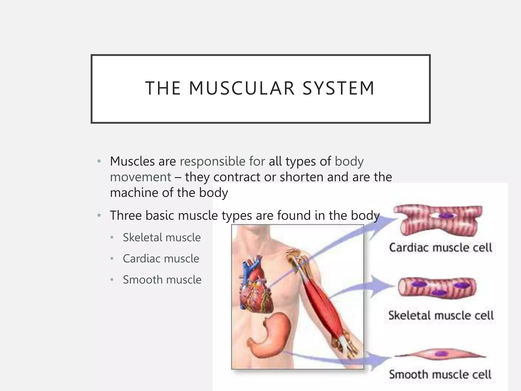
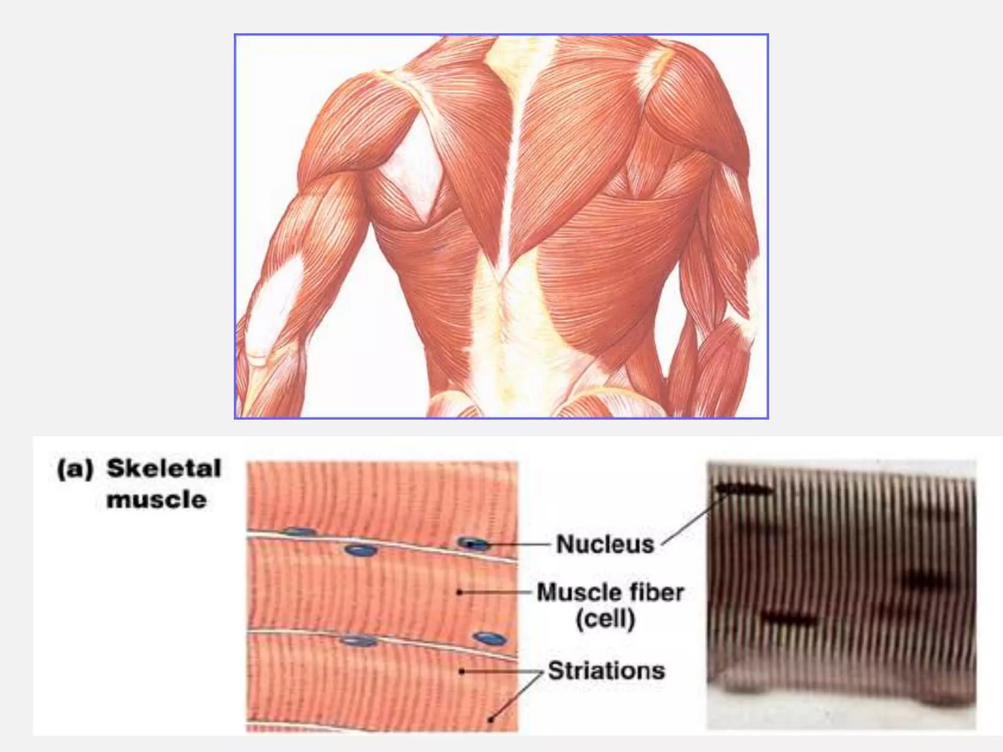
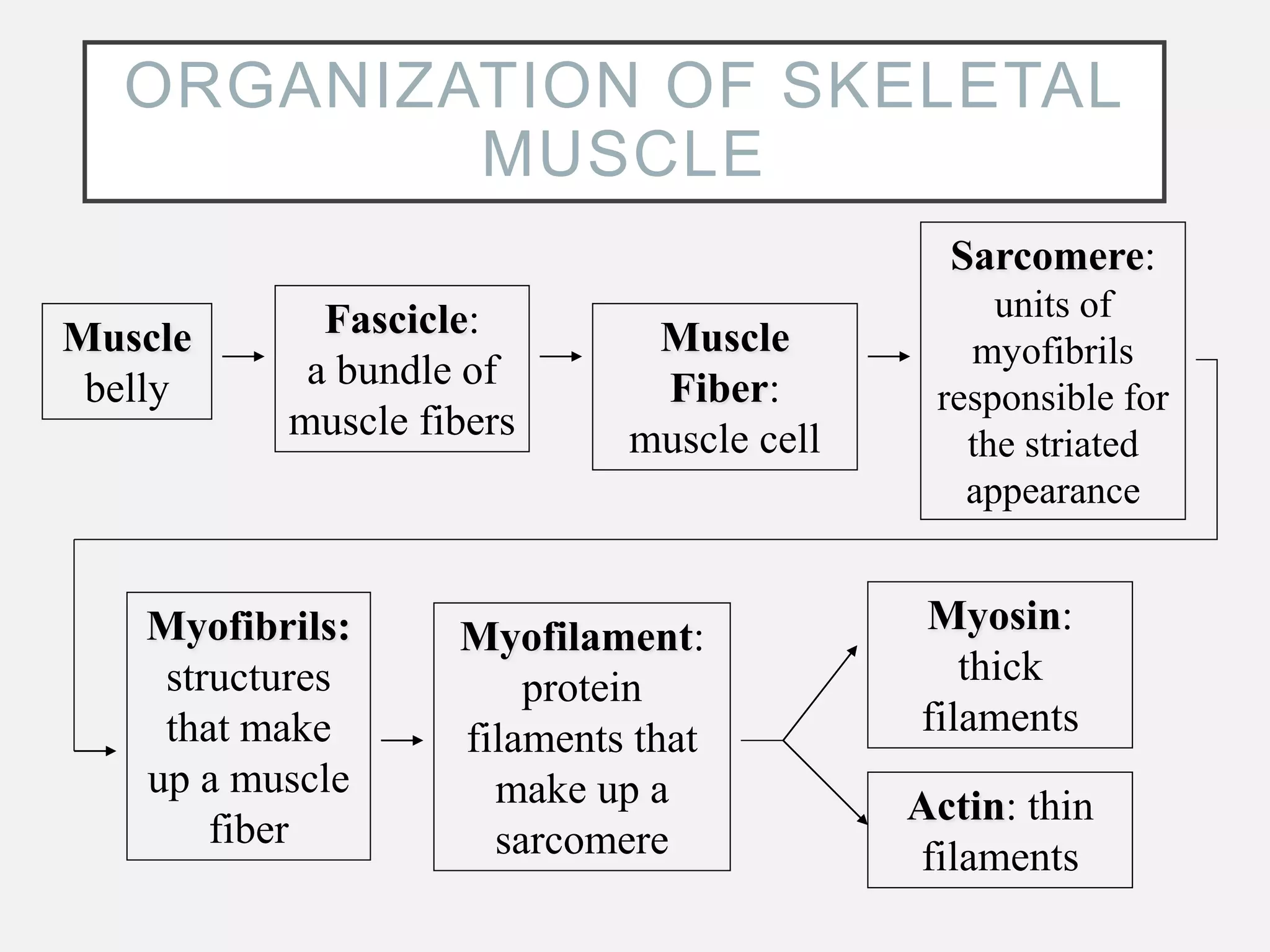
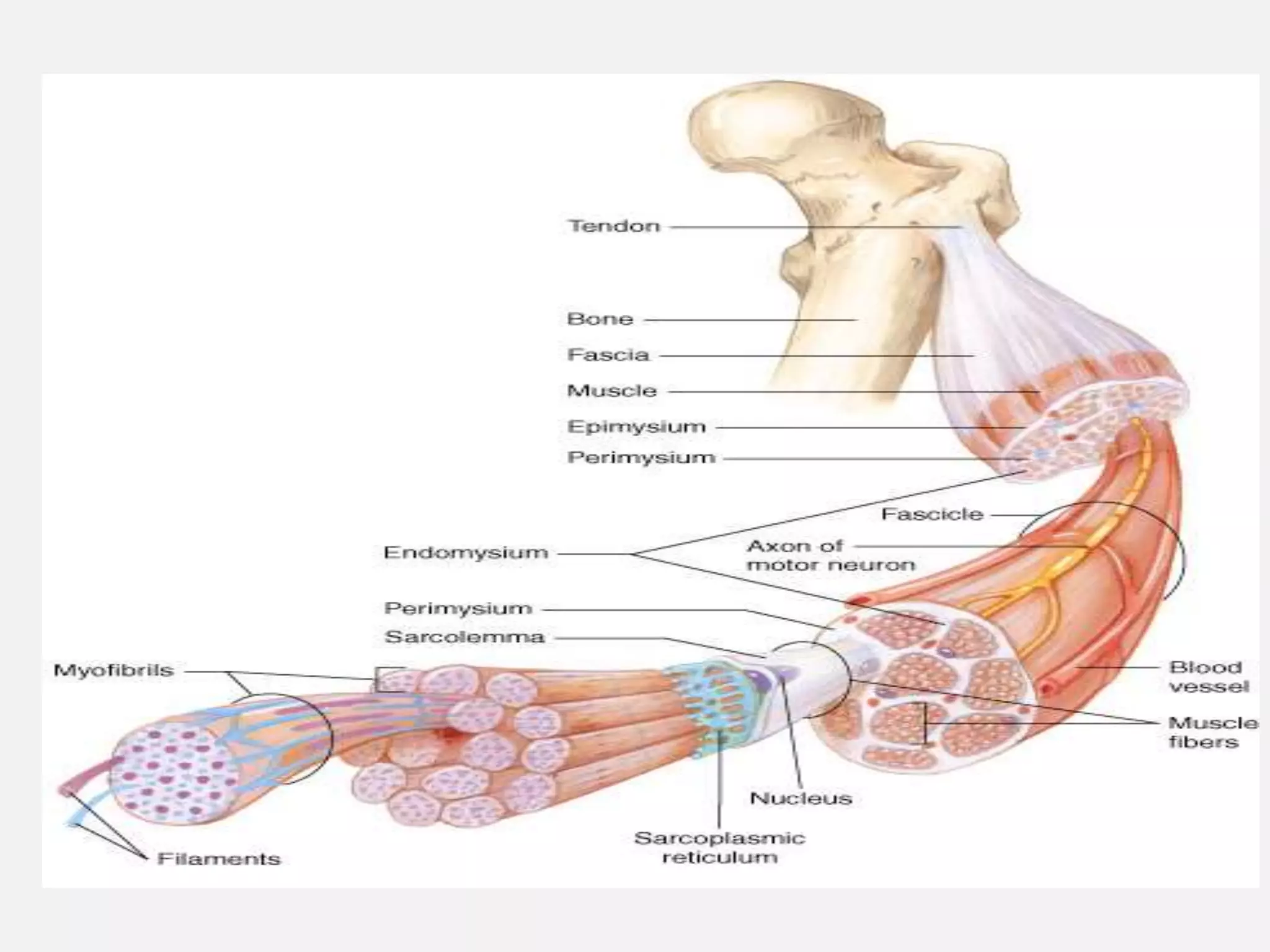
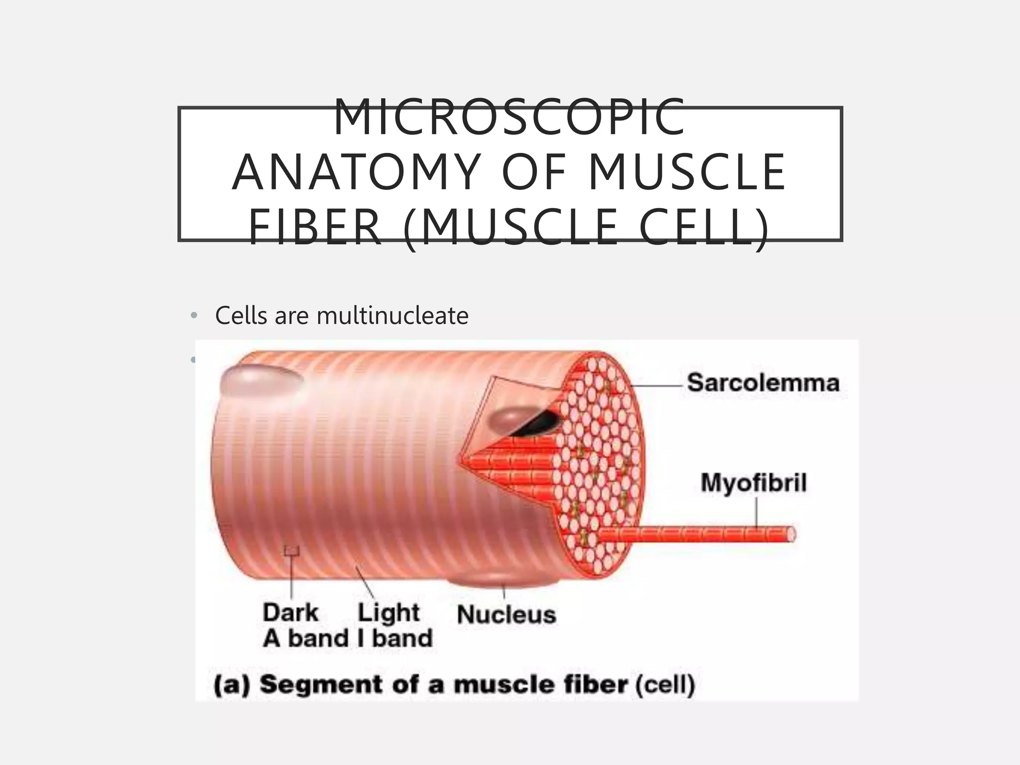
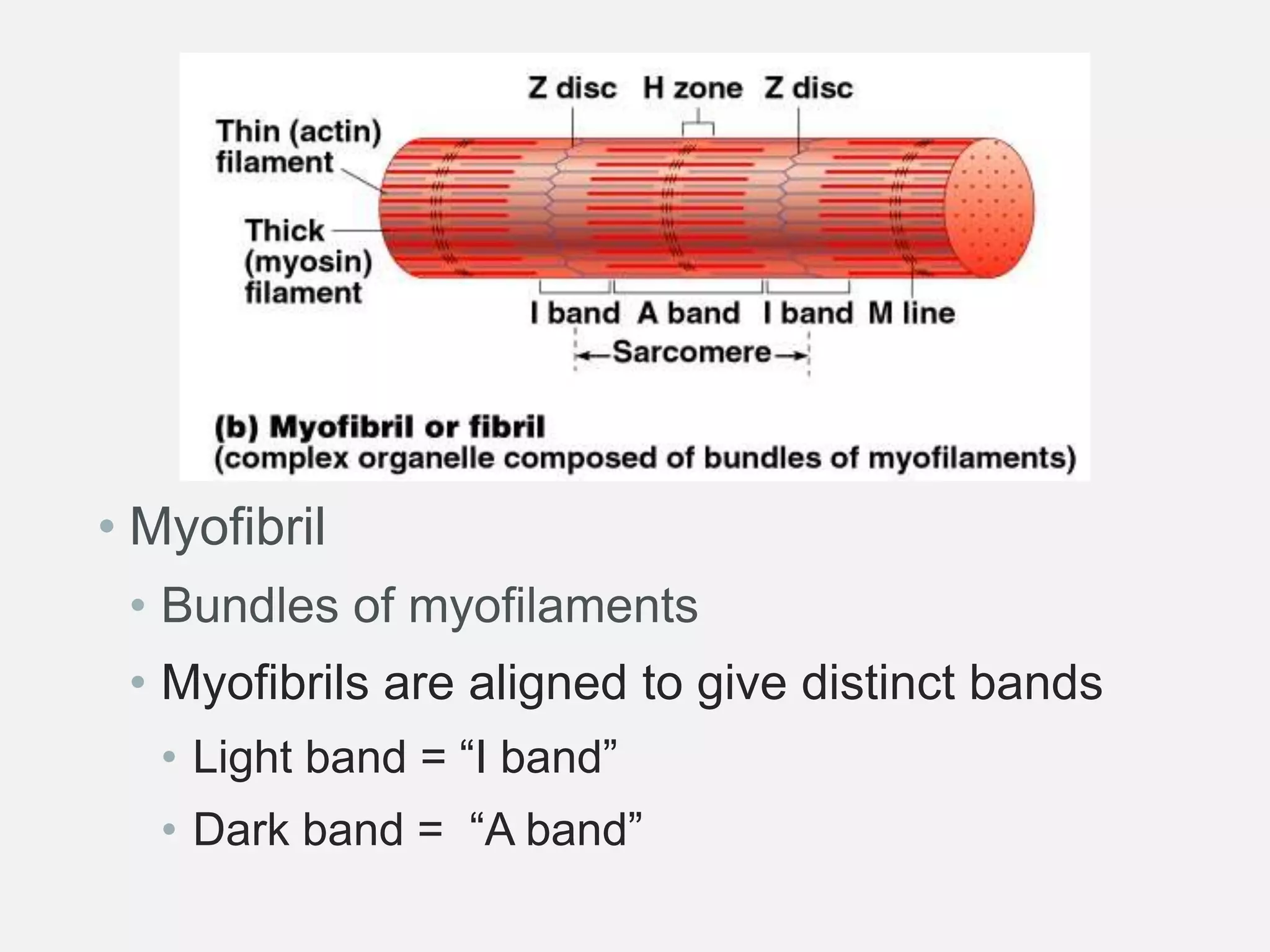
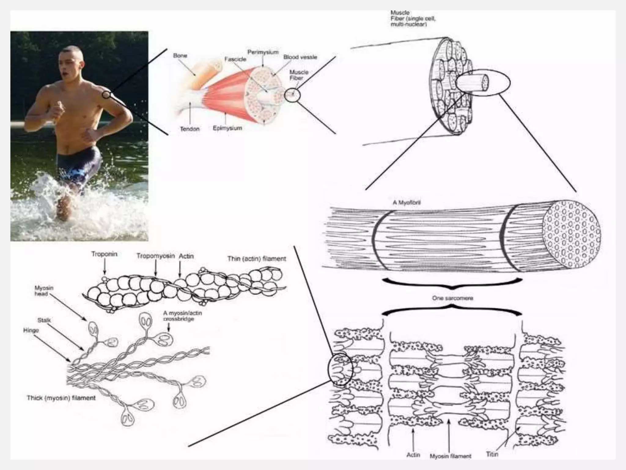
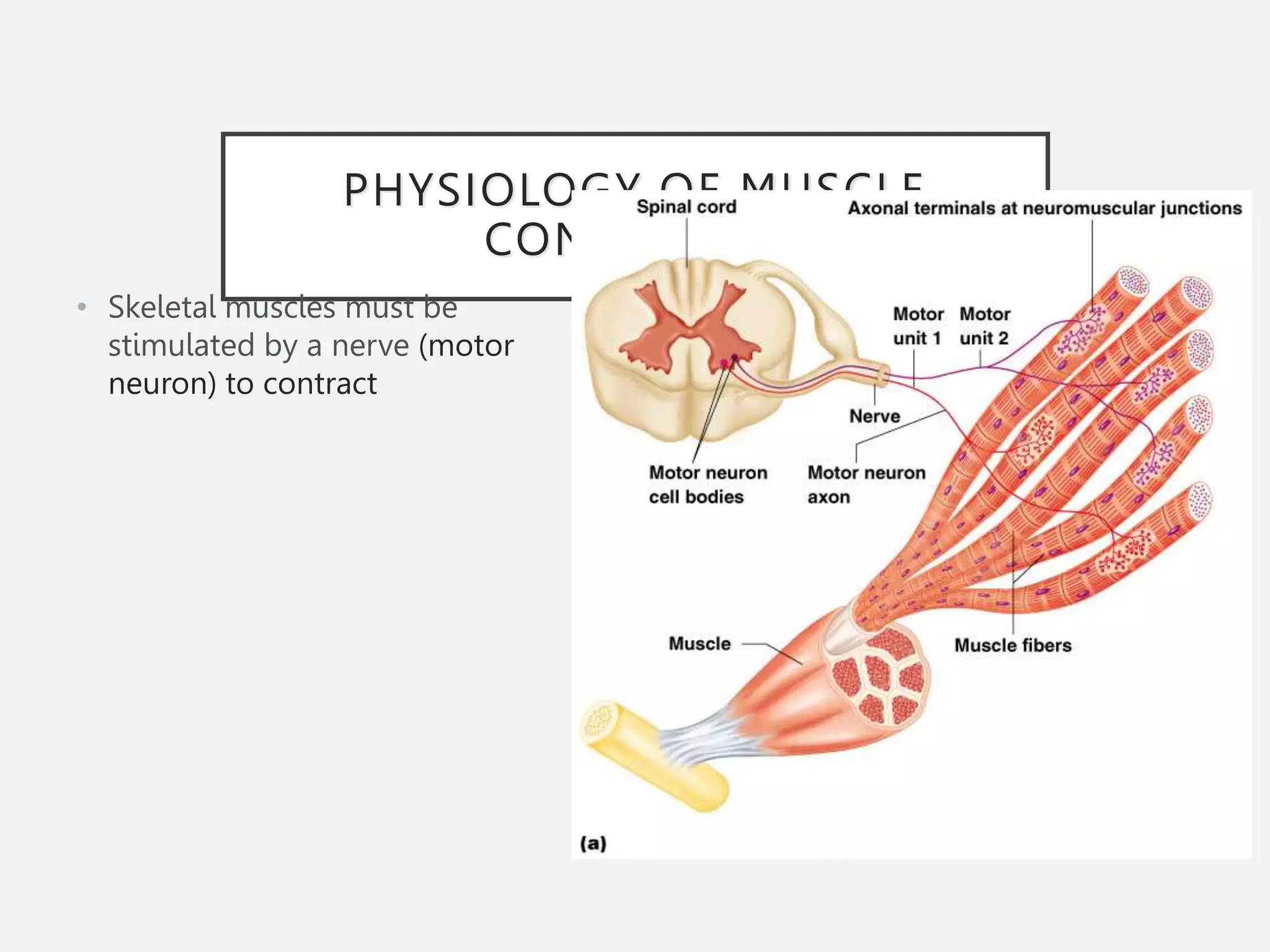
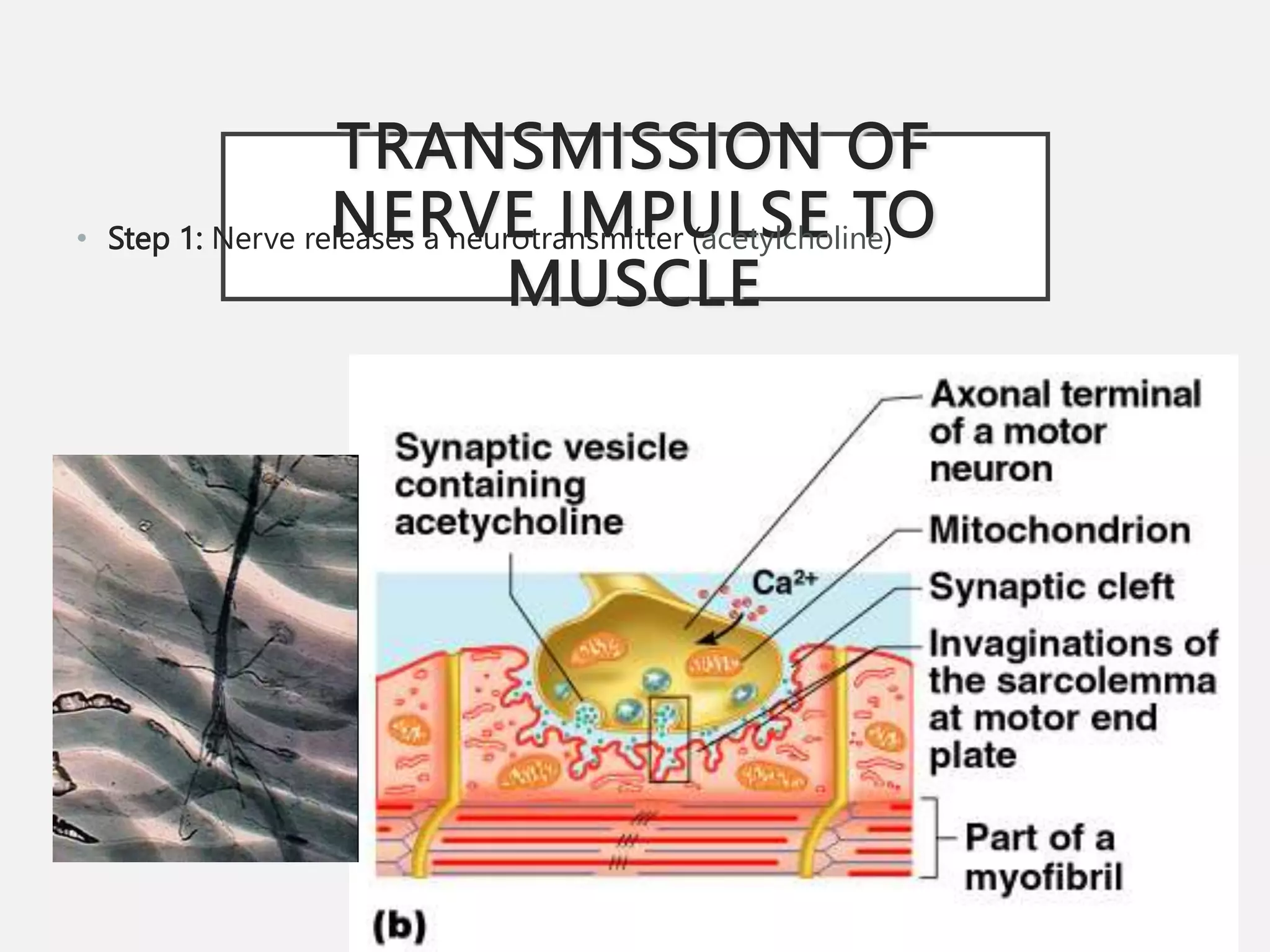
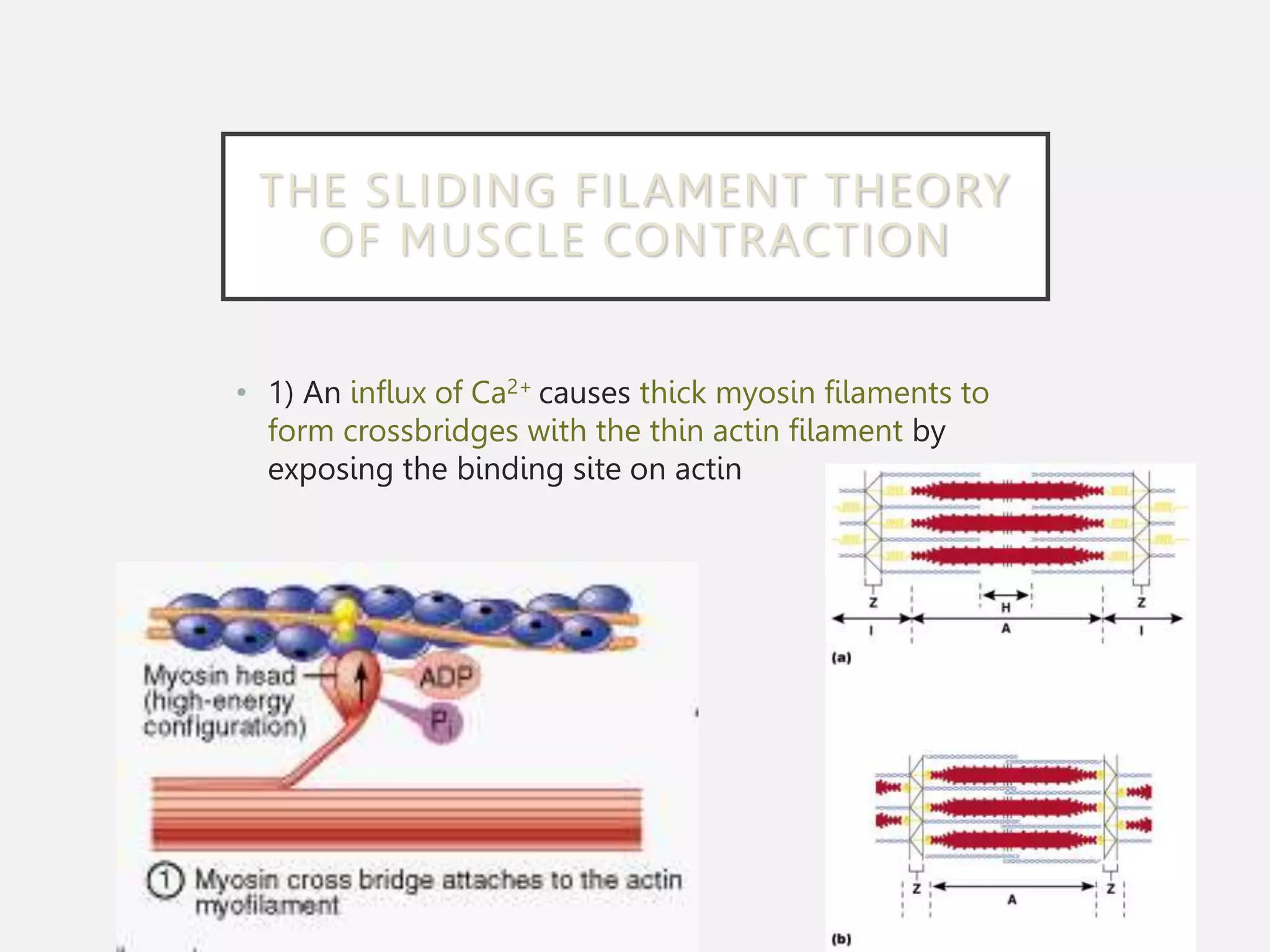
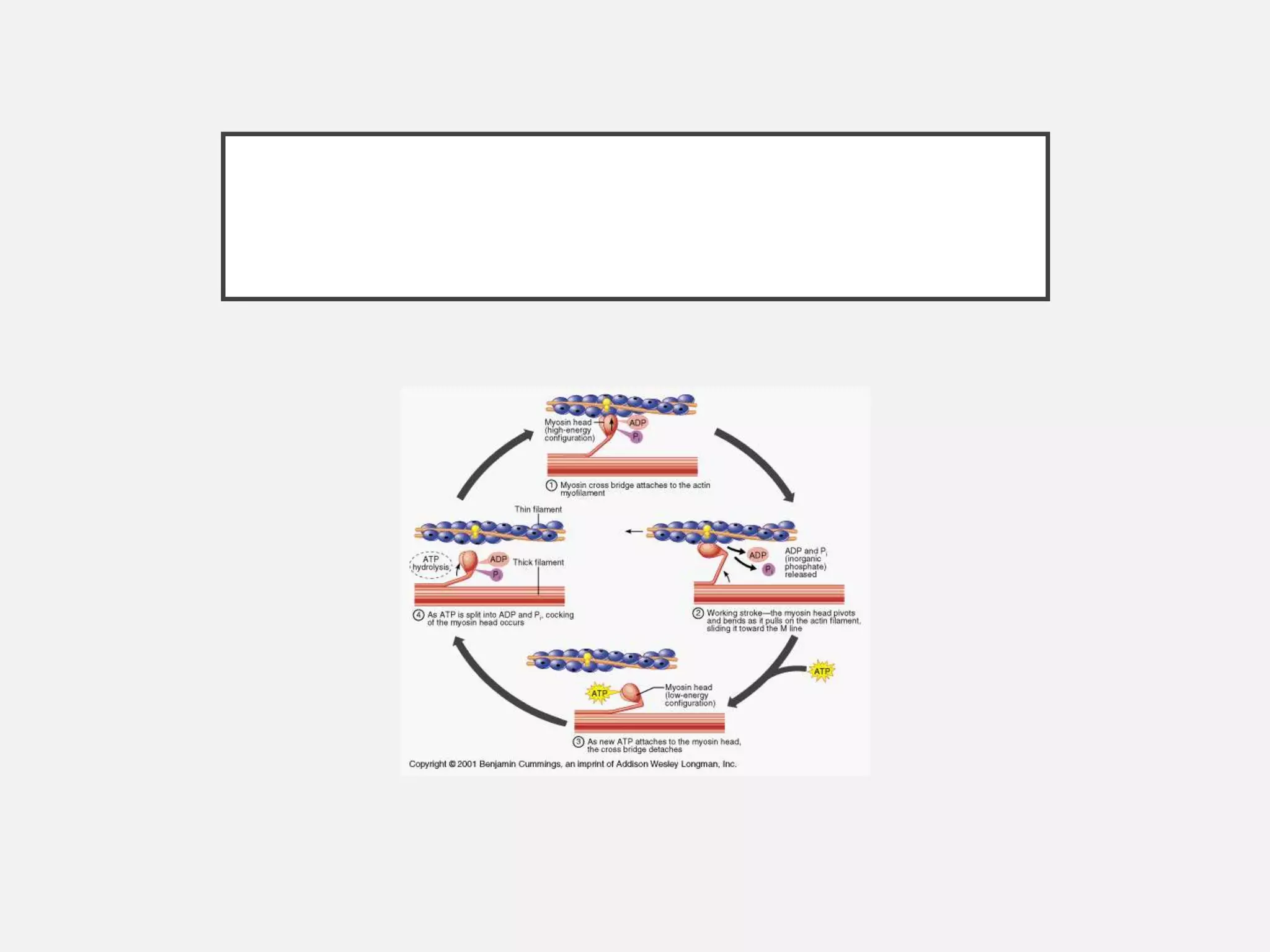
MUSCULAR SYSTEM
Chapter 7
THE MUSCULAR SYSTEM
• Muscles are responsible for all types of body
movement – they contract or shorten and are the
machine of the body
• Three basic muscle types are found in the body
• Skeletal muscle
• Cardiac muscle
• Smooth muscle
FUNCTION OF MUSCLES
• 1. Support the body
• 2. Allow for movement by making
bones and other body parts move
• 3. Maintain constant body
temperature
• 4. Assist in movement of
cardiovascular veins and lymph
• 5. Protect internal organs and
stabilize joints
ORGANIZATION OF SKELETAL
MUSCLE
Muscle
belly
Fascicle:
a bundle of
muscle fibers
Muscle
Fiber:
muscle cell
Sarcomere:
units of
myofibrils
responsible for
the striated
appearance
Myofibrils:
structures
that make
up a muscle
fiber
Myofilament:
protein
filaments that
make up a
sarcomere
Myosin:
thick
filaments
Actin: thin
filaments
COVERINGS OF A
SKELETAL MUSCLE
• Skeletal muscles are organs
• They contain muscle fibers, nerves, and blood
vessels
• Connective tissue membranes separate each
muscle structure
• Fascia – layer of fibrous tissue that separates
muscles from each other and from the skin
COVERINGS FROM LARGEST TO
SMALLEST
• Epimysium – covers the entire
skeletal muscle
• Perimysium – surrounds a bundle
of muscle fibers (fascicle)
• Endomysium – surrounds a single
muscle fiber (cell)
SKELETAL MUSCLE
ATTACHMENTS
• Epimysium blends into a connective tissue attachment
• Tendon – cord-like structure
• Sites of muscle attachment
• Bones
• Cartilages
• Connective tissue coverings
MICROSCOPIC
ANATOMY OF MUSCLE
FIBER (MUSCLE CELL)
• Cells are multinucleate
• Nuclei are just beneath the membrane
• Sarcolemma – specialized plasma membrane
• Sarcoplasmic reticulum – specialized smooth
endoplasmic reticulum involved in muscle contraction
• Myofibril
• Bundles of myofilaments
• Myofibrils are aligned to give distinct bands
• Light band = “I band”
• Dark band = “A band”
• Sarcomere
• Contractile unit of a muscle fiber
• Organization of the sarcomere
• Thick filaments = myosin protein
• Thin filaments = actin protein
• Myosin and actin overlap somewhat
in the sarcomere
• Myosin filaments have heads
(extensions) that can ‘grab’ onto
actin forming a crossbridge
PHYSIOLOGY OF MUSCLE
CONTRACTION
• Skeletal muscles must be
stimulated by a nerve (motor
neuron) to contract
TRANSMISSION OF
NERVE IMPULSE TO
MUSCLE
• Step 1: Nerve releases a neurotransmitter (acetylcholine)
• Step 2: Neurotransmitter causes the muscle cell membrane gates to open
• Step 3: Ions (Na+ & K+) exchange places causing the sarcoplasmic reticulum
to release Ca2+
• Step 4: This release of Ca+ starts the muscle contraction as the actin
slide past the myosin filaments
THE SLIDING FILAMENT
THEORY OF MUSCLE
CONTRACTION
• Sliding Filament Model - a muscle contracts when the thin filament in the
muscle fiber slides over the thick filament
• Activated by ATP and calcium (Ca+) ions
THE SLIDING FILAMENT THEORY
OF MUSCLE CONTRACTION
• 1) An influx of Ca2+ causes thick myosin filaments to
form crossbridges with the thin actin filament by
exposing the binding site on actin
THE SLIDING FILAMENT
THEORY OF MUSCLE
CONTRACTION
• 2) The crossbridges change shape as it pulls on filaments which slides towards the
center of the sacromere in the power stroke
• The distance between the Z line decreases, shortening the muscle.
• 3) The crossbridges detach from the actin filament when ATP bonds to
myosin head.
THE SLIDING FILAMENT THEORY
• 4) The myosin head gets ready to bond to actin again
using ATP energy
• The cycle is repeated on another site of actin filament
using the stored ATP energy
MUSCLE CONTRACTION
• Physiology of Muscle Contraction
CONTRACTION OF A SKELETAL
MUSCLE
• Muscle fiber contraction is “all or none”
• Within a skeletal muscle, not all fibers may be
stimulated during the same interval
• Different combinations of muscle fiber contractions
may give differing responses
• Graded responses – different degrees of skeletal
muscle shortening
• Rapid stimulus = constant contraction or tetanus
• Muscle force depends upon the number of fibers stimulated
• More fibers contracting results in greater muscle tension
• Muscles can continue to contract unless they run out of ATP
or Ca2+
• One molecule of ATP supplies enough energy for one actin and
myosin cross-bridge
ENERGY FOR MUSCLE
CONTRACTION
• Muscles use stored ATP for energy
• Bonds of ATP are broken to release energy
• Only 4-6 seconds worth of ATP is stored by muscles
• Three ways for muscle to make energy (ATP)
ATP production
for Muscle
Contraction
Creatine
Phosphate
Cellular
Respiration
Fermentation
(Anaerobic
Respiration)
1. CREATINE PHOSPHATE
• Creatine phosphate is a high-energy compound and is
the fastest way to make ATP available for muscles
• Used for activities lasting < 15 seconds
• Anaerobic (no oxygen needed)
• Reaction:
• Creatine phosphate + ADP ↔ creatine + ATP
• Creatine phosphate is made when a muscle is at rest
2. CELLULAR RESPIRATION
• Mitochondria use glucose molecules to make ATP in
the presence of oxygen
• Provides most of a muscle’s ATP
• Aerobic (needs oxygen)
• Used for activities lasting hours
• Reaction
• C6H12O6 + 6O2  6CO2 + 6H2O + ATP energy
• 1 glucose = 36 ATP
3. ANAEROBIC
RESPIRATION/
FERMENTATION
• Reaction that breaks down glucose without oxygen
• Used for activities lasting 30 – 60 seconds
• Anaerobic (no oxygen)
• Reaction
• Glucose  pyruvic acid + 2 ATP  lactic acid
• Lactic acid is also produced and causes pain in the
muscle
• Heavy breathing after exercise is a sign of
oxygen deficiency
• A marathon runner is exhausted after
crossing the finish line because they have
depleted not only their oxygen but their
glucose as well
• It takes up to two days to replace all of the
glucose in the muscles and glycogen in the
liver
MUSCLES AND BODY
MOVEMENTS
• Movement is attained due to a muscle
moving an attached bone
• Muscles are attached to at least two
points
• Insertion – attachment to a moveable
bone
• Origin – attachment to an immovable
bone
TYPES OF ORDINARY BODY
MOVEMENTS
• Flexion – decreases angle of joint
and brings two bones closer
together
• Extension- increases angle of
joint
• Rotation- movement of a bone in longitudinal
axis, shaking head “no”
• Abduction – moving away from the midline
• Adduction - moving toward the midline
• Circumduction - cone-shaped movement,
proximal end doesn’t move, while distal end
moves in a circle.
TYPES OF MUSCLES
• Muscles work in opposing pairs
• Ex. Biceps (flexion of arm) and Triceps (extension of arm)
• Prime mover – muscle that does most of the work
• Synergist – muscle that helps a prime mover in a
movement
• Antagonist – muscle that opposes or reverses a prime
mover
NAMING OF SKELETAL MUSCLES
• Direction of muscle fibers
• Example: rectus (straight), orbicularis (circular)
• Relative size of the muscle
• Example: maximus (largest), minimus (smallest), longus (long), brevis (short)
• Location of the muscle
• Example: pectoralis (chest), external (outside), frontalis (frontal)
• Number of origins
• Example: triceps (three heads)
• Location of the muscles origin and insertion
• Example: sterno (on the sternum)
• Shape of the muscle
• Example: deltoid (triangular)
• Action of the muscle
• Example: flexor and extensor (flexes or extends a bone)
AFFECTS OF AGING ON
MUSCLES
1. Muscles that are not used are replaced by
connective tissue then by fat
2. With age comes degeneration of mitochondria
due to exposure to oxygen and free radicals
3. Changes in the nervous system and endocrine
system also effect structure and function of
muscles
4. Muscles become weaker as we age but exercise
can stimulate muscle build-up
• She is 86 years young and a body builder.
• He is 80, and the oldest Iron man triathlon participant.
(1.2 mile swim, a 56-mile bike and a 13.1 mile run = 70.3
miles.)
DISORDERS RELATING TO THE
MUSCULAR SYSTEM
• Muscular Dystrophy: inherited, muscle enlarge due
to increased fat and connective tissue, but fibers
degenerate and atrophy
• Duchenne MD: lacking a protein to maintain the
sarcolemma
• Myasthemia Gravis: progressive weakness due to a
shortage of acetylcholine receptors
SPRAIN VERSES STRAIN
• Strain – overstretching of a
muscle near a joint
• Sprain – twisting of a joint
leading to swelling and injury
to ligaments, tendons, blood
vessels and nerves
MYALGIA AND TENDINITIS
• Myalgia – inflammation of
muscle tissue (arthritis on
previous slide)
• Tendinitis – inflammation
of the tendon due to
strain of repeated activity

muscle_system.ppt

  • 1.
  • 2.
    THE MUSCULAR SYSTEM •Muscles are responsible for all types of body movement – they contract or shorten and are the machine of the body • Three basic muscle types are found in the body • Skeletal muscle • Cardiac muscle • Smooth muscle
  • 4.
    FUNCTION OF MUSCLES •1. Support the body • 2. Allow for movement by making bones and other body parts move • 3. Maintain constant body temperature
  • 5.
    • 4. Assistin movement of cardiovascular veins and lymph • 5. Protect internal organs and stabilize joints
  • 6.
    ORGANIZATION OF SKELETAL MUSCLE Muscle belly Fascicle: abundle of muscle fibers Muscle Fiber: muscle cell Sarcomere: units of myofibrils responsible for the striated appearance Myofibrils: structures that make up a muscle fiber Myofilament: protein filaments that make up a sarcomere Myosin: thick filaments Actin: thin filaments
  • 9.
    COVERINGS OF A SKELETALMUSCLE • Skeletal muscles are organs • They contain muscle fibers, nerves, and blood vessels • Connective tissue membranes separate each muscle structure • Fascia – layer of fibrous tissue that separates muscles from each other and from the skin
  • 10.
    COVERINGS FROM LARGESTTO SMALLEST • Epimysium – covers the entire skeletal muscle • Perimysium – surrounds a bundle of muscle fibers (fascicle) • Endomysium – surrounds a single muscle fiber (cell)
  • 11.
    SKELETAL MUSCLE ATTACHMENTS • Epimysiumblends into a connective tissue attachment • Tendon – cord-like structure • Sites of muscle attachment • Bones • Cartilages • Connective tissue coverings
  • 12.
    MICROSCOPIC ANATOMY OF MUSCLE FIBER(MUSCLE CELL) • Cells are multinucleate • Nuclei are just beneath the membrane
  • 13.
    • Sarcolemma –specialized plasma membrane • Sarcoplasmic reticulum – specialized smooth endoplasmic reticulum involved in muscle contraction
  • 14.
    • Myofibril • Bundlesof myofilaments • Myofibrils are aligned to give distinct bands • Light band = “I band” • Dark band = “A band”
  • 15.
    • Sarcomere • Contractileunit of a muscle fiber • Organization of the sarcomere • Thick filaments = myosin protein • Thin filaments = actin protein
  • 16.
    • Myosin andactin overlap somewhat in the sarcomere • Myosin filaments have heads (extensions) that can ‘grab’ onto actin forming a crossbridge
  • 18.
    PHYSIOLOGY OF MUSCLE CONTRACTION •Skeletal muscles must be stimulated by a nerve (motor neuron) to contract
  • 19.
    TRANSMISSION OF NERVE IMPULSETO MUSCLE • Step 1: Nerve releases a neurotransmitter (acetylcholine)
  • 20.
    • Step 2:Neurotransmitter causes the muscle cell membrane gates to open • Step 3: Ions (Na+ & K+) exchange places causing the sarcoplasmic reticulum to release Ca2+ • Step 4: This release of Ca+ starts the muscle contraction as the actin slide past the myosin filaments
  • 21.
    THE SLIDING FILAMENT THEORYOF MUSCLE CONTRACTION • Sliding Filament Model - a muscle contracts when the thin filament in the muscle fiber slides over the thick filament • Activated by ATP and calcium (Ca+) ions
  • 22.
    THE SLIDING FILAMENTTHEORY OF MUSCLE CONTRACTION • 1) An influx of Ca2+ causes thick myosin filaments to form crossbridges with the thin actin filament by exposing the binding site on actin
  • 23.
    THE SLIDING FILAMENT THEORYOF MUSCLE CONTRACTION • 2) The crossbridges change shape as it pulls on filaments which slides towards the center of the sacromere in the power stroke • The distance between the Z line decreases, shortening the muscle.
  • 24.
    • 3) Thecrossbridges detach from the actin filament when ATP bonds to myosin head.
  • 25.
    THE SLIDING FILAMENTTHEORY • 4) The myosin head gets ready to bond to actin again using ATP energy • The cycle is repeated on another site of actin filament using the stored ATP energy
  • 28.
    MUSCLE CONTRACTION • Physiologyof Muscle Contraction
  • 29.
    CONTRACTION OF ASKELETAL MUSCLE • Muscle fiber contraction is “all or none” • Within a skeletal muscle, not all fibers may be stimulated during the same interval • Different combinations of muscle fiber contractions may give differing responses • Graded responses – different degrees of skeletal muscle shortening • Rapid stimulus = constant contraction or tetanus
  • 30.
    • Muscle forcedepends upon the number of fibers stimulated • More fibers contracting results in greater muscle tension • Muscles can continue to contract unless they run out of ATP or Ca2+ • One molecule of ATP supplies enough energy for one actin and myosin cross-bridge
  • 31.
    ENERGY FOR MUSCLE CONTRACTION •Muscles use stored ATP for energy • Bonds of ATP are broken to release energy • Only 4-6 seconds worth of ATP is stored by muscles • Three ways for muscle to make energy (ATP) ATP production for Muscle Contraction Creatine Phosphate Cellular Respiration Fermentation (Anaerobic Respiration)
  • 32.
    1. CREATINE PHOSPHATE •Creatine phosphate is a high-energy compound and is the fastest way to make ATP available for muscles • Used for activities lasting < 15 seconds • Anaerobic (no oxygen needed) • Reaction: • Creatine phosphate + ADP ↔ creatine + ATP • Creatine phosphate is made when a muscle is at rest
  • 33.
    2. CELLULAR RESPIRATION •Mitochondria use glucose molecules to make ATP in the presence of oxygen • Provides most of a muscle’s ATP • Aerobic (needs oxygen) • Used for activities lasting hours • Reaction • C6H12O6 + 6O2  6CO2 + 6H2O + ATP energy • 1 glucose = 36 ATP
  • 34.
    3. ANAEROBIC RESPIRATION/ FERMENTATION • Reactionthat breaks down glucose without oxygen • Used for activities lasting 30 – 60 seconds • Anaerobic (no oxygen) • Reaction • Glucose  pyruvic acid + 2 ATP  lactic acid • Lactic acid is also produced and causes pain in the muscle
  • 35.
    • Heavy breathingafter exercise is a sign of oxygen deficiency • A marathon runner is exhausted after crossing the finish line because they have depleted not only their oxygen but their glucose as well • It takes up to two days to replace all of the glucose in the muscles and glycogen in the liver
  • 36.
    MUSCLES AND BODY MOVEMENTS •Movement is attained due to a muscle moving an attached bone • Muscles are attached to at least two points • Insertion – attachment to a moveable bone • Origin – attachment to an immovable bone
  • 37.
    TYPES OF ORDINARYBODY MOVEMENTS • Flexion – decreases angle of joint and brings two bones closer together • Extension- increases angle of joint
  • 38.
    • Rotation- movementof a bone in longitudinal axis, shaking head “no” • Abduction – moving away from the midline • Adduction - moving toward the midline • Circumduction - cone-shaped movement, proximal end doesn’t move, while distal end moves in a circle.
  • 39.
    TYPES OF MUSCLES •Muscles work in opposing pairs • Ex. Biceps (flexion of arm) and Triceps (extension of arm) • Prime mover – muscle that does most of the work • Synergist – muscle that helps a prime mover in a movement • Antagonist – muscle that opposes or reverses a prime mover
  • 40.
    NAMING OF SKELETALMUSCLES • Direction of muscle fibers • Example: rectus (straight), orbicularis (circular) • Relative size of the muscle • Example: maximus (largest), minimus (smallest), longus (long), brevis (short) • Location of the muscle • Example: pectoralis (chest), external (outside), frontalis (frontal) • Number of origins • Example: triceps (three heads)
  • 41.
    • Location ofthe muscles origin and insertion • Example: sterno (on the sternum) • Shape of the muscle • Example: deltoid (triangular) • Action of the muscle • Example: flexor and extensor (flexes or extends a bone)
  • 42.
    AFFECTS OF AGINGON MUSCLES 1. Muscles that are not used are replaced by connective tissue then by fat 2. With age comes degeneration of mitochondria due to exposure to oxygen and free radicals 3. Changes in the nervous system and endocrine system also effect structure and function of muscles 4. Muscles become weaker as we age but exercise can stimulate muscle build-up
  • 43.
    • She is86 years young and a body builder. • He is 80, and the oldest Iron man triathlon participant. (1.2 mile swim, a 56-mile bike and a 13.1 mile run = 70.3 miles.)
  • 44.
    DISORDERS RELATING TOTHE MUSCULAR SYSTEM • Muscular Dystrophy: inherited, muscle enlarge due to increased fat and connective tissue, but fibers degenerate and atrophy • Duchenne MD: lacking a protein to maintain the sarcolemma • Myasthemia Gravis: progressive weakness due to a shortage of acetylcholine receptors
  • 45.
    SPRAIN VERSES STRAIN •Strain – overstretching of a muscle near a joint • Sprain – twisting of a joint leading to swelling and injury to ligaments, tendons, blood vessels and nerves
  • 46.
    MYALGIA AND TENDINITIS •Myalgia – inflammation of muscle tissue (arthritis on previous slide) • Tendinitis – inflammation of the tendon due to strain of repeated activity