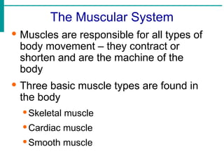
The Muscular System
·Muscles are responsible for all types of
body movement – they contract or
shorten and are the machine of the
body
· Three basic muscle types are found in
the body
·Skeletal muscle
·Cardiac muscle
·Smooth muscle
3.
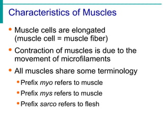
Characteristics of Muscles
·Muscle cells are elongated
(muscle cell = muscle fiber)
· Contraction of muscles is due to the
movement of microfilaments
· All muscles share some terminology
·Prefix myo refers to muscle
·Prefix mys refers to muscle
·Prefix sarco refers to flesh
4.
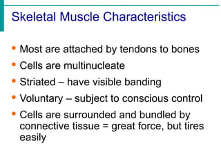
Skeletal Muscle Characteristics
·Most are attached by tendons to bones
· Cells are multinucleate
· Striated – have visible banding
· Voluntary – subject to conscious control
· Cells are surrounded and bundled by
connective tissue = great force, but tires
easily
5.
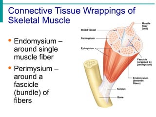
Connective Tissue Wrappingsof
Skeletal Muscle
· Endomysium –
around single
muscle fiber
· Perimysium –
around a
fascicle
(bundle) of
fibers
6.
6
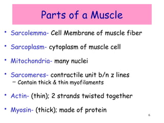
Parts of aMuscle
• Sarcolemma- Cell Membrane of muscle fiber
• Sarcoplasm- cytoplasm of muscle cell
• Mitochondria- many nuclei
• Sarcomeres- contractile unit b/n z lines
– Contain thick & thin myofilaments
• Actin- (thin); 2 strands twisted together
• Myosin- (thick); made of protein
7.
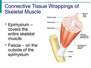
Connective Tissue Wrappingsof
Skeletal Muscle
· Epimysium –
covers the
entire skeletal
muscle
· Fascia – on the
outside of the
epimysium
8.
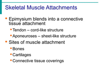
Skeletal Muscle Attachments
·Epimysium blends into a connective
tissue attachment
·Tendon – cord-like structure
·Aponeuroses – sheet-like structure
· Sites of muscle attachment
·Bones
·Cartilages
·Connective tissue coverings
9.
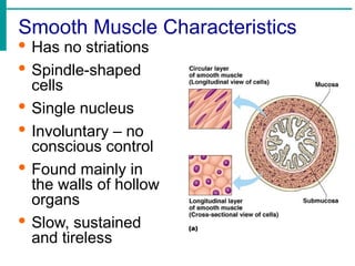
Smooth Muscle Characteristics
·Has no striations
· Spindle-shaped
cells
· Single nucleus
· Involuntary – no
conscious control
· Found mainly in
the walls of hollow
organs
· Slow, sustained
and tireless
10.
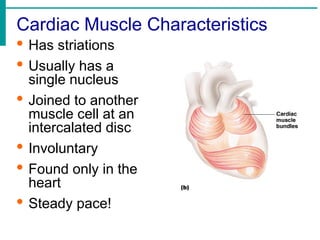
Cardiac Muscle Characteristics
·Has striations
· Usually has a
single nucleus
· Joined to another
muscle cell at an
intercalated disc
· Involuntary
· Found only in the
heart
· Steady pace!
11.
Types of Muscles
·Prime mover – muscle with the major
responsibility for a certain movement
· Antagonist – muscle that opposes or
reverses a prime mover
· Synergist – muscle that aids a prime
mover in a movement and helps prevent
rotation
12.
Function of Muscles
·Produce movement
· Maintain posture
· Stabilize joints
· Generate heat
13.
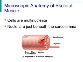
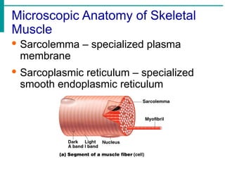
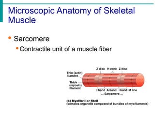
Microscopic Anatomy ofSkeletal
Muscle
· Cells are multinucleate
· Nuclei are just beneath the sarcolemma
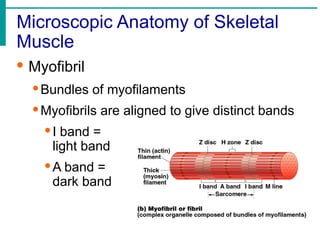
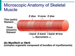
Microscopic Anatomy ofSkeletal
Muscle
· Myofibril
·Bundles of myofilaments
·Myofibrils are aligned to give distinct bands
·I band =
light band
·A band =
dark band
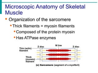
Microscopic Anatomy ofSkeletal
Muscle
· Organization of the sarcomere
·Thick filaments = myosin filaments
·Composed of the protein myosin
·Has ATPase enzymes
19.
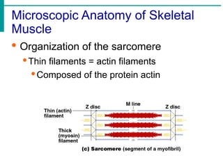
Microscopic Anatomy ofSkeletal
Muscle
· Organization of the sarcomere
·Thin filaments = actin filaments
·Composed of the protein actin
20.
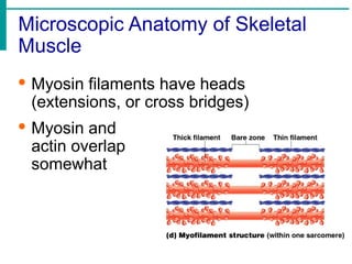
Microscopic Anatomy ofSkeletal
Muscle
· Myosin filaments have heads
(extensions, or cross bridges)
· Myosin and
actin overlap
somewhat
21.
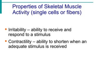
Properties of SkeletalMuscle
Activity (single cells or fibers)
· Irritability – ability to receive and
respond to a stimulus
· Contractility – ability to shorten when an
adequate stimulus is received
22.
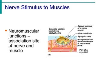
Nerve Stimulus toMuscles
· Skeletal
muscles must
be stimulated
by a nerve to
contract (motor
neruron)
· Motor unit
·One neuron
·Muscle cells
stimulated by
that neuron
23.
Nerve Stimulus toMuscles
· Neuromuscular
junctions –
association site
of nerve and
muscle
24.
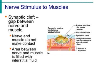
Nerve Stimulus toMuscles
· Synaptic cleft –
gap between
nerve and
muscle
·Nerve and
muscle do not
make contact
·Area between
nerve and muscle
is filled with
interstitial fluid
25.
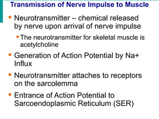
Transmission of NerveImpulse to Muscle
· Neurotransmitter – chemical released
by nerve upon arrival of nerve impulse
·The neurotransmitter for skeletal muscle is
acetylcholine
· Generation of Action Potential by Na+
Influx
· Neurotransmitter attaches to receptors
on the sarcolemma
· Entrance of Action Potential to
Sarcoendoplasmic Reticulum (SER)
26.
Transmission of NerveImpulse to
Muscle
· Release of Ca++ from SER
· Formation of Complex with Troponin
and Tropomysin (Covering of Actin and
Myosin)
· Exposure of G-Actin Active site
· Binding of G-Actin Active site with
Myosin Active site (Actin-Myosin
complex formation)
27.
Transmission of NerveImpulse to
Muscle
· Utilization of ATP by breakdown of into
ADP + Pi + Energy
· CONTRACTION OF MUSCLE
· Again Utilization of ATP for separation of
Actin-Myosin Complex
· Reuptake of Ca++ by SER
· Degenration of Action Potential and
Muscle Relaxation
28.
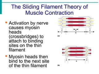
The Sliding FilamentTheory of
Muscle Contraction
· Activation by nerve
causes myosin
heads
(crossbridges) to
attach to binding
sites on the thin
filament
· Myosin heads then
bind to the next site
of the thin filament
29.
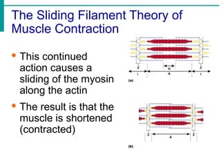
The Sliding FilamentTheory of
Muscle Contraction
· This continued
action causes a
sliding of the myosin
along the actin
· The result is that the
muscle is shortened
(contracted)
30.
Contraction of aSkeletal Muscle
· Muscle fiber contraction is “all or none”
· Within a skeletal muscle, not all fibers
may be stimulated during the same
interval
· Different combinations of muscle fiber
contractions may give differing
responses
· Graded responses – different degrees
of skeletal muscle shortening, rapid
stimulus = constant contraction or
tetanus
31.
Types of MuscleContractions
· Isotonic contractions
·Myofilaments are able to slide past each
other during contractions
·The muscle shortens
· Isometric contractions
·Tension in the muscles increases
·The muscle is unable to shorten
32.
Muscle Response toStrong Stimuli
· Muscle force depends upon the number
of fibers stimulated
· More fibers contracting results in greater
muscle tension
· Muscles can continue to contract unless
they run out of energy
33.
Energy for MuscleContraction
· Initially, muscles used stored ATP for
energy
·Bonds of ATP are broken to release energy
·Only 4-6 seconds worth of ATP is stored by
muscles
· After this initial time, other pathways
must be utilized to produce ATP
34.
Energy for MuscleContraction
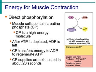
· Direct phosphorylation
· Muscle cells contain creatine
phosphate (CP)
· CP is a high-energy
molecule
· After ATP is depleted, ADP is
left
· CP transfers energy to ADP,
to regenerate ATP
· CP supplies are exhausted in
about 20 seconds
35.
Energy for MuscleContraction
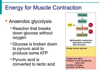
· Anaerobic glycolysis
·Reaction that breaks
down glucose without
oxygen
·Glucose is broken down
to pyruvic acid to
produce some ATP
·Pyruvic acid is
converted to lactic acid
36.
Energy for MuscleContraction
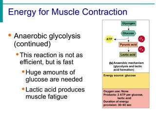
· Anaerobic glycolysis
(continued)
·This reaction is not as
efficient, but is fast
·Huge amounts of
glucose are needed
·Lactic acid produces
muscle fatigue
37.
Energy for MuscleContraction
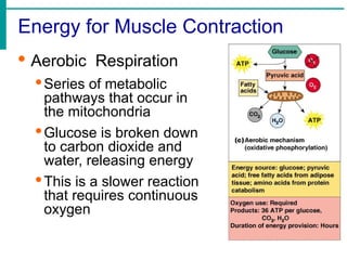
· Aerobic Respiration
·Series of metabolic
pathways that occur in
the mitochondria
·Glucose is broken down
to carbon dioxide and
water, releasing energy
·This is a slower reaction
that requires continuous
oxygen
38.
Muscle Fatigue andOxygen Debt
· When a muscle is fatigued, it is unable to
contract
· The common reason for muscle fatigue is
oxygen debt
·Oxygen must be “repaid” to tissue to remove
oxygen debt
·Oxygen is required to get rid of accumulated
lactic acid
· Increasing acidity (from lactic acid) and lack
of ATP causes the muscle to contract less
39.
Muscle Tone
· Somefibers are contracted even in a
relaxed muscle
· Different fibers contract at different
times to provide muscle tone
· The process of stimulating various fibers
is under involuntary control
40.
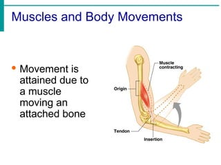
Muscles and BodyMovements
· Movement is
attained due to
a muscle
moving an
attached bone
41.
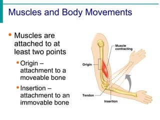
Muscles and BodyMovements
· Muscles are
attached to at
least two points
·Origin –
attachment to a
moveable bone
·Insertion –
attachment to an
immovable bone
42.
Effects of Exerciseon Muscle
· Results of increased muscle use
·Increase in muscle size
·Increase in muscle strength
·Increase in muscle efficiency
·Muscle becomes more fatigue resistant
43.
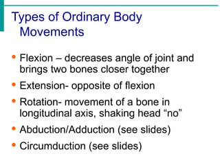
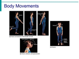
Types of OrdinaryBody
Movements
· Flexion – decreases angle of joint and
brings two bones closer together
· Extension- opposite of flexion
· Rotation- movement of a bone in
longitudinal axis, shaking head “no”
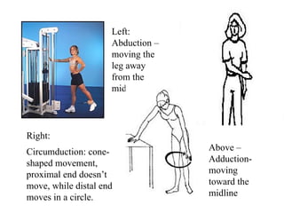
· Abduction/Adduction (see slides)
· Circumduction (see slides)
Left:
Abduction –
moving the
legaway
from the
midline
Above –
Adduction-
moving
toward the
midline
Right:
Circumduction: cone-
shaped movement,
proximal end doesn’t
move, while distal end
moves in a circle.
46.
46
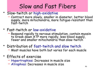
Slow and FastFibers
• Slow-twitch or high-oxidative
– Contract more slowly, smaller in diameter, better blood
supply, more mitochondria, more fatigue-resistant than
fast-twitch
• Fast-twitch or low-oxidative
– Respond rapidly to nervous stimulation, contain myosin
to break down ATP more rapidly, less blood supply,
fewer and smaller mitochondria than slow-twitch
• Distribution of fast-twitch and slow twitch
– Most muscles have both but varies for each muscle
• Effects of exercise
– Hypertrophies: Increases in muscle size
– Atrophies: Decreases in muscle size
47.
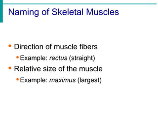
Naming of SkeletalMuscles
· Direction of muscle fibers
·Example: rectus (straight)
· Relative size of the muscle
·Example: maximus (largest)
48.
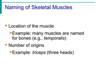
Naming of SkeletalMuscles
· Location of the muscle
·Example: many muscles are named
for bones (e.g., temporalis)
· Number of origins
·Example: triceps (three heads)
49.
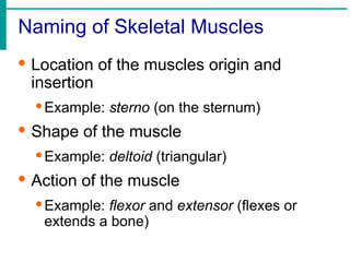
Naming of SkeletalMuscles
· Location of the muscles origin and
insertion
·Example: sterno (on the sternum)
· Shape of the muscle
·Example: deltoid (triangular)
· Action of the muscle
·Example: flexor and extensor (flexes or
extends a bone)
57
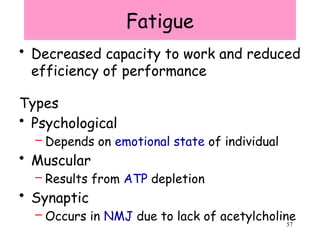
Fatigue
• Decreased capacityto work and reduced
efficiency of performance
Types
• Psychological
– Depends on emotional state of individual
• Muscular
– Results from ATP depletion
• Synaptic
– Occurs in NMJ due to lack of acetylcholine
58.
58
Effects of Agingon Skeletal
Muscle
• Reduced muscle mass
• Increased time for muscle to contract in
response to nervous stimuli
• Reduced stamina
• Increased recovery time
• Loss of muscle fibers
• Decreased density of capillaries in muscle
59.
Disorders relating tothe
Muscular System
• Muscular Dystrophy: inherited, muscle
enlarge due to increased fat and connective
tissue, but fibers degenerate and atrophy
• Duchenne MD: lacking a protein to
maintain the sarcolemma
• Myasthemia Gravis: progressive weakness
due to a shortage of acetylcholine receptors