Downloaded 667 times
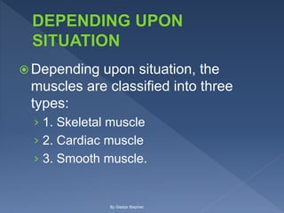

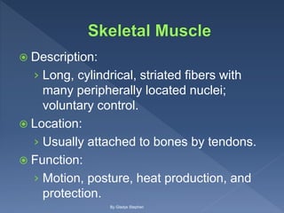
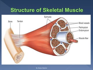
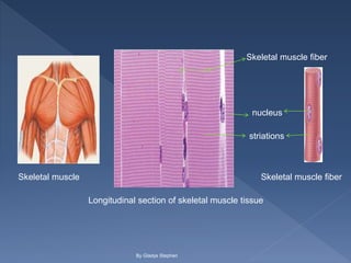
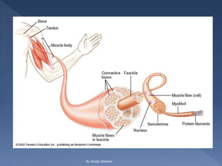
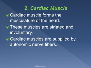
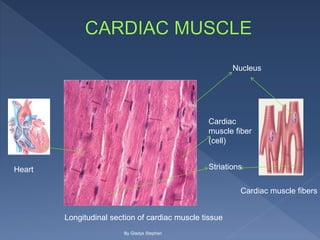
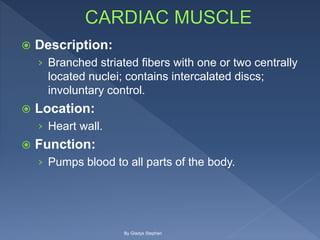

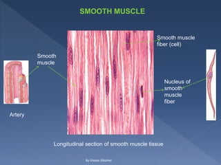

This document discusses the three types of muscle tissue: skeletal, cardiac, and smooth muscle. It provides details on their characteristics, such as whether they are striated or not, voluntary or involuntary, and their locations and functions in the body. Skeletal muscle is voluntary, attached to bones, and enables movement. Cardiac muscle is involuntary and makes up the heart wall. Smooth muscle is involuntary and located in organs like the stomach.


![CASE_PRESENTATION_ON_subdural_hematoma(SDH)[1 FINAL PPT]-1.pptx](https://cdn.slidesharecdn.com/ss_thumbnails/casepresentationonsubduralhematomasdh1finalppt-1-260129172522-d405d375-thumbnail.jpg?width=640&height=640&fit=bounds)


