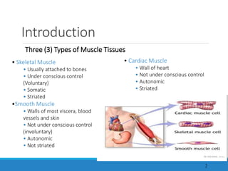
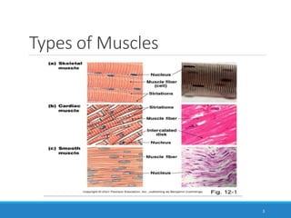
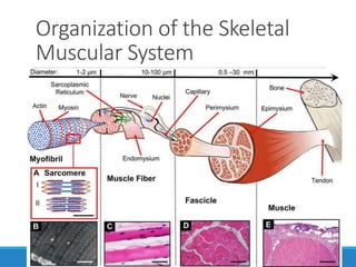
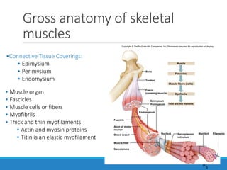
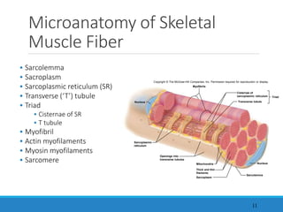
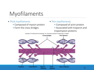
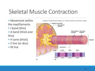
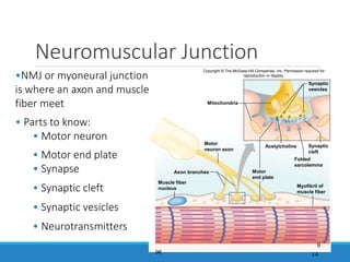
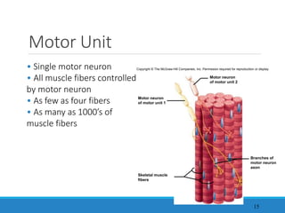
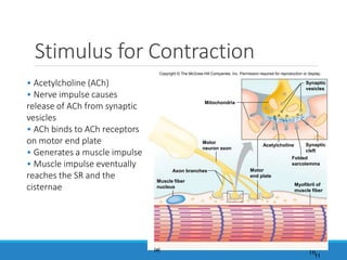
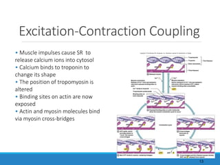
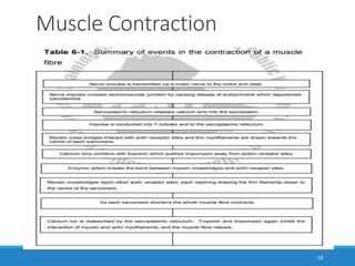
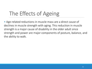
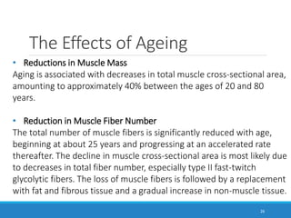
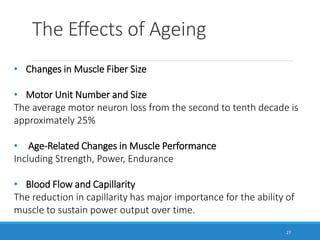
Anatomy
and
Physiology
Dr. Ali Jibbawi
5 July 2023 1
CHAPTER
2
THE MUSCULAR SYSTEM
Copyright © The McGraw-Hill Companies, Inc. Permission required for reproduction or display.
Introduction
2
Three (3) Types of Muscle Tissues
• Skeletal Muscle
• Usually attached to bones
• Under conscious control
(Voluntary)
• Somatic
• Striated
•Smooth Muscle
• Walls of most viscera, blood
vessels and skin
• Not under conscious control
(involuntary)
• Autonomic
• Not striated
• Cardiac Muscle
• Wall of heart
• Not under conscious control
• Autonomic
• Striated
Types of Muscles
3
Functions of the Muscular
System
Through sustained contraction or alternating contraction and relaxation,
muscle tissue has three key functions:
1. Producing motion
2. Providing stabilization (standing or sitting)
3. Generating heat (generation of heat)
4
1. Motion: Motion is obvious in movements such as walking and running,
and in localized movements, such as grasping a pencil or nodding the
head. These movements rely on the integrated functioning of bones,
joints, and skeletal muscles.
2. Stabilizing body positions and regulating the volume of cavities in the
body: Postural muscles display sustained contractions when a person is
awake, for example, partially contracted neck muscles hold the head
upright. In addition, the volumes of the body cavities are regulated
through the contractions of skeletal muscles. For example muscles of
respiration regulate the volume of the thoracic cavity during the process
of breathing.
3. Thermo genesis : As skeletal muscle contracts to perform work, a by-
product is heat. Much of the heat released by muscle is used to maintain
normal body temperature. Muscle contractions are thought to generate
as much as 85% of all body heat.
5
Functions of the Muscular
System
Physiologic Characteristics of
Muscle Tissue
Muscle tissue has four principal characteristics that enable it to carry
out its functions and thus contribute to homeostasis.
1. Excitability (irritability)
2. Contractility
3. Extensibility
4. Elasticity
6
Structure of Skeletal Muscle
7
• Skeletal Muscle
• Organ of the muscular system
• Skeletal muscle tissue
• Nervous tissue
• Blood
• Connective tissues
• Fascia
• Tendons
• Aponeuroses
Copyright © The McGraw-Hill Companies, Inc. Permission required for reproduction or display.
Aponeuroses
Skeletal muscles
Tendons
8
Organization of the Skeletal
Muscular System
9
Gross anatomy of skeletal
muscles
10
•Connective Tissue Coverings:
• Epimysium
• Perimysium
• Endomysium
Bone
Muscle
Epimysium
Perimysium
Endomysium
Fascicle
Fascicles
Muscle fibers (cells)
Myofibrils
Thick and thin filaments
Blood vessel
Muscle fiber
Myofibril
Sarcolemma
Nucleus Filaments
Tendon
Fascia
(covering muscle)
Axon of motor
neuron
Sarcoplasmic
reticulum
Copyright © The McGraw-Hill Companies, Inc. Permission required for reproduction or display.
• Muscle organ
• Fascicles
• Muscle cells or fibers
• Myofibrils
• Thick and thin myofilaments
• Actin and myosin proteins
• Titin is an elastic myofilament
5
Microanatomy of Skeletal
Muscle Fiber
11
• Sarcolemma
• Sacroplasm
• Sarcoplasmic reticulum (SR)
• Transverse (‘T’) tubule
• Triad
• Cisternae of SR
• T tubule
• Myofibril
• Actin myofilaments
• Myosin myofilaments
• Sarcomere
Copyright © The McGraw-Hill Companies, Inc. Permission required for reproduction or display.
Nucleus
Mitochondria
Sarcolemma
Sarcoplasm
Nucleus
Myofibrils
Sarcoplasmic
reticulum
Openings into
transverse tubules
Thick and thin
filaments
Cisternae of
sarcoplasmic reticulum
Transverse tubule
Triad
Myofilaments
12
• Thick myofilaments
• Composed of myosin protein
• Form the cross-bridges
• Thin myofilaments
• Composed of actin protein
• Associated with troponin and
tropomyosin proteins
Copyright © The McGraw-Hill Companies, Inc. Permission required for reproduction or display.
Cross-bridges
Actin molecule
Thin filament
Myosin
molecule
Thick
filament
Troponin Tropomyosin
Skeletal Muscle Contraction
13
• Movement within
the myofilaments
• I band (thin)
• A band (thick and
thin)
• H zone (thick)
• Z line (or disc)
• M line
Copyright © The McGraw-Hill Companies, Inc. Permission required for reproduction or display.
Myofibril
Sarcomere
(a)
Skeletal muscle fiber
Z line
H zone M line
I band I band
(b)
Z line
Sarcoplasmic
reticulum
Thick (myosin)
filaments
Thin (actin)
filaments
A band
A band
Neuromuscular Junction
14
•NMJ or myoneural junction
is where an axon and muscle
fiber meet
• Parts to know:
• Motor neuron
• Motor end plate
• Synapse
• Synaptic cleft
• Synaptic vesicles
• Neurotransmitters
Copyright © The McGraw-Hill Companies, Inc. Permission required for
reproduction or display.
Axon branches
Mitochondria
Acetylcholine
(a)
Synaptic
vesicles
Synaptic
cleft
Folded
sarcolemma
Motor
end plate
Myofibril of
muscle fiber
Muscle fiber
nucleus
Motor
neuron axon
9
Motor Unit
15
• Single motor neuron
• All muscle fibers controlled
by motor neuron
• As few as four fibers
• As many as 1000’s of
muscle fibers
Copyright © The McGraw-Hill Companies, Inc. Permission required for reproduction or display.
Motor neuron
of motor unit 2
Motor neuron
of motor unit 1
Skeletal muscle
fibers
Branches of
motor neuron
axon
Stimulus for Contraction
16
• Acetylcholine (ACh)
• Nerve impulse causes
release of ACh from synaptic
vesicles
• ACh binds to ACh receptors
on motor end plate
• Generates a muscle impulse
• Muscle impulse eventually
reaches the SR and the
cisternae
Copyright © The McGraw-Hill Companies, Inc. Permission required for reproduction or display.
Axon branches
Mitochondria
Acetylcholine
(a)
Synaptic
vesicles
Synaptic
cleft
Folded
sarcolemma
Motor
end plate
Myofibril of
muscle fiber
Muscle fiber
nucleus
Motor
neuron axon
11
Excitation-Contraction Coupling
• Muscle impulses cause SR to
release calcium ions into cytosol
• Calcium binds to troponin to
change its shape
• The position of tropomyosin is
altered
• Binding sites on actin are now
exposed
• Actin and myosin molecules bind
via myosin cross-bridges
Copyright © The McGraw-Hill Companies, Inc. Permission required for reproduction or display.
Actin monomers
Release of Ca+2 from sarcoplasmic
reticulum exposes binding sites on
actin:
Muscle contraction Muscle relaxation
Active transport of Ca+2 into sarcoplasmic
reticulum, which requires ATP, makes
myosin binding sites unavailable.
Ca+2 binds to troponin
Tropomyosin pulled aside
ATP
Contraction cycle
P
ADP +
P
ADP + P
ADP +
Ca+2
Ca+2
P
ADP +
P
ADP + P
ADP +
P
ADP
P
ADP
ATP ATP ATP
P
ADP + P
ADP +
ATP
Tropomyosin
Troponin Thin filament
Thick filament
Ca+2
Binding sites on
actin exposed
1 Relaxed muscle
Ca+2
P
ADP +
Ca+2
2 Exposed binding sites on actin molecules
allow the muscle contraction cycle to occur
6 ATP splits, which
provides power to
“cock” the myosin
cross-bridges
3 Cross-bridges
bind actin to
myosin
5 New ATP binds to myosin, releasing linkages 4 Cross-bridges pull thin filament (power stroke),
ADP and P released from myosin
13
Muscle Contraction
18
Types of contractions
19
• Isotonic: when a muscle contracts and its ends are
pulled closer together.
• Isometric: when a muscle contracts but attachments do
net move.
• Isokinetic: when the force a muscle generates is less
than that required to move or lift an object, the
contraction is called isokinetic.
20
Energy Sources for Contraction
• Creatine phosphate – stores energy that quickly converts
ADP to ATP
1) Creatine phosphate
2) Cellular respiration
Copyright © The McGraw-Hill Companies, Inc. Permission required for reproduction or display.
ADP
ATP
ATP
P
When cellular
Creatine
Creatine
ADP
ATP
ATP
P
is low
Creatine
Creatine
When cellular is low
17
Aerobic Respiration
21
22
Oxygen Supply and
Cellular Respiration
• Cellular respiration:
• Anaerobic Phase
• Glycolysis
• Occurs in cytoplasm
• Produces little ATP
• Aerobic Phase
• Citric acid cycle
• Electron transport system
• Occurs in the mitochondria
• Produces most ATP
• Myoglobin stores extra oxygen
Copyright © The McGraw-Hill Companies, Inc. Permission required for reproduction or display.
ATP
2
Energy
Lactic acid
Glucose
ATP
Synthesis of 34
CO2 + H2O + Energy
Pyruvic acid
Heat
2
1
Mitochondria
Cytosol
In the absence of
sufficient oxygen,
glycolysis leads to
lactic acid
accumulation.
Oxygen carried from
the lungs by
hemoglobin in red
blood cells is stored
in muscle cells by
myoglobin and is
available to support
aerobic respiration.
Citric acid
cycle
Electron
transport
chain
23
Oxygen Debt
• Oxygen not available
• Glycolysis continues
• Pyruvic acid converted to
lactic acid
• Liver converts lactic acid to
glucose
• Oxygen debt – amount of oxygen needed by liver cells to use
the accumulated lactic acid to produce glucose
Copyright © The McGraw-Hill Companies, Inc. Permission required for reproduction or display.
ATP
Glucose
Glycogen
Lactic acid
Pyruvic acid
Energy to
synthesize
Energy
from
Glycolysis and
lactic acid formation
(in muscle)
Synthesis of glucose
from lactic acid
(in liver)
ATP
24
Muscle Fatigue
• Inability to contract muscle
• Commonly caused from:
• Decreased blood flow
• Ion imbalances across the sarcolemma
• Accumulation of lactic acid
• Cramp – sustained, involuntary muscle contraction
• Physiological vs. psychological fatigue
The Effects of Ageing
25
• Age-related reductions in muscle mass are a direct cause of
declines in muscle strength with aging. This reduction in muscle
strength is a major cause of disability in the older adult since
strength and power are major components of posture, balance, and
the ability to walk.
The Effects of Ageing
26
• Reductions in Muscle Mass
Aging is associated with decreases in total muscle cross-sectional area,
amounting to approximately 40% between the ages of 20 and 80
years.
• Reduction in Muscle Fiber Number
The total number of muscle fibers is significantly reduced with age,
beginning at about 25 years and progressing at an accelerated rate
thereafter. The decline in muscle cross-sectional area is most likely due
to decreases in total fiber number, especially type II fast-twitch
glycolytic fibers. The loss of muscle fibers is followed by a replacement
with fat and fibrous tissue and a gradual increase in non-muscle tissue.
The Effects of Ageing
27
• Changes in Muscle Fiber Size
• Motor Unit Number and Size
The average motor neuron loss from the second to tenth decade is
approximately 25%
• Age-Related Changes in Muscle Performance
Including Strength, Power, Endurance
• Blood Flow and Capillarity
The reduction in capillarity has major importance for the ability of
muscle to sustain power output over time.
28

the muscular system.ppt

  • 1.
    Anatomy and Physiology Dr. Ali Jibbawi 5July 2023 1 CHAPTER 2 THE MUSCULAR SYSTEM Copyright © The McGraw-Hill Companies, Inc. Permission required for reproduction or display.
  • 2.
    Introduction 2 Three (3) Typesof Muscle Tissues • Skeletal Muscle • Usually attached to bones • Under conscious control (Voluntary) • Somatic • Striated •Smooth Muscle • Walls of most viscera, blood vessels and skin • Not under conscious control (involuntary) • Autonomic • Not striated • Cardiac Muscle • Wall of heart • Not under conscious control • Autonomic • Striated
  • 3.
  • 4.
    Functions of theMuscular System Through sustained contraction or alternating contraction and relaxation, muscle tissue has three key functions: 1. Producing motion 2. Providing stabilization (standing or sitting) 3. Generating heat (generation of heat) 4
  • 5.
    1. Motion: Motionis obvious in movements such as walking and running, and in localized movements, such as grasping a pencil or nodding the head. These movements rely on the integrated functioning of bones, joints, and skeletal muscles. 2. Stabilizing body positions and regulating the volume of cavities in the body: Postural muscles display sustained contractions when a person is awake, for example, partially contracted neck muscles hold the head upright. In addition, the volumes of the body cavities are regulated through the contractions of skeletal muscles. For example muscles of respiration regulate the volume of the thoracic cavity during the process of breathing. 3. Thermo genesis : As skeletal muscle contracts to perform work, a by- product is heat. Much of the heat released by muscle is used to maintain normal body temperature. Muscle contractions are thought to generate as much as 85% of all body heat. 5 Functions of the Muscular System
  • 6.
    Physiologic Characteristics of MuscleTissue Muscle tissue has four principal characteristics that enable it to carry out its functions and thus contribute to homeostasis. 1. Excitability (irritability) 2. Contractility 3. Extensibility 4. Elasticity 6
  • 7.
    Structure of SkeletalMuscle 7 • Skeletal Muscle • Organ of the muscular system • Skeletal muscle tissue • Nervous tissue • Blood • Connective tissues • Fascia • Tendons • Aponeuroses Copyright © The McGraw-Hill Companies, Inc. Permission required for reproduction or display. Aponeuroses Skeletal muscles Tendons
  • 8.
  • 9.
    Organization of theSkeletal Muscular System 9
  • 10.
    Gross anatomy ofskeletal muscles 10 •Connective Tissue Coverings: • Epimysium • Perimysium • Endomysium Bone Muscle Epimysium Perimysium Endomysium Fascicle Fascicles Muscle fibers (cells) Myofibrils Thick and thin filaments Blood vessel Muscle fiber Myofibril Sarcolemma Nucleus Filaments Tendon Fascia (covering muscle) Axon of motor neuron Sarcoplasmic reticulum Copyright © The McGraw-Hill Companies, Inc. Permission required for reproduction or display. • Muscle organ • Fascicles • Muscle cells or fibers • Myofibrils • Thick and thin myofilaments • Actin and myosin proteins • Titin is an elastic myofilament 5
  • 11.
    Microanatomy of Skeletal MuscleFiber 11 • Sarcolemma • Sacroplasm • Sarcoplasmic reticulum (SR) • Transverse (‘T’) tubule • Triad • Cisternae of SR • T tubule • Myofibril • Actin myofilaments • Myosin myofilaments • Sarcomere Copyright © The McGraw-Hill Companies, Inc. Permission required for reproduction or display. Nucleus Mitochondria Sarcolemma Sarcoplasm Nucleus Myofibrils Sarcoplasmic reticulum Openings into transverse tubules Thick and thin filaments Cisternae of sarcoplasmic reticulum Transverse tubule Triad
  • 12.
    Myofilaments 12 • Thick myofilaments •Composed of myosin protein • Form the cross-bridges • Thin myofilaments • Composed of actin protein • Associated with troponin and tropomyosin proteins Copyright © The McGraw-Hill Companies, Inc. Permission required for reproduction or display. Cross-bridges Actin molecule Thin filament Myosin molecule Thick filament Troponin Tropomyosin
  • 13.
    Skeletal Muscle Contraction 13 •Movement within the myofilaments • I band (thin) • A band (thick and thin) • H zone (thick) • Z line (or disc) • M line Copyright © The McGraw-Hill Companies, Inc. Permission required for reproduction or display. Myofibril Sarcomere (a) Skeletal muscle fiber Z line H zone M line I band I band (b) Z line Sarcoplasmic reticulum Thick (myosin) filaments Thin (actin) filaments A band A band
  • 14.
    Neuromuscular Junction 14 •NMJ ormyoneural junction is where an axon and muscle fiber meet • Parts to know: • Motor neuron • Motor end plate • Synapse • Synaptic cleft • Synaptic vesicles • Neurotransmitters Copyright © The McGraw-Hill Companies, Inc. Permission required for reproduction or display. Axon branches Mitochondria Acetylcholine (a) Synaptic vesicles Synaptic cleft Folded sarcolemma Motor end plate Myofibril of muscle fiber Muscle fiber nucleus Motor neuron axon 9
  • 15.
    Motor Unit 15 • Singlemotor neuron • All muscle fibers controlled by motor neuron • As few as four fibers • As many as 1000’s of muscle fibers Copyright © The McGraw-Hill Companies, Inc. Permission required for reproduction or display. Motor neuron of motor unit 2 Motor neuron of motor unit 1 Skeletal muscle fibers Branches of motor neuron axon
  • 16.
    Stimulus for Contraction 16 •Acetylcholine (ACh) • Nerve impulse causes release of ACh from synaptic vesicles • ACh binds to ACh receptors on motor end plate • Generates a muscle impulse • Muscle impulse eventually reaches the SR and the cisternae Copyright © The McGraw-Hill Companies, Inc. Permission required for reproduction or display. Axon branches Mitochondria Acetylcholine (a) Synaptic vesicles Synaptic cleft Folded sarcolemma Motor end plate Myofibril of muscle fiber Muscle fiber nucleus Motor neuron axon 11
  • 17.
    Excitation-Contraction Coupling • Muscleimpulses cause SR to release calcium ions into cytosol • Calcium binds to troponin to change its shape • The position of tropomyosin is altered • Binding sites on actin are now exposed • Actin and myosin molecules bind via myosin cross-bridges Copyright © The McGraw-Hill Companies, Inc. Permission required for reproduction or display. Actin monomers Release of Ca+2 from sarcoplasmic reticulum exposes binding sites on actin: Muscle contraction Muscle relaxation Active transport of Ca+2 into sarcoplasmic reticulum, which requires ATP, makes myosin binding sites unavailable. Ca+2 binds to troponin Tropomyosin pulled aside ATP Contraction cycle P ADP + P ADP + P ADP + Ca+2 Ca+2 P ADP + P ADP + P ADP + P ADP P ADP ATP ATP ATP P ADP + P ADP + ATP Tropomyosin Troponin Thin filament Thick filament Ca+2 Binding sites on actin exposed 1 Relaxed muscle Ca+2 P ADP + Ca+2 2 Exposed binding sites on actin molecules allow the muscle contraction cycle to occur 6 ATP splits, which provides power to “cock” the myosin cross-bridges 3 Cross-bridges bind actin to myosin 5 New ATP binds to myosin, releasing linkages 4 Cross-bridges pull thin filament (power stroke), ADP and P released from myosin 13
  • 18.
  • 19.
    Types of contractions 19 •Isotonic: when a muscle contracts and its ends are pulled closer together. • Isometric: when a muscle contracts but attachments do net move. • Isokinetic: when the force a muscle generates is less than that required to move or lift an object, the contraction is called isokinetic.
  • 20.
    20 Energy Sources forContraction • Creatine phosphate – stores energy that quickly converts ADP to ATP 1) Creatine phosphate 2) Cellular respiration Copyright © The McGraw-Hill Companies, Inc. Permission required for reproduction or display. ADP ATP ATP P When cellular Creatine Creatine ADP ATP ATP P is low Creatine Creatine When cellular is low 17
  • 21.
  • 22.
    22 Oxygen Supply and CellularRespiration • Cellular respiration: • Anaerobic Phase • Glycolysis • Occurs in cytoplasm • Produces little ATP • Aerobic Phase • Citric acid cycle • Electron transport system • Occurs in the mitochondria • Produces most ATP • Myoglobin stores extra oxygen Copyright © The McGraw-Hill Companies, Inc. Permission required for reproduction or display. ATP 2 Energy Lactic acid Glucose ATP Synthesis of 34 CO2 + H2O + Energy Pyruvic acid Heat 2 1 Mitochondria Cytosol In the absence of sufficient oxygen, glycolysis leads to lactic acid accumulation. Oxygen carried from the lungs by hemoglobin in red blood cells is stored in muscle cells by myoglobin and is available to support aerobic respiration. Citric acid cycle Electron transport chain
  • 23.
    23 Oxygen Debt • Oxygennot available • Glycolysis continues • Pyruvic acid converted to lactic acid • Liver converts lactic acid to glucose • Oxygen debt – amount of oxygen needed by liver cells to use the accumulated lactic acid to produce glucose Copyright © The McGraw-Hill Companies, Inc. Permission required for reproduction or display. ATP Glucose Glycogen Lactic acid Pyruvic acid Energy to synthesize Energy from Glycolysis and lactic acid formation (in muscle) Synthesis of glucose from lactic acid (in liver) ATP
  • 24.
    24 Muscle Fatigue • Inabilityto contract muscle • Commonly caused from: • Decreased blood flow • Ion imbalances across the sarcolemma • Accumulation of lactic acid • Cramp – sustained, involuntary muscle contraction • Physiological vs. psychological fatigue
  • 25.
    The Effects ofAgeing 25 • Age-related reductions in muscle mass are a direct cause of declines in muscle strength with aging. This reduction in muscle strength is a major cause of disability in the older adult since strength and power are major components of posture, balance, and the ability to walk.
  • 26.
    The Effects ofAgeing 26 • Reductions in Muscle Mass Aging is associated with decreases in total muscle cross-sectional area, amounting to approximately 40% between the ages of 20 and 80 years. • Reduction in Muscle Fiber Number The total number of muscle fibers is significantly reduced with age, beginning at about 25 years and progressing at an accelerated rate thereafter. The decline in muscle cross-sectional area is most likely due to decreases in total fiber number, especially type II fast-twitch glycolytic fibers. The loss of muscle fibers is followed by a replacement with fat and fibrous tissue and a gradual increase in non-muscle tissue.
  • 27.
    The Effects ofAgeing 27 • Changes in Muscle Fiber Size • Motor Unit Number and Size The average motor neuron loss from the second to tenth decade is approximately 25% • Age-Related Changes in Muscle Performance Including Strength, Power, Endurance • Blood Flow and Capillarity The reduction in capillarity has major importance for the ability of muscle to sustain power output over time.
  • 28.

Editor's Notes

  • #7 Excitability (irritability)a property of both muscle and nerve cells (neurons), is the ability to respond to certain stimuli by producing electrical signal called action potentials (impulses). For example, the stimuli that trigger action potentials are chemicals-neurotransmitters, released by neurons, hormones distributed by the blood. 2. Contractility is the ability of muscle tissue to shorten and thicken (contract), thus generating force to do work. Muscles contract in response to one or more muscle action potentials. 3. Extensibility means that the muscle can be extended (stretched) without damaging the tissue. Most skeletal muscles are arranged in opposing pairs. While one is contracting, the other not only relaxed but also usually is being stretched. 4. Elasticity means that muscle tissue tends to return to its original shape after contraction or extension.
  • #11 A. Connective Tissue Component A skeletal muscle is an organ composed mainly of striated muscle cells and connective tissue. Each skeletal muscle has two parts; the connective tissue sheath that extend to form specialized structures that aid in attaching the muscle to bone and the fleshy part the belly or gaster. The extended specialized structure may take the form of a cord, called a tendon; alternatively, a broad sheet called an aponeurosis may attach muscles to bones or to other muscles, as in the abdomen or across the top of the skull. A connective tissue sheath called facia surrounds and separates muscles (Figure 6-1). Connective tissue also extends into the muscle and divides it into numerous muscle bundles (fascicles). There are three connective tissue components that cover a skeletal muscle tissue. These are: 1. Epimysium─a connective tissue sheath that surrounds and separates muscle. 2. Perimysium─a connective tissue that surrounds and holds fascicles together. 3. Endomysium─a connective tissue that surrounds each muscle fibre
  • #12 Microscopic structures The muscle bundles are composed of many elongated muscle cells called muscle fibres. Each muscle fibre is a cylindrical cell containing several nuclei located immediately beneath the cell membrane (sarcolemma). The cytoplasm of each muscle fibre (sarcoplasm) is filled with myofibrils. Each myofibril is a thread-like structure that extends from one end of the muscle fibre to the other. Myofibrils consist of two major kinds of protein fibres: actins or thin myofilaments, and myosin or thick myofilaments. The actins and myosin myofilaments form highly ordered units called sarcomers, which are joined end-to-end to form the myofibrils (see Figure 6-1). Sarcomere is a structural and functional unit of muscle tissue. The ends of a sarcomere are a network of protein fibres, which form the Z-lines when the sarcomere is viewed from side. The Z-lines form an attachment site for actins myofilaments. The arrangement of the actin and myosin myofilaments in a sarcomere gives the myofibril a banded appearance because the myofibril appears darker where the actin and myosin myofilaments overlap. The alternating light (I-band) and dark (A-band) areas of the sarcomers are responsible for striation (banding pattern) seen in skeletal muscle cells observed through the microscope. The sarcolemma of a muscle fibre forms blind-ended sacs or T-tubules which penetrate the cell and lie in the spaces between the myofibrils. The T-tubules contain interstitial fluid and do not open into the interior of the muscle fibre. Within the sarcoplasm of the muscle fibre there is an extensive network of branching and anastomosing channels, which forms the sarcoplasmic reticulum (this structure is a modified endoplasmic reticulum). The channels of the sarcoplasmic reticulum lay in close contact around the ends of T-tubules, and contain stores of calcium.
  • #14 Muscle contractions The thick myofilaments are composed of a protein called myosin. Each myosin filament has small regular projections known as crossbridges. The crossbridges lie in a radial fashion around the long axis of the myofilament. The rounded heads of the crossbridges lie in apposition to the thin myofilaments. The thin myofilaments are composed of a complex protein called actin, arranged in a double stranded coil. The actin filaments also contain two additional proteins called troponin and tropomysinIn a resting muscle fibre the myosin crossbridges are prevented from combining with the actin filaments by the presence of troponin and tropomysin. When a nerve impulse reaches a muscle fibre it is conducted over the sarcolemma and in to the T-tubules, then to the sarcoplasmic reticulum. The sarcoplasmic reticulum releases calcium ions into the sacrcoplasm. The liberated calcium ions combine with troponin causing it to push tropomysin away from the receptor sites on the actins filaments. The myosin crossbridges interact with the actin receptor sites and pull the actins myofilaments toward the centre (H-zone) of each sarcomere. The bond between the myosin crossbridges and actin breaks down under the influence of enzymes and the crossbridges are then free to rejoin with other actin receptor sites. The actin filaments do not shorten but slide past the myosin filaments overlapping them so that the Z lines are drawn toward each other, shortening the sarcomere. As each sarcomere shortens the whole muscle fibre contracts. Relaxation of the muscle fibres occurs when the calcium ions are actively reabsorbed by the sarcoplasmic reticulum thus allowing troponin and tropomysin to again inhibit the interaction of the actins and myosin filaments (see Table 6-1 for summary of events in the contraction of a muscle fibre).
  • #21 Energy Requirements for Muscle Contraction Contraction o skeletal muscle requires adenosine triphosphate (ATP). The ATP releases energy when it breaks down to adenosine diphosphate (ADP) and a phosphate (P), some of the energy is used to move the crossbridges and some of the energy is released as heat. ATP→ADP + P + Energy (for crossbridge movement) + Heat The ATP required for muscle contraction is produced primarily in numerous mitochondria located with in the muscle fibres. Because ATP is a very short-lived molecule and rapidly degenerates to the more stable ADP, it is necessary for muscle cells to constantly produce ATP. ATP is produced by anaerobic or aerobic respiration. Anaerobic respiration, which occurs in the absence of oxygen, results in the breakdown of glucose to yield ATP and lactic acid. Aerobic respiration requires oxygen and breaks down glucose to produce ATP, carbon dioxide, and water (figure 6-2). Compared with anaerobic respiration, aerobic respiration is much more efficient. The breakdown of glucose molecule by aerobic respiration theoretically can produce 19 times as much ATP as is produced by anaerobic respiration. In addition, aerobic respiration can utilize a greater variety of nutrient molecules to produce ATP than can anaerobic respiration. For example, aerobic respiration can use fatty acids to generate ATP. Although anaerobic respiration is less efficient than aerobic respiration, it can produce ATP when lack of oxygen limits aerobic respiration. By utilizing many glucose molecules, anaerobic respiration can rapidly produce much ATP, but only for a short period. Resting muscles or muscles undergoing long-term exercise such as a long-distance bicycling on level ground depend primarily on aerobic respiration for ATP synthesis. Although some glucose is used as an energy source, fatty acids are a more important energy source during sustained exercise as well as during resting conditions. On the other hand, during intense exercise such as riding a bicycle up a steep hill, anaerobic respiration provides enough ATP to support intense muscle Contractions for approximately 1 to 2 minutes. Anaerobic respiration is ultimately limited by depletion of glucose and a build up of lactic acid within the muscle fibre. Lactic acid can also irritate muscle fibres, causing short-term muscle pain. Muscle pain that lasts for a couple of days following exercise, however, results from damage to connective tissue and muscle fibres within the muscle. Muscle fatigue results when ATP is used during muscle contraction faster than it can be produced in the muscle cells, and lactic acid builds up faster than it can be removed. As a consequence, ATP levels are too low to sustain crossbridge movement and the contractions become weaker and weaker. For most of us, however, complete muscle fatigue is rarely the reason we stop exercising. Instead, we stop because of psychological fatigue, the feeling that the muscles have tired. A burst of activity in a tired athlete as a result of encouragement from spectators is an example of how psychological fatigue can be overcome. After intense exercise, the respiration rate remains elevated for a period. Even though oxygen is not needed for anaerobic production of ATP molecules for contraction, oxygen is needed to convert the lactic acid produced by anaerobic respiration back to glucose. The increased amount of oxygen needed in chemical reactions to convert lactic acid to glucose is the oxygen debt. After the oxygen debt is paid, respiration rate returns to normal.