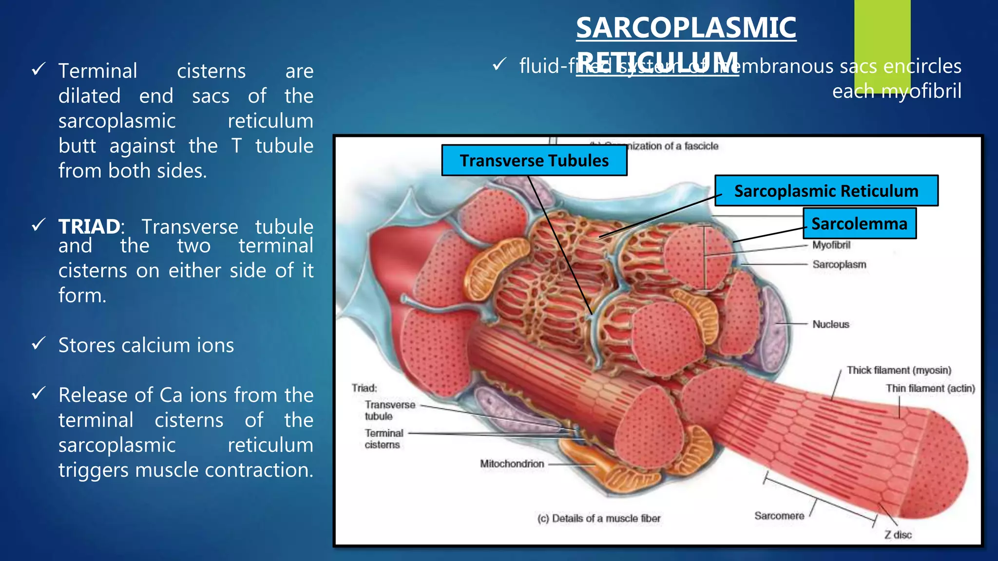
Describes the overview of the skeletal muscles, its description, functons, and properties. It also inccludes the gross organization of the skeletal system.
Describes the action potential occuring in the muscle. It includes the cellular and molecular organization of the muscle particularly on the myosin and actin myofilaments. Describes likewise the steps of muscle contraction.
Skeletal muscle is one of the three significant muscle tissues in the human body. Each skeletal muscle consists of thousands of muscle fibers wrapped together by connective tissue sheaths. The individual bundles of muscle fibers in a skeletal muscle are known as fasciculi.
Muscle is one of the four primary tissue types of the body, and the body contains three types of muscle tissue: skeletal muscle, cardiac muscle, and smooth muscle.
i've used this note before this for my first year medicine in egypt. Fot those who taking this course(medicine) , I hope it'll give some ideas to you to study about this subject.
Goodluck :) !
AI and Machine Learning Demystified by Carol Smith at Midwest UX 2017Carol Smith
What is machine learning? Is UX relevant in the age of artificial intelligence (AI)? How can I take advantage of cognitive computing? Get answers to these questions and learn about the implications for your work in this session. Carol will help you understand at a basic level how these systems are built and what is required to get insights from them. Carol will present examples of how machine learning is already being used and explore the ethical challenges inherent in creating AI. You will walk away with an awareness of the weaknesses of AI and the knowledge of how these systems work.
Power Point covering the Functions of the Muscular System from the Physiological Point of study from a Collegiate Anatomy and Physiology Class (super basic and easy to understand).
Muscles is a contractile tissue which brings about movement.
Muscle cell responsible for our movement both visible and invisible, example walking, talking, bowel movement ,urination, breathing, heartbeats, the dilation and constriction of the pupils of our eyes and many other.
When we are still sitting or standing muscle cells keep us erect.
CONT...Muscles can be regarded as motors of the body.Muscles comprises about 40% to 50% (approximate) of body weight.There are approximate 650 muscles in body.Alternating contraction and relaxation of cells
skeletal, cardiac & smooth Muscles by Thiru Murugan.pptxthiru murugan
Unit III – The Muscular System - Anatomy
Types and structure of muscles
Muscle groups
Alterations in disease
Applications and implications in nursing
Muscle:
Muscle is a soft tissue and it is one of the 4 basic tissues, along with nervous tissue, epithelium, and connective tissue.
Muscles helps in movement, support and protection of internal organs.
Muscles can perform variety of functions
Muscles tissue is made up of cells called “MYOCYTES” or muscle fibers.
There are more than 600 muscles in the human body. A kind of elastic tissue makes up each muscle, which consists of thousands, or tens of thousands, of small muscle fibers.
Types of Muscles: There are 3 main types of muscles
Skeletal muscle
Cardiac muscle
Smooth muscle
Skeletal muscle:
These are having close relationship to the bone or skeleton, so called Skeletal muscles
It present in limbs and related body parts & It form about 40% of body weight.
Under microscope the skeletal muscles fibers shows prominent striations, so called “Striated Muscles” & It is also known as “Voluntary Muscles” (movements are under our control)
Structure of Skeletal muscle:
Muscle fibers shows transverse striations under light microscope so it is called “striated muscles”
The nucleus is located peripherally.
Each skeletal muscle is an organ that consists of numerous cells called muscle fibers.
Each muscle fibers surrounded by “ Endomysium”
Inside each skeletal muscle, muscle fibers are organized into bundles, called fascicles, each fascicle surrounded by perimysium.
The whole muscle is covered by “epimysium”
Each skeletal muscle has three layers: endomysium, perimysium and epimysium
Muscle fibers:
Muscle is composed of many long cylindrical-shaped elongated fibres called muscle fibers
Length varies according to the size and shape of the muscles.
The actual arrangement of the fibres depending on the function of the muscle.
Each muscle fibers covered by a membrane is called the sarcolemma.
The cytoplasm of a muscle fiber is called Sarcoplasm
In sarcoplasm there are many mitochondria and bundles of fine longitudinal thread like part is called “myofibrils”
Microscopic structure of myofibrils:
A myofibril (also known as a muscle fibril or sarcostyle) is a basic rod-like part of a muscle cell.
Muscles are composed of tubular cells called myocytes, known as muscle fibres in striated muscle, and these cells in turn contain many chains of myofibrils.
They are created during embryonic development in a process known as myogenesis.
Under light microscope each myofibril consist of 2 bands:
Light band or “I” Band and Dark band or “A” Band
The alternating pattern of these bands results in the striated appearance of skeletal muscle.
Light band or “I” Band:
The I-bands (isotropic in polarized light) appear light in color.
I band divided into 2 portions by a narrow dark line called “Z” line or “Z” Disc.
This “Z” line is formed by protein which does not permit the light.
The part in between 2 “Z” lines called “sarc
Lung Cancer: Artificial Intelligence, Synergetics, Complex System Analysis, S...Oleg Kshivets
RESULTS: Overall life span (LS) was 2252.1±1742.5 days and cumulative 5-year survival (5YS) reached 73.2%, 10 years – 64.8%, 20 years – 42.5%. 513 LCP lived more than 5 years (LS=3124.6±1525.6 days), 148 LCP – more than 10 years (LS=5054.4±1504.1 days).199 LCP died because of LC (LS=562.7±374.5 days). 5YS of LCP after bi/lobectomies was significantly superior in comparison with LCP after pneumonectomies (78.1% vs.63.7%, P=0.00001 by log-rank test). AT significantly improved 5YS (66.3% vs. 34.8%) (P=0.00000 by log-rank test) only for LCP with N1-2. Cox modeling displayed that 5YS of LCP significantly depended on: phase transition (PT) early-invasive LC in terms of synergetics, PT N0—N12, cell ratio factors (ratio between cancer cells- CC and blood cells subpopulations), G1-3, histology, glucose, AT, blood cell circuit, prothrombin index, heparin tolerance, recalcification time (P=0.000-0.038). Neural networks, genetic algorithm selection and bootstrap simulation revealed relationships between 5YS and PT early-invasive LC (rank=1), PT N0—N12 (rank=2), thrombocytes/CC (3), erythrocytes/CC (4), eosinophils/CC (5), healthy cells/CC (6), lymphocytes/CC (7), segmented neutrophils/CC (8), stick neutrophils/CC (9), monocytes/CC (10); leucocytes/CC (11). Correct prediction of 5YS was 100% by neural networks computing (area under ROC curve=1.0; error=0.0).
CONCLUSIONS: 5YS of LCP after radical procedures significantly depended on: 1) PT early-invasive cancer; 2) PT N0--N12; 3) cell ratio factors; 4) blood cell circuit; 5) biochemical factors; 6) hemostasis system; 7) AT; 8) LC characteristics; 9) LC cell dynamics; 10) surgery type: lobectomy/pneumonectomy; 11) anthropometric data. Optimal diagnosis and treatment strategies for LC are: 1) screening and early detection of LC; 2) availability of experienced thoracic surgeons because of complexity of radical procedures; 3) aggressive en block surgery and adequate lymph node dissection for completeness; 4) precise prediction; 5) adjuvant chemoimmunoradiotherapy for LCP with unfavorable prognosis.
ARTIFICIAL INTELLIGENCE IN HEALTHCARE.pdfAnujkumaranit
Artificial intelligence (AI) refers to the simulation of human intelligence processes by machines, especially computer systems. It encompasses tasks such as learning, reasoning, problem-solving, perception, and language understanding. AI technologies are revolutionizing various fields, from healthcare to finance, by enabling machines to perform tasks that typically require human intelligence.
Knee anatomy and clinical tests 2024.pdfvimalpl1234
This includes all relevant anatomy and clinical tests compiled from standard textbooks, Campbell,netter etc..It is comprehensive and best suited for orthopaedicians and orthopaedic residents.
Title: Sense of Smell
Presenter: Dr. Faiza, Assistant Professor of Physiology
Qualifications:
MBBS (Best Graduate, AIMC Lahore)
FCPS Physiology
ICMT, CHPE, DHPE (STMU)
MPH (GC University, Faisalabad)
MBA (Virtual University of Pakistan)
Learning Objectives:
Describe the primary categories of smells and the concept of odor blindness.
Explain the structure and location of the olfactory membrane and mucosa, including the types and roles of cells involved in olfaction.
Describe the pathway and mechanisms of olfactory signal transmission from the olfactory receptors to the brain.
Illustrate the biochemical cascade triggered by odorant binding to olfactory receptors, including the role of G-proteins and second messengers in generating an action potential.
Identify different types of olfactory disorders such as anosmia, hyposmia, hyperosmia, and dysosmia, including their potential causes.
Key Topics:
Olfactory Genes:
3% of the human genome accounts for olfactory genes.
400 genes for odorant receptors.
Olfactory Membrane:
Located in the superior part of the nasal cavity.
Medially: Folds downward along the superior septum.
Laterally: Folds over the superior turbinate and upper surface of the middle turbinate.
Total surface area: 5-10 square centimeters.
Olfactory Mucosa:
Olfactory Cells: Bipolar nerve cells derived from the CNS (100 million), with 4-25 olfactory cilia per cell.
Sustentacular Cells: Produce mucus and maintain ionic and molecular environment.
Basal Cells: Replace worn-out olfactory cells with an average lifespan of 1-2 months.
Bowman’s Gland: Secretes mucus.
Stimulation of Olfactory Cells:
Odorant dissolves in mucus and attaches to receptors on olfactory cilia.
Involves a cascade effect through G-proteins and second messengers, leading to depolarization and action potential generation in the olfactory nerve.
Quality of a Good Odorant:
Small (3-20 Carbon atoms), volatile, water-soluble, and lipid-soluble.
Facilitated by odorant-binding proteins in mucus.
Membrane Potential and Action Potential:
Resting membrane potential: -55mV.
Action potential frequency in the olfactory nerve increases with odorant strength.
Adaptation Towards the Sense of Smell:
Rapid adaptation within the first second, with further slow adaptation.
Psychological adaptation greater than receptor adaptation, involving feedback inhibition from the central nervous system.
Primary Sensations of Smell:
Camphoraceous, Musky, Floral, Pepperminty, Ethereal, Pungent, Putrid.
Odor Detection Threshold:
Examples: Hydrogen sulfide (0.0005 ppm), Methyl-mercaptan (0.002 ppm).
Some toxic substances are odorless at lethal concentrations.
Characteristics of Smell:
Odor blindness for single substances due to lack of appropriate receptor protein.
Behavioral and emotional influences of smell.
Transmission of Olfactory Signals:
From olfactory cells to glomeruli in the olfactory bulb, involving lateral inhibition.
Primitive, less old, and new olfactory systems with different path
Flu Vaccine Alert in Bangalore Karnatakaaddon Scans
As flu season approaches, health officials in Bangalore, Karnataka, are urging residents to get their flu vaccinations. The seasonal flu, while common, can lead to severe health complications, particularly for vulnerable populations such as young children, the elderly, and those with underlying health conditions.
Dr. Vidisha Kumari, a leading epidemiologist in Bangalore, emphasizes the importance of getting vaccinated. "The flu vaccine is our best defense against the influenza virus. It not only protects individuals but also helps prevent the spread of the virus in our communities," he says.
This year, the flu season is expected to coincide with a potential increase in other respiratory illnesses. The Karnataka Health Department has launched an awareness campaign highlighting the significance of flu vaccinations. They have set up multiple vaccination centers across Bangalore, making it convenient for residents to receive their shots.
To encourage widespread vaccination, the government is also collaborating with local schools, workplaces, and community centers to facilitate vaccination drives. Special attention is being given to ensuring that the vaccine is accessible to all, including marginalized communities who may have limited access to healthcare.
Residents are reminded that the flu vaccine is safe and effective. Common side effects are mild and may include soreness at the injection site, mild fever, or muscle aches. These side effects are generally short-lived and far less severe than the flu itself.
Healthcare providers are also stressing the importance of continuing COVID-19 precautions. Wearing masks, practicing good hand hygiene, and maintaining social distancing are still crucial, especially in crowded places.
Protect yourself and your loved ones by getting vaccinated. Together, we can help keep Bangalore healthy and safe this flu season. For more information on vaccination centers and schedules, residents can visit the Karnataka Health Department’s official website or follow their social media pages.
Stay informed, stay safe, and get your flu shot today!
These lecture slides, by Dr Sidra Arshad, offer a quick overview of physiological basis of a normal electrocardiogram.
Learning objectives:
1. Define an electrocardiogram (ECG) and electrocardiography
2. Describe how dipoles generated by the heart produce the waveforms of the ECG
3. Describe the components of a normal electrocardiogram of a typical bipolar leads (limb II)
4. Differentiate between intervals and segments
5. Enlist some common indications for obtaining an ECG
Study Resources:
1. Chapter 11, Guyton and Hall Textbook of Medical Physiology, 14th edition
2. Chapter 9, Human Physiology - From Cells to Systems, Lauralee Sherwood, 9th edition
3. Chapter 29, Ganong’s Review of Medical Physiology, 26th edition
4. Electrocardiogram, StatPearls - https://www.ncbi.nlm.nih.gov/books/NBK549803/
5. ECG in Medical Practice by ABM Abdullah, 4th edition
6. ECG Basics, http://www.nataliescasebook.com/tag/e-c-g-basics
Explore natural remedies for syphilis treatment in Singapore. Discover alternative therapies, herbal remedies, and lifestyle changes that may complement conventional treatments. Learn about holistic approaches to managing syphilis symptoms and supporting overall health.
HOT NEW PRODUCT! BIG SALES FAST SHIPPING NOW FROM CHINA!! EU KU DB BK substit...GL Anaacs
Contact us if you are interested:
Email / Skype : kefaya1771@gmail.com
Threema: PXHY5PDH
New BATCH Ku !!! MUCH IN DEMAND FAST SALE EVERY BATCH HAPPY GOOD EFFECT BIG BATCH !
Contact me on Threema or skype to start big business!!
Hot-sale products:
NEW HOT EUTYLONE WHITE CRYSTAL!!
5cl-adba precursor (semi finished )
5cl-adba raw materials
ADBB precursor (semi finished )
ADBB raw materials
APVP powder
5fadb/4f-adb
Jwh018 / Jwh210
Eutylone crystal
Protonitazene (hydrochloride) CAS: 119276-01-6
Flubrotizolam CAS: 57801-95-3
Metonitazene CAS: 14680-51-4
Payment terms: Western Union,MoneyGram,Bitcoin or USDT.
Deliver Time: Usually 7-15days
Shipping method: FedEx, TNT, DHL,UPS etc.Our deliveries are 100% safe, fast, reliable and discreet.
Samples will be sent for your evaluation!If you are interested in, please contact me, let's talk details.
We specializes in exporting high quality Research chemical, medical intermediate, Pharmaceutical chemicals and so on. Products are exported to USA, Canada, France, Korea, Japan,Russia, Southeast Asia and other countries.
Ozempic: Preoperative Management of Patients on GLP-1 Receptor Agonists Saeid Safari
Preoperative Management of Patients on GLP-1 Receptor Agonists like Ozempic and Semiglutide
ASA GUIDELINE
NYSORA Guideline
2 Case Reports of Gastric Ultrasound
micro teaching on communication m.sc nursing.pdfAnurag Sharma
Microteaching is a unique model of practice teaching. It is a viable instrument for the. desired change in the teaching behavior or the behavior potential which, in specified types of real. classroom situations, tends to facilitate the achievement of specified types of objectives.
4. DESCRIPTION:
• Skeletal muscle tissue: consists of
long, cylindrical, striated fibers
(striations are alternating light and
dark bands within fibers)
• Skeletal muscle fiber: roughly
cylindrical, multinucleated cell with
nuclei at the periphery
• Skeletal muscle: voluntary ( it can
be made to contract or relax by
conscious control)
LOCATION: attached to bones and
tendons
FUNCTION: Motion, Posture, Heat
production, Protection
5. Functions of Muscular Tissue
1. Producing body movements.
Movements of the whole body such as
walking and running, and localized
movements such as grasping a pencil,
keyboarding, or nodding the head as a
result of muscular contractions, rely on the
integrated functioning of skeletal muscles,
bones, and joints.
2. Stabilizing body positions.
Skeletal muscle contractions stabilize joints
and help maintain body positions, such as
standing or sitting. Postural muscles
contract continuously when you are awake;
for example, sustained contractions of your
neck muscles hold your head upright when
you are listening intently to your anatomy
and physiology lecture.
3. Storing and moving substances within the
body.
Storage is accomplished by sustained
contractions
o Cardiac muscle contractions
o Smooth muscle contractions also
move food and substances
4. Generating heat.
As muscular tissue contracts, it
produces heat, a process known as
thermogenesis.
heat generated by muscle is used to
maintain normal body temperature.
Involuntary contractions of skeletal
muscles, known as shivering, can
increase the rate of heat production.
6. Properties of Muscular Tissue
1. Electrical excitability
is the ability to respond to certain stimuli
by producing electrical signals called
action potentials (impulses).
Action potentials in muscles are referred
to as muscle action potentials; those in
nerve cells are called nerve action
potentials.
two main types of stimuli trigger action
potentials:
i. Auto-rhythmic electrical signals
arising in the muscular tissue itself,
as in the heart’s pacemaker.
ii. Chemical stimuli, such as
neurotransmitters released by
neurons, hormones distributed by
the blood, or even local changes in
pH.
2. Contractility
is the ability of muscular tissue to contract
forcefully when stimulated by an action
potential.
When a skeletal muscle contracts, it
generates tension (force of contraction) while
pulling on its attachment points.
3. Extensibility
is the ability of muscular tissue to stretch,
within limits, without being damaged.
The connective tissue within the muscle
limits the range of extensibility and keeps it
within the contractile range of the muscle
cells.
4. Elasticity
is the ability of muscular tissue to return to
its original length and shape after
contraction or extension.
7. Layers of connective tissue:
• extend from the fascia to protect and
strengthen skeletal muscle
1. Epimysium (Muscle)
• dense irregular connective tissue.
2. Perimysium (Muscle fascicle/ bundle)
• dense irregular connective tissue
• surrounds groups of 10 to 100 or more
muscle fibers, separating them into
bundles called fascicles.
3. Endomysium (muscle fiber)
• penetrates the interior of each fascicle and
separates individual muscle fibers from one
another
• mostly reticular fibers.
Epimysium
.
Perimysium
.
Endomysium
.
9. 1. SKELETAL MUSCLE
o Organ made up of fascicles
that contain muscle fibers
(cells), BV, and nerves
o Wrapped in epimysium
2. FASCICLE
o Bundles of muscle fibers
o Wrapped in perimysium
10. 3. MUSCLE FIBER (CELL)
o Long cylindrical cell
o Wrapped in endomysium and
sarcolemma
o Straited appearance
o each fiber extends the entire
length of the muscle except for
about 2 per cent of the fibers
o each fiber is usually innervated
by only one nerve ending,
located near the middle of the
fiber.4. MYOFIBRIL
o Threadlike contractile elements
within a sarcoplasm of muscle fiber
that extend entire length of fiber
o Composed of filaments
11. 5. FILAMENTS
o Contractile proteins within
myofibrils that are two types:
thick and thin
o Sliding of thin filaments
produces muscle shortening
12. Sarcolemma
Sarcoplasmic Reticulum
Transverse Tubules
SARCOLEMMA:
- the cell membrane of the muscle fiber
-where muscle action potentials travel along
SARCOPLASMIC RETICULUM
-wraps around each myofibril
-where muscle action potentials quickly
spread throughout the muscle fiber
TRANSVERSE TUBULES
- Filled with extracellular fluid
-invaginate from the sarcolemma toward the
of the muscle fiber
MUSCLE FIBER (CELL)
This arrangement ensures
that an action potential
excites all parts of the muscle
fiber at essentially the same
instant.
• single cell
• multinucleated
• surrounded by the sarcolemma
13. Intracellular fluid containing large quantities of
potassium, magnesium, and phosphate, plus multiple
protein enzymes.
Presence of tremendous numbers of mitochondria that
lie parallel to the myofibrils which supply the
contracting myofibrils with large amounts of energy in
the form of adenosine triphosphate (ATP)
includes a substantial amount of glycogen which can be
used for synthesis of ATP
contains a red-colored protein called myoglobin
• which binds oxygen molecules that diffuse into
muscle fibers from interstitial fluid
• releases oxygen when it is needed by the
mitochondria for ATP production
SARCOPLASM
14. Invaginations of the sarcolemma
filled with extracellular fluid
• Penetrate the muscle fiber,
branch and form networks
• Transmit AP’s deep into the
muscle fiber
TRANSVERSE TUBULE
(T- TUBULES)
15. Terminal cisterns are
dilated end sacs of the
sarcoplasmic reticulum
butt against the T tubule
from both sides.
TRIAD: Transverse tubule
and the two terminal
cisterns on either side of it
form.
Stores calcium ions
Release of Ca ions from the
terminal cisterns of the
sarcoplasmic reticulum
triggers muscle contraction.
Sarcolemma
Sarcoplasmic Reticulum
Transverse Tubules
fluid-filled system of membranous sacs encircles
each myofibril
SARCOPLASMIC
RETICULUM
16. Embryonic development of skeletal muscle
MYOBRILS- The contractile elements of muscle fibers which
contain overlapping thick and thin filaments
embryonic development
myoblasts fuse to form one
skeletal muscle fiber
fusion has occurred
skeletal muscle fiber loses the
ability to undergo cell