2. In knee jerk elicitation extension of leg
instead of returning to normal position
swings forwards and backwards several
times before coming to rest.
These are due to hypotonia and lack of
restrictive effect.
These are seen in cerebellar lesion.
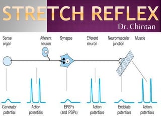
3. THE STRETCH REFLEX – Best
studied monosynaptic reflexes in the
body.
When a skeletal muscle with an
intact nerve supply is stretched, it
contracts.
This response is called the stretch
reflex.
4. The stimulus that initiates the reflex is
stretch of the muscle, and the
response is contraction of the muscle being
stretched.
The sense organ is the muscle spindle.
Center - spinal cord
The impulses originating in the spindle are
conducted in the CNS by fast sensory fibers
that pass directly to the motor neurons which
supply the same muscle.
The neurotransmitter at the central synapse is
glutamate.
5.
6. Tapping the patellar tendon elicits the
knee jerk, a stretch reflex of the
quadriceps femoris muscle, because the
tap on the tendon stretches the muscle.
Tapping on the tendon of the triceps
brachii, for example, causes an extensor
response at the elbow as a result of
reflex contraction of the triceps;
Tapping on the Achilles tendon causes
an ankle jerk due to reflex contraction
gastrocnemius;
7. Monosynaptic reflex
1) One synapse
between afferent and
efferent neurons.
2) Eg stretch reflex –
knee jerk.
3) No after discharge
and irradiation of
impulses.
4) Latency of response
shorter.
Polysynaptic reflex
1) Many synapses
between afferent and
efferent neurons.
2) Eg withdrawal
reflexes.
3) They show after
discharge and
irradiation
4) Latency of response
longer.
8. 1) Both reflexes are basically protective
reflexes.
2) Both are characterized by reciprocal
innervation.
3) Both show integration at the level of
α- motor neurons which serve as the
final common pathway.
9. The cord gray matter is the integrative
area for the cord reflexes.
Sensory signals enter the cord almost
entirely through the sensory (posterior)
roots.
After entering the cord, every sensory
signal travels to two separate
destinations:
10. (1) One branch of the sensory nerve
terminates almost immediately in the
gray matter of the cord and elicits local
segmental cord reflexes and other local
effects.
(2) Another branch transmits signals to
higher levels of the nervous system—
to higher levels in the cord itself,
to the brain stem, or
to the cerebral Cortex.
11. A) Anterior motor neurons – two
types
1) Alpha motor neuron
2) Gamma motor neuron
B) Interneuron
C) Renshaw cells
12. Several thousand neurons that are 50 to 100
per cent larger than most of the others and are
called anterior motor neurons.
They give rise to the nerve fibers that leave
the cord by way of the anterior roots and
directly innervate the skeletal muscle fibers.
The neurons are of two types, alpha motor
neurons and gamma motor neurons.
13. 1) large type A alpha (Aα) motor nerve fibers,
2) averaging 14 micrometers in diameter;
3) these fibers branch many times after they
enter the muscle and innervate the large
skeletal muscle fibers.
4) Stimulation of a single alpha nerve fiber
excites anywhere from three to several
hundred skeletal muscle fibers, which are
collectively called the motor unit.
14. Gamma motor neurons transmit impulses
through much
1) smaller type A gamma motor nerve fibers,
2) averaging 5 micrometers in diameter,
3) which go to small, special skeletal muscle
fibers called intrafusal fibers.
4) These fibers constitute the middle of the
muscle spindle, which helps control basic
muscle “tone”.
15. 1) Interneurons are present in all areas of the
cord gray matter - in the dorsal horns, the
anterior horns, and the intermediate areas
between them.
2) These cells are about 30 times as numerous
as the anterior motor neurons.
3) They are small and highly excitable, often
exhibiting spontaneous activity and capable of
firing as rapidly as 1500 times per second.
16. Also located in the anterior horns of the
spinal cord, in close association with the motor
neurons, are a large number of small neurons
called Renshaw cells.
Almost immediately after the anterior motor
neuron axon leaves the body of the neuron,
collateral branches from the axon pass to
adjacent Renshaw cells.
These are inhibitory cells that transmit
inhibitory signals to the surrounding motor
neurons.
17.
18. 1) Each spindle is 3 to 10
millimeters long.
2) It is built around 3 to 12
very small intrafusal
muscle fibers
that are pointed at their
ends and attached to the
glycocalyx of the
surrounding large extrafusal
skeletal muscle fibers
19. 1) the central region of each
of these fibers—that is, the
area midway between its two
ends - has few or no actin and
myosin filaments.
2) Therefore, this central
portion does not contract
when the ends do.
3) Instead, it functions as a
sensory receptor.
20. (1) nuclear bag muscle
fibers (one to three in
each spindle),
in which several muscle
fiber nuclei are
assembled in expanded
“bags” in the central
portion of the receptor
area.
(2) nuclear chain fibers
(three to nine),
have nuclei aligned in a
chain throughout the
receptor area.
21. Each muscle spindle
has 2 nuclear bags
and 4 or more
nuclear chains.
One nuclear bag has
low levels of myosin
ATPase activity,
and second has high
levels of it.
22. 1) Lengthening the whole muscle
stretches the midportion of the spindle
and, therefore, excites the receptor.
2) Even if the length of the entire muscle
does not change, contraction of the
end portions of the spindle’s intrafusal
fibers stretches the midportion of the
spindle and therefore excites the
receptor.
23. The end portions that do
contract are excited by small
gamma motor nerve fibers
that originate from small
gamma motor neurons in the
anterior horns of the spinal
cord,
These gamma motor nerve
fibers are also called gamma
efferent fibers,
the large alpha efferent
fibers (type A alpha nerve
fibers) that innervate the
extrafusal skeletal muscle
24. Primary Ending.
1) In the center of the
receptor area, a large sensory
nerve fiber encircles the
central portion of each
intrafusal fiber.
2) Type Ia fiber averaging 17
µm in diameter,
3) Transmits sensory signals to
the spinal cord at a velocity of
70 to 120 m/sec.
4) They supply both nuclear
bag and nuclear chain fibres
25. 1) type II fibers with an
average diameter of 8 µm
innervate the receptor
region on one or both sides of
the primary Endings.
2) sometimes it encircles
the intrafusal fibers in the
same way that the type Ia
fiber does,
3) but often it spreads like
branches on a bush - Flower
spray endings.
4) These mainly innervate
the nuclear chain type of
intrafusal fibres.
26.
27. Response of Both the Primary and the Secondary
Endings to the Length of the Receptor — “Static”
Response.
When the receptor portion of the muscle spindle is
stretched slowly,
the number of impulses transmitted from both the
primary and the secondary endings increases almost
directly in proportion to the degree of stretching,
and the endings continue to transmit these impulses
for several minutes if the muscle spindle itself
remains stretched.
The nuclear chain is supplied by both types of nerves
so they are responsible for static response.
28. Response of the Primary Ending (but
Not the Secondary Ending) to Rate of
Change of Receptor Length —“Dynamic”
Response.
When the length of the spindle receptor
increases suddenly, the primary ending
(but not the secondary ending) is
stimulated especially powerfully.
29. Control of Intensity of the Static and
Dynamic Responses by the Gamma Motor
Nerves.
The gamma motor nerves to the muscle
spindle can be divided into two types:
(1)gamma-dynamic (gamma-d) - Nuclear
bag
(2)gamma-static (gamma-s) - Nuclear
chain
30. When the gamma-d fibers excite the
nuclear bag fibers, the dynamic response
of the muscle spindle becomes
tremendously enhanced,
Conversely, stimulation more of the
gamma-s fibers, which excite the nuclear
chain fibers, enhances the static
response.
These two types of muscle spindle
responses are important in different types
of muscle control.
31. Normally, there is some degree of gamma nerve
excitation, and the muscle spindles emit sensory
nerve impulses continuously.
Stretching the muscle spindles increases the rate of
firing, whereas shortening the spindle decreases the
rate of firing.
Thus, the spindles can send to the spinal cord either
positive signals—that is, increased numbers of
impulses to indicate stretch of a muscle
negative signals—below-normal numbers of impulses
to indicate that the muscle is unstretched.
32. 1) Type Ia proprioceptor nerve fiber
originating in a muscle spindle and
entering a dorsal root of the spinal cord.
2) A branch of this fiber then goes
directly to the anterior horn of the cord
gray matter and synapses with anterior
motor neurons
3) that send motor nerve fibers back to
the same muscle from which the muscle
spindle fiber originated.
33. Most type II fibers (as well as many
collaterals of Ia) from the muscle
spindle terminate on multiple
interneurons in the cord gray matter,
and
these transmit delayed signals to the
anterior motor neurons or serve other
functions.
Eg for static reflex activity to
maintain tone.
34. Stretch reflex is well developed in antigravity
muscles such as extensors of legs & flexors
of arm.
Reflex arc has
1) Afferent limb - muscle spindle and
afferent nerve. Nerve supply of spindle is
sensory 1a & II nerves and motor by gamma
fibers both static and dynamic.
2) Centre - ventral grey horn where Ia
synapses with alpha motor neuron which
supplies extrafusal fibres.
3) Efferent limb - Axon of alpha motor neuron
& effecter organ is muscle (extensor or flexor)
35.
36. stimulation causes the contractile ends of
the intrafusal fibers to shorten and therefore
stretches the nuclear bag portion of the
spindles,
deforming the annulospiral endings and
initiating impulses in the Ia fibers - lead to
reflex contraction of the muscle.
Thus, muscle can be made to contract via
stimulation of the α motor neurons that
innervate the extrafusal fibers
or the γ efferent neurons that initiate
contraction indirectly via the stretch reflex.
37. The dynamic stretch reflex is elicited by the
potent dynamic signal transmitted from the
primary sensory endings of the muscle
spindles, caused by rapid stretch
That is, when a muscle is suddenly stretched a
strong signal is transmitted to the spinal cord;
this causes an instant strong reflex
contraction of the same muscle from which
the signal originated.
Thus, the reflex functions to oppose sudden
changes in muscle length.
38. The dynamic stretch reflex is over within a
fraction of a second after the muscle has
been stretched to its new length,
but then a weaker static stretch reflex
continues for a prolonged period thereafter.
This reflex is elicited by the continuous static
receptor signals transmitted by both primary
and secondary endings.
The importance of the static stretch reflex is
that it continues to causes the muscle
contraction as long as the muscle is
maintained at excessive length
39. Upon shortening of the muscle –
Decrease nerve impulses from spindle -
both dynamic and static reflex - muscle
inhibition rather than reflex excitation
This is negative stretch reflex which
opposes shortening in the same way that
the positive stretch reflex opposes
lengthening of the muscle.
40. 31 per cent of all the motor nerve fibers to the muscle
are the small type A gamma efferent fibers rather
than large type A alpha motor fibers.
Whenever signals are transmitted from the motor
cortex or from any other area of the brain to the
alpha motor neurons, in most instances the gamma
motor neurons are stimulated simultaneously,
an effect called coactivation of the alpha and gamma
motor neurons.
This causes both the extrafusal skeletal muscle fibers
and the muscle spindle intrafusal muscle fibers to
contract at the same time.
41. 1)For tone and posture:
Stimulation of γ motor fibers initiates
stretch reflex and tone increases.
Maintenance of our posture - standing or
changing the posture are effected by
appropriate adjustments of γ motor
neuron activities of different groups of
muscles.
42. 2) Stimulation of gamma efferent by
causing stretch of spindle keeps muscle
fiber sensitive for stretch reflex.
In some diseases like in pyramidal tract
lesions this sensitivity increases and
produces exaggerated tendon jerks.
3) For effective and smooth skeletal
muscle contraction gamma efferent
activity is very important.
43. The gamma efferent system is excited
specifically by
signals from the bulboreticular
facilitatory region of the brain stem
and,
secondarily, by impulses transmitted
into the bulboreticular area from
(1) the cerebellum, (2) the basal
ganglia, and (3) the cerebral cortex.
44. Facilitatory Reticular formation - excites
gamma and stretch reflex becomes
hyperactive.
Inhibitory Reticular formation - inhibits
gamma and decreases spindle activity.
Other factors - Anxiety increases gamma
discharge, so hyperactive reflexes in anxious
pts.
Stimulation of skin by pull pressure or
noxious agents also increases gamma
discharge eg Jendrassik’s maneuver.
45. 1) Cutting or destruction of afferent or
efferent nerve to the muscle.
2) Stimulation of inhibitory reticular
formation which inhibits γ efferent
activity and thus the spindle.
3) Inhibition of facilitatory areas.
4) Hypothyroidism
46. 1) Destruction of inhibitory areas.
2) Stimulation of facilitatory areas.
3) Any factors which increase γ efferent
discharge like anxiety, stimulation of skin
by pull pressure or noxious agents,
Jendrassik’s maneuver.
4) Hyperthyroidism
47.
48. It is the resistance offered by a
muscle to stretch in a resting
condition due to a state of
partial contraction
resulting from low frequency,
asynchronous discharge of
gamma motor neurons.
49. Certain amount of tension is present in
resting muscle due to γ efferent
discharge which produces resistance to
stretch.
Viscosity and elasticity of muscle fibers
also contributes to it.
It is tested by passive movements of
limbs at the joints of a subject by an
examiner.
50. Hypertonia
Muscle tone is increased in some conditions,
and the muscle becomes rigid and stiff and
spastic.
Here the resistance offered to stretch is very
high.
Hypotonia
Muscle tone is reduced eg if motor nerve is cut
and the muscle becomes flaccid or lax and
offers very less resistance to stretch.
51. The normal tone is between the
states of flaccidity and spasticity
which gives the muscle a firm
feeling.
The resting length of a muscle is
maintained due to this muscle tone.
The muscles are generally hypotonic
when gamma efferent discharge is
low and hypertonic when high.
52. 1) Destruction of local
reflex arc anywhere in
its pathway, eg
destruction of efferent
(motor) nerve by injury
or poliomyelitis,
or destruction of dorsal
columns as seen in
syphilis.
Syphilis causes
constrictive fibrosis
around the dorsal nerve
roots fibers where they
enter the spinal cord.
This condition is called
Tabes dorsalis.
2) Stimulation of
inhibitory area or
destruction of facilitatory
area in brain.
3) Sleep - decreases
gamma activity
4) Drugs like barbiturates,
tranquilizers in high doses
by their action on CNS.
5) LMN paralysis the
muscles are soft, flaccid
and hypotonic
53. 1) Stimulation of facilitatory area or
destruction of inhibitory areas of
brain.
2) Factors increasing gamma activity
like anxiety.
3) Complete section of spinal cord-
muscle tone below the section
increases as supraspinal inhibitory
control is lost.
54. 4) Decerebrate animals – decerebrate
rigidity in all four limbs as loss of
inhibition from higher areas (cerebral
cortex, basal ganglia, cerebellum) thus
facilitation is predominant so more tone.
Also free operation of vestibular nucleus.
Thus more gamma activity causes this.
Posterior rhizotomy disrupts alpha-
gamma linkage and this rigidity
disappears.
This affects antigravity muscles of all
four limbs.
55. 5) Decortication - Decorticate
rigidity-increased tone in flexors
of upper limbs and extensors of
lower limbs
Seen after hemiplegia due to
loss of inhibition by cortex.
6) UMNL- more tone.
56. Spasticity – If hypertonia is confined
to only one group of muscles eg
UMNL and lesion of internal capsule.
Rigidity - If hypertonia involves
both flexors as well as extensors
equally. eg basal ganglia lesion.
57.
58. It is an encapsulated
sensory receptor through
which muscle tendon fibers
pass.
About 10 to 15 muscle
fibers are usually
connected to each Golgi
tendon organ,
and the organ is stimulated
when this small bundle of
muscle fibers is “tensed” by
contracting or stretching the
muscle.
59. The tendon organ, like the primary receptor of the
muscle spindle, has both a dynamic response and a
static response.
responding intensely when the muscle tension or
force suddenly increases (the dynamic response) but
settling down within a fraction of a second to a lower
level of steady-state firing that is almost directly
proportional to the muscle tension (the static
response).
Thus, Golgi tendon organs provide the nervous system
with instant information on the degree of tension in
each small segment of each muscle.
60. Signals from the tendon organ are
transmitted through large, rapidly
conducting type I b nerve fibers that
average 16 micrometers in diameter.
These fiber transmit signals both into
1) local areas of the cord and, after
synapsing in a dorsal horn of the cord,
2) through long fiber pathways such as
the Spinocerebellar tracts into the
cerebellum and through still other
tracts to the cerebral cortex.
61. The local cord signal excites a single
inhibitory interneuron which
secretes Glycine that inhibits the
anterior motor neuron.
This local circuit directly inhibits
the individual muscle without
affecting adjacent muscles.
62.
63. When the Golgi tendon organs of a muscle
tendon are stimulated by increased tension in
the connecting muscle, signals are
transmitted to the spinal cord to cause reflex
inhibitory effects in the respective muscle
Thus, this reflex provides a negative feedback
mechanism that prevents the development of
too much tension on the muscle.
64. When tension on the muscle and, therefore, on
the tendon becomes extreme, the inhibitory
effect from the tendon organ can be so great
that it leads to a sudden reaction in the spinal
cord that causes instant relaxation of the
entire muscle.
This effect is called the lengthening reaction;
it is probably a protective mechanism to
prevent tearing of the muscle or rupture of
the tendon from its attachments to the bones.
65. Those fibers that exert excess tension
become inhibited by the reflex,
whereas
those that exert too little tension
become more excited because of
absence of reflex inhibition.
This spreads the muscle load over all
the fibers and prevents damage in
isolated areas of a muscle where small
numbers of fibers might be overloaded.
66. These two sensory organs apprise the higher motor
control centers of instant changes taking place in the
muscles.
the dorsal Spinocerebellar tracts carry instant
information from both the muscle spindles and the
Golgi tendon organs directly to the cerebellum at
conduction velocities approaching 120 m/sec,
the most rapid conduction anywhere in the brain or
spinal cord.
Additional pathways transmit similar information into
the reticular regions of the brain stem and, to a lesser
extent, all the way to the motor areas of the cerebral
cortex.
67.
68. Cutaneous sensory stimulus from a
limb is likely to cause the flexor
muscles of the limb to contract,
thereby withdrawing the limb from
the stimulating object.
This is called the flexor reflex.
69. In its classic form, the flexor reflex is
elicited most powerfully by stimulation
of pain endings, such as by a pinprick,
heat, or a wound, for which reason it is
also called a nociceptive reflex, or
simply a pain reflex.
Stimulation of touch receptors can also
elicit a weaker and less prolonged flexor
reflex.
70. If some part of the body other than one
of the limbs is painfully stimulated, that
part will similarly be withdrawn from the
stimulus,
but the reflex may not be confined to
flexor muscles, even though it is
basically the same type of reflex.
Therefore, the many patterns of these
reflexes in the different areas of the
body are called withdrawal reflexes.
71. After a pain nerve begins to be stimulated, the flexor
response appears.
Then, in the next few seconds, the reflex begins to
fatigue
Finally, after the stimulus is over, the contraction of
the muscle returns toward the baseline, but because
of after discharge, it takes many milliseconds for this
to occur.
The duration of after discharge depends on the
intensity of the sensory stimulus that elicited the
reflex;
A weak tactile stimulus causes almost no after
discharge, but after a strong pain stimulus, the after
discharge may last for a second or more.
72. Because of after discharge, the
reflex can hold the irritated part
away from the stimulus for 0.1 to
3 seconds after the irritation is
over.
During this time, other reflexes and
actions of the central nervous
system can move the entire body
away from the painful stimulus.
73. Thus, a pain stimulus on the inward side of
the arm elicits not only contraction of the
flexor muscles of the arm but also
contraction of abductor muscles to pull the
arm outward.
Thus the integrative centers of the cord
cause those muscles to contract that can most
effectively remove the pained part of the
body away from the object causing the pain.
74. About 0.2 to 0.5 second after a stimulus
elicits a flexor reflex in one limb, the
opposite limb begins to extend.
This is called the crossed extensor
reflex.
Extension of the opposite limb can
push the entire body away from the
object causing the painful stimulus in
withdrawn limb.
75.
76.
77. 1) Positive supporting reaction.
2) Cord righting reflexes.
3) Stepping and walking movements.
a) Rhythmic stepping movements of
single limb.
b) Reciprocal stepping of opposite limbs.
c) Diagonal stepping of all four limbs -
Mark Time reflex.
d) Galloping reflex.
78. Pressure on the footpad of a decerebrate
animal causes the limb to extend against the
pressure applied to the foot.
this reflex is so strong that if an animal whose
spinal cord has been transected for several
months is placed on its feet,
the reflex often stiffens the limbs sufficiently
to support the weight of the body.
This reflex is called the positive supportive
reaction.
79. The positive supportive reaction involves a
complex circuit in the interneurons similar to
the circuits responsible for the flexor and cross
extensor reflexes.
The locus of the pressure on the pad of the
foot determines the direction in which the limb
will extend;
pressure on one side causes extension in that
direction, an effect called the magnet
reaction.
This helps keep an animal from falling to that
side.
80. When a spinal animal is
laid on its side, it will
raise itself to the standing
position.
This is called the cord
righting reflex.
81. Rhythmical stepping movements are frequently
observed in the limbs of spinal animals.
even when the lumbar portion of the spinal cord is
separated from the remainder of the cord and a
longitudinal section is made down the center of the
cord to block neuronal connections between the two
sides of the cord and between the two limbs,
each hind limb can still perform individual stepping
functions.
Forward flexion of the limb is followed a second or so
later by backward extension.
Then flexion occurs again, and the cycle is repeated
over and over.
82. The sensory signals from the footpads and from the
position sensors around the joints play a strong role
in controlling foot pressure and frequency of
stepping when the foot is allowed to walk along a
surface.
if the top of the foot encounters an obstruction
during forward thrust, the forward thrust will stop
temporarily;
then, in rapid sequence, the foot will be lifted
higher and proceed forward to be placed over the
obstruction.
This is the stumble reflex - the cord is an intelligent
walking controller.
83. every time stepping occurs in
the forward direction in one
limb, the opposite limb ordinarily
moves backward.
This effect results from
reciprocal innervation between
the two limbs.
84. Stepping occurs diagonally between the
forelimbs and hind limbs.
This diagonal response is another
manifestation of reciprocal innervation,
occurring the entire distance up and
down the cord between the forelimbs and
hind limbs.
Such a walking pattern is called a mark
time reflex.
85. Another type of reflex that occasionally
develops in a spinal animal is the galloping
reflex, in which both forelimbs move backward
in unison while both hind limbs move forward.
This often occurs when almost equal stretch or
pressure stimuli are applied to the limbs on
both sides of the body at the same time;
unequal stimulation elicits the diagonal walking
reflex.
86. 1) Muscle spasm due to broken bones.
2) Abdominal muscle spasm in peritonitis.
3) Muscle cramps.
Any local irritating factor or metabolic
abnormality of a muscle, such as severe cold,
lack of blood flow, or over exercise,
can elicit pain or other sensory signals
transmitted from the muscle to the spinal
cord, which in turn cause reflex feedback
muscle contraction.
87. In a spinal animal or human being, the spinal
cord suddenly becomes excessively active,
causing massive discharge in large portions of
the cord.
The usual stimulus that causes this is a strong
pain stimulus to the skin or excessive filling
of a viscus, such as over distention of the
bladder or the gut.
Regardless of the type of stimulus, the
resulting reflex, called the mass reflex,
involves large portions or even all of the
cord.
88. (1) a major portion of the body’s skeletal
muscles goes into strong flexor spasm;
(2) the colon and bladder are likely to
evacuate;
(3) the arterial pressure often rises to
maximal values, sometimes to a systolic
pressure well over 200 mm Hg;
(4) large areas of the body break out into
profuse sweating.
89. Because the mass reflex can last for
minutes, it results from activation of
great numbers of reverberating
circuits that excite large areas of
the cord at once.
This is similar to the mechanism of
epileptic seizures, which involve
reverberating circuits that occur in
the brain instead of in the cord.