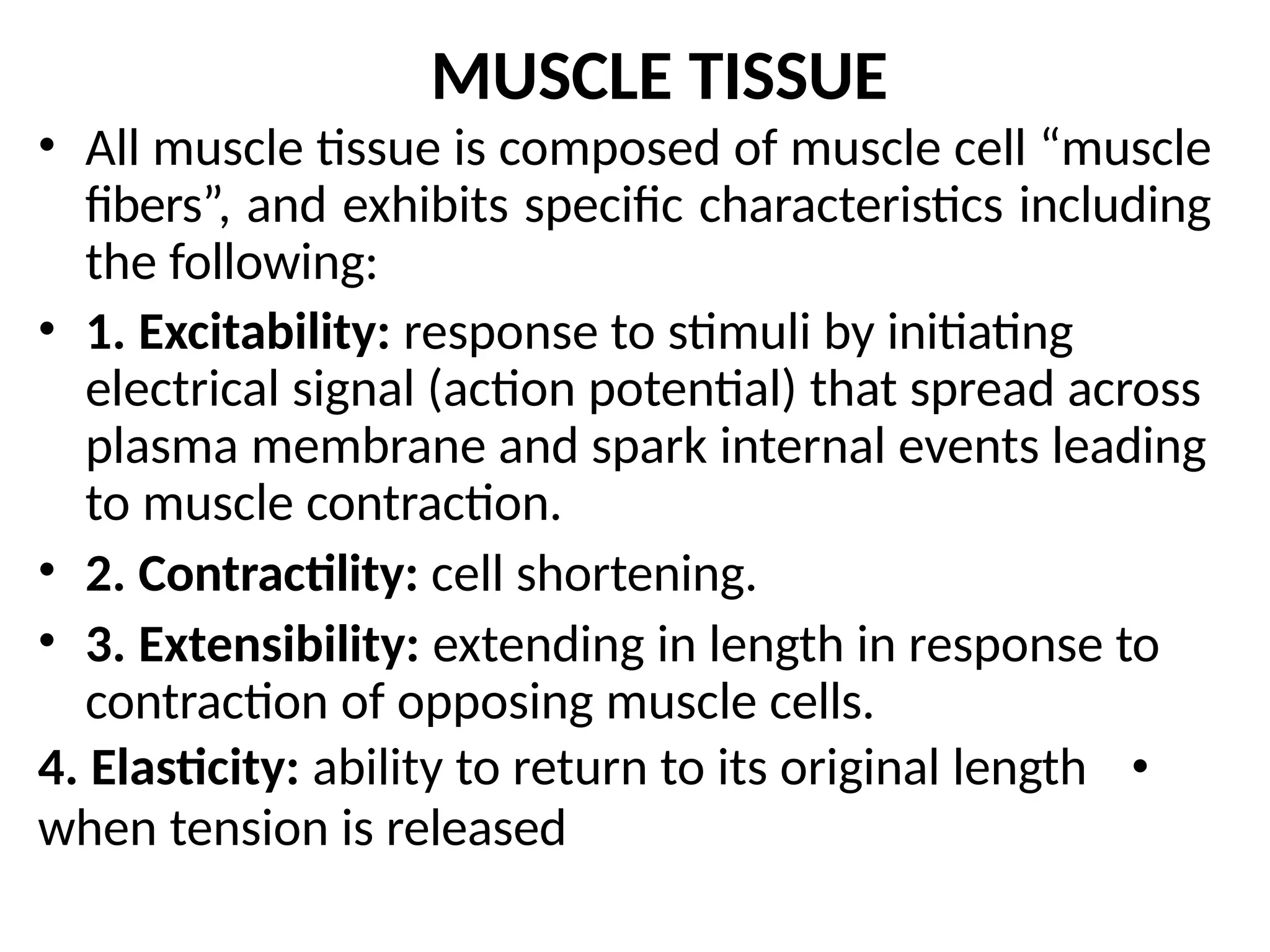
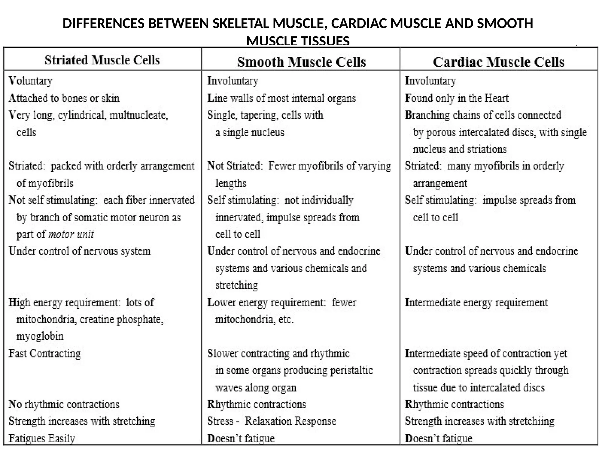
MUSCLE TISSUE
• Allmuscle tissue is composed of muscle cell “muscle
fibers”, and exhibits specific characteristics including
the following:
• 1. Excitability: response to stimuli by initiating
electrical signal (action potential) that spread across
plasma membrane and spark internal events leading
to muscle contraction.
• 2. Contractility: cell shortening.
• 3. Extensibility: extending in length in response to
contraction of opposing muscle cells.
4. Elasticity: ability to return to its original length •
when tension is released
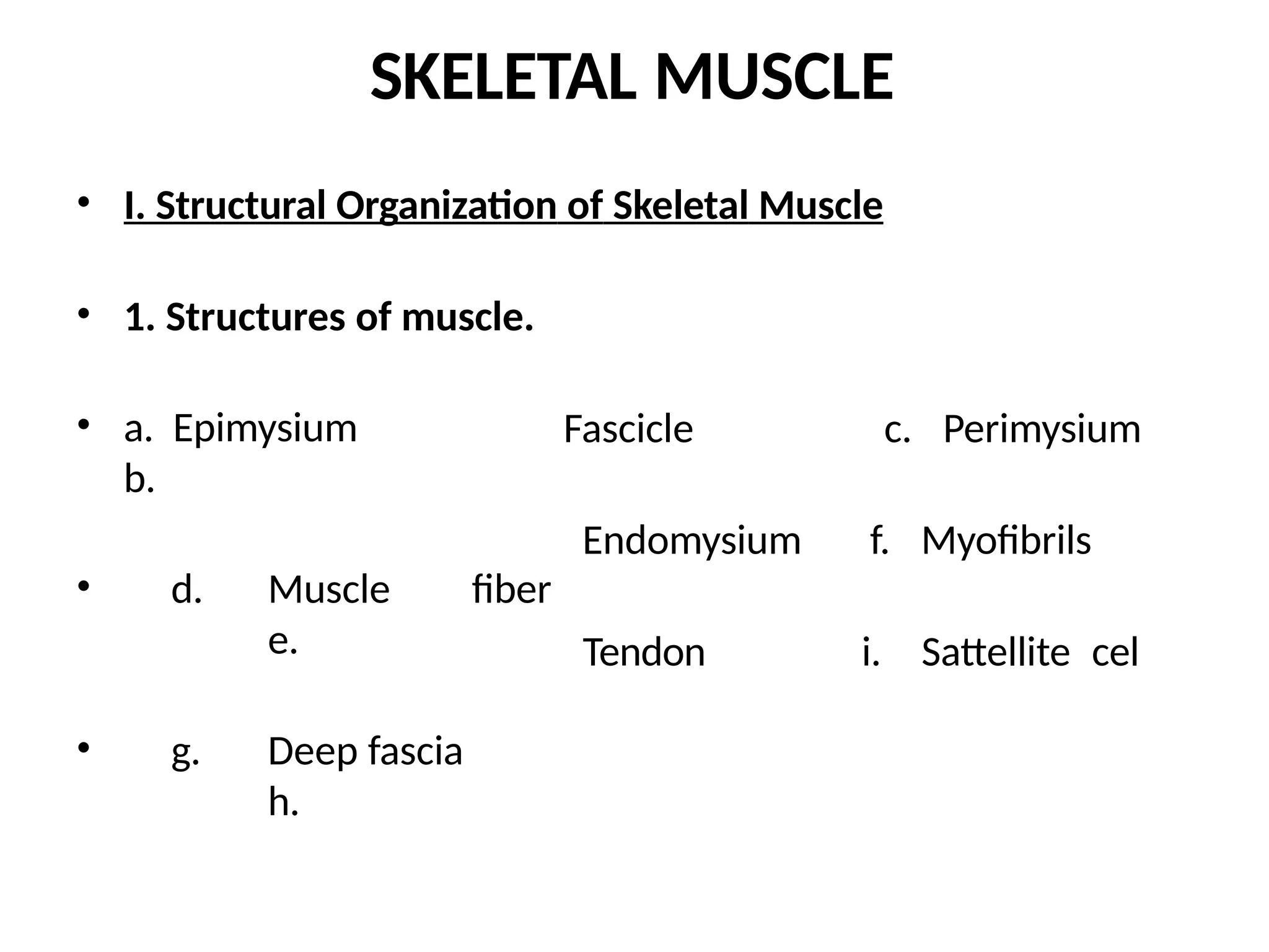
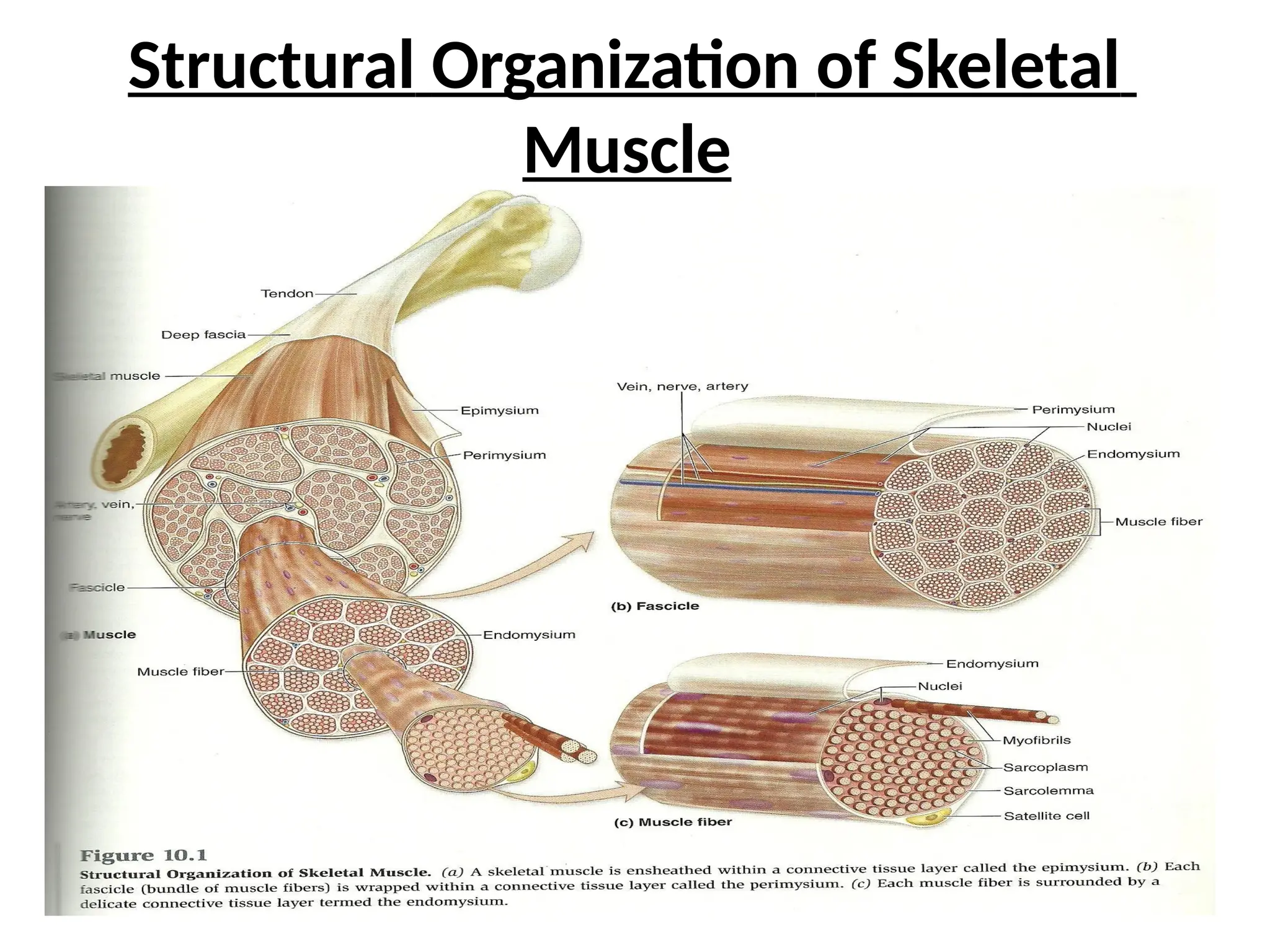
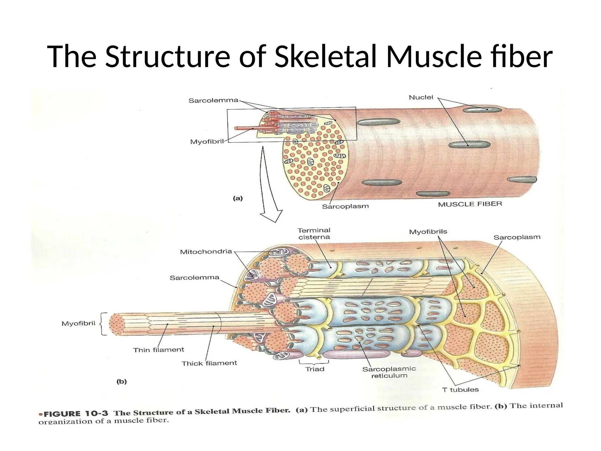
SKELETAL MUSCLE
• I.Structural Organization of Skeletal Muscle
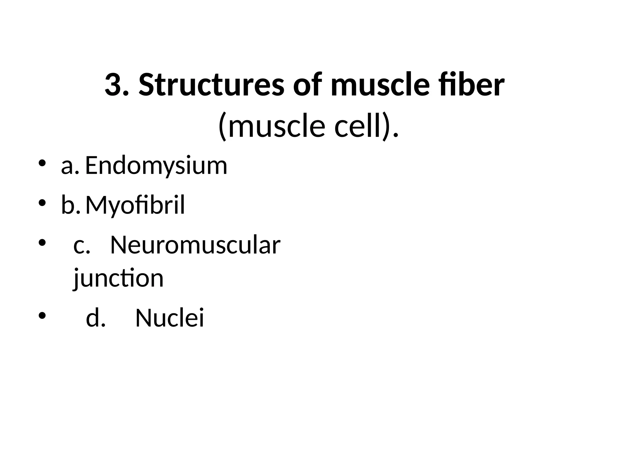
• 1. Structures of muscle.
• a. Epimysium
b.
• d. Muscle fiber
e.
• g. Deep fascia
h.
Fascicle c. Perimysium
Endomysium f. Myofibrils
Tendon i. Sattellite cel
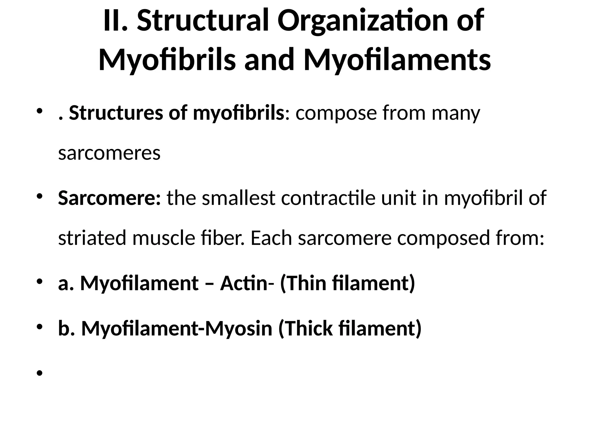
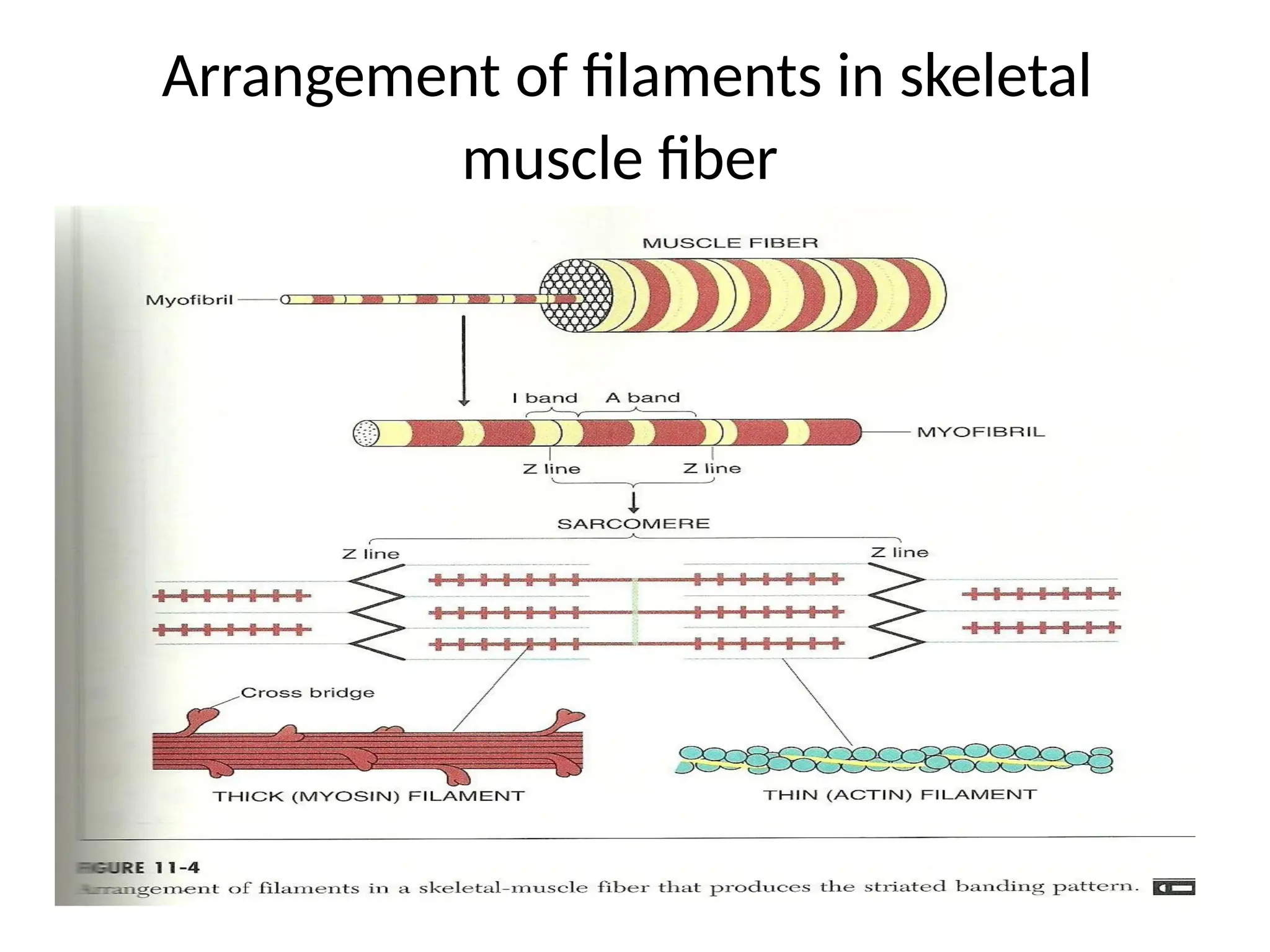
II. Structural Organizationof
Myofibrils and Myofilaments
• . Structures of myofibrils: compose from many
sarcomeres
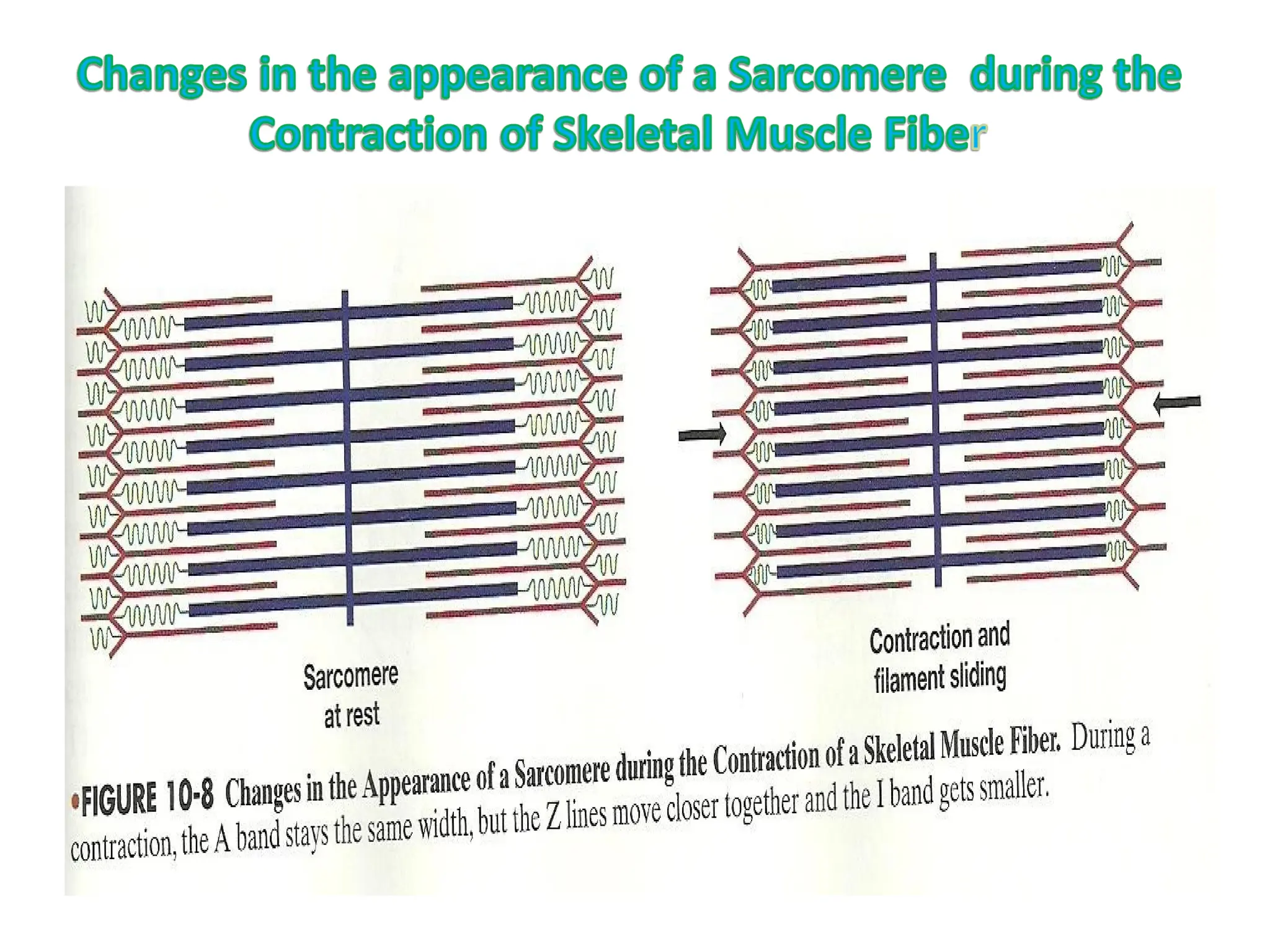
• Sarcomere: the smallest contractile unit in myofibril of
striated muscle fiber. Each sarcomere composed from:
• a. Myofilament – Actin- (Thin filament)
• b. Myofilament-Myosin (Thick filament)
•
CONTRACTION OF SKELETALMUSCLE
Contraction: is the sliding of actin over myosin
• in the presence of Ca++ (calcium ion). “Skeletal
muscle are attached to bones by tendons, and
contraction of skeletal muscle exerts a pull on
bone and movement”, most skeleton muscles
extend between bones. The less movable
point of attachment of the muscle called
“Origin”, and the more movable called
“Insertion”.
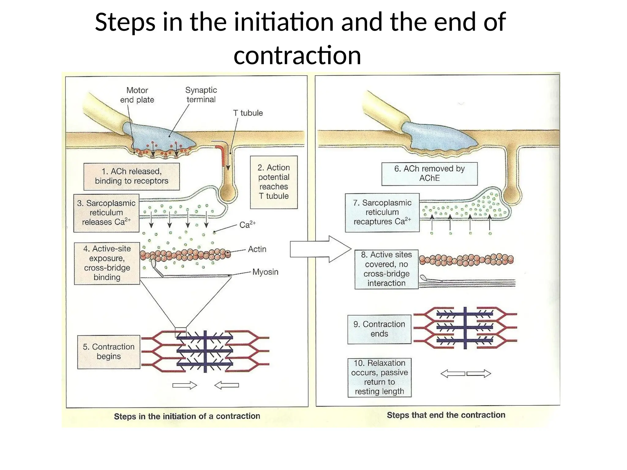
I. Steps Involvedin the Mechanisms of
Sliding Theory:
• 1. Excitable tissues: nerve and muscle (nerve
impulse in axons cause muscle impulses in
sarcolemma).
• 2. Excitable cell: cell that is capable to create and
conduct action potential
• 3. Action potential: Changes in membrane
potential of excitable cells .also defined as
electrical activity or electrical signal.
• 4. Motor end plate produces neurotransmeter at
the neuromuscular junction, to stimulate the cell
membrane (sarcolema) to produce Action
potential (electrical signal).
17.
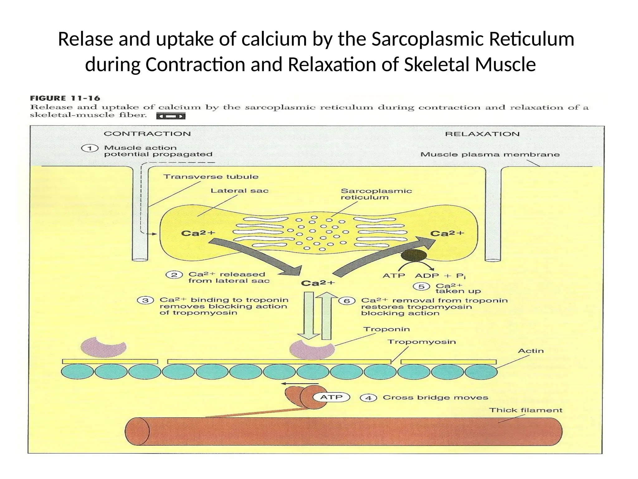
• 5. Actionpotential (electrical signal) spread
through cell membrane to “T –tube” and then
to sarcoplasmic reticulum to release calcium ion.
• 6. Calcium ion triggers the process of sliding.
• 7. Relaxation: the period after a contraction when
the tension in the muscle fiber return to resting
levels, and this done by:
• a. Active cytosolic calcium (Ca++) transported
across the cell membrane into the extracellular
fluid.
b. Active cytosolic calcium (Ca++) transported into
•
the sarcoplasmic reticulum.
CONTROL OF SKELETALMUSCLE
CONTRACTION
Skeletal muscle fibers contract only under the •
control of the nervous system.
Communication between the nervous system
and the skeletal muscle fiber occurs at a
specialized intercellular connection known as
neuromuscular junction
22.
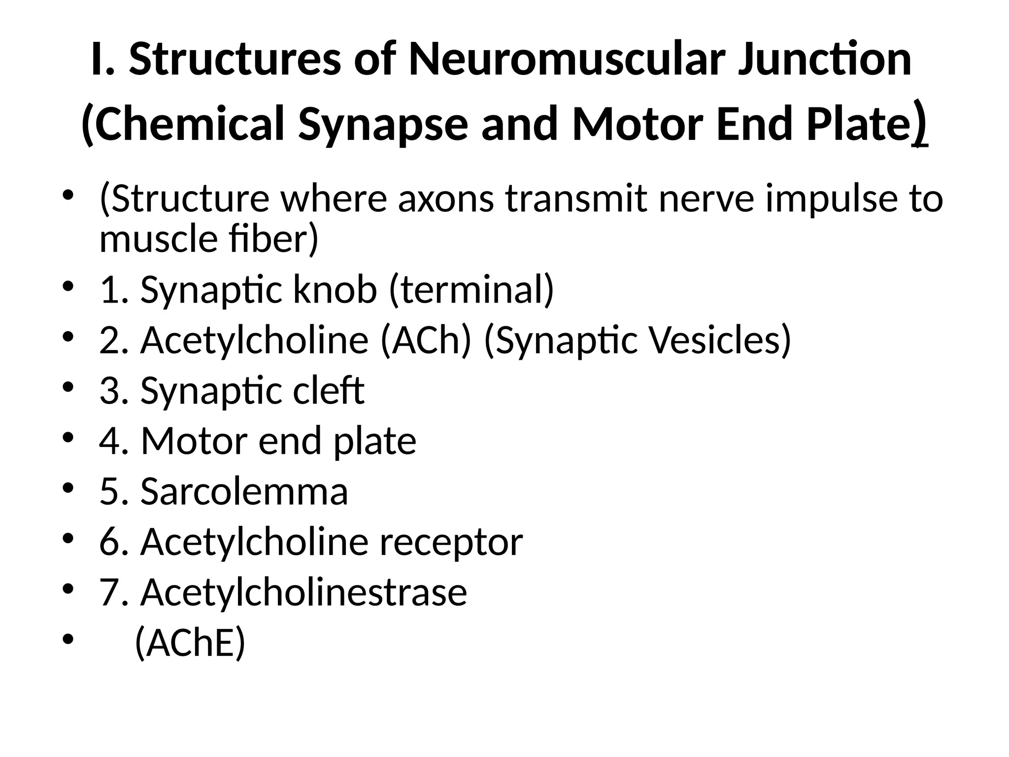
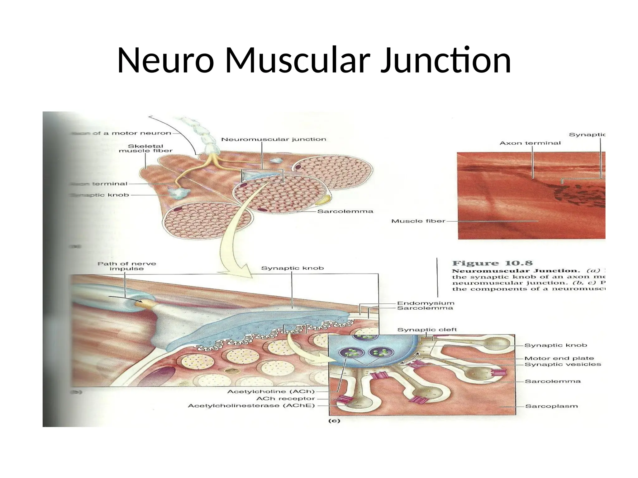
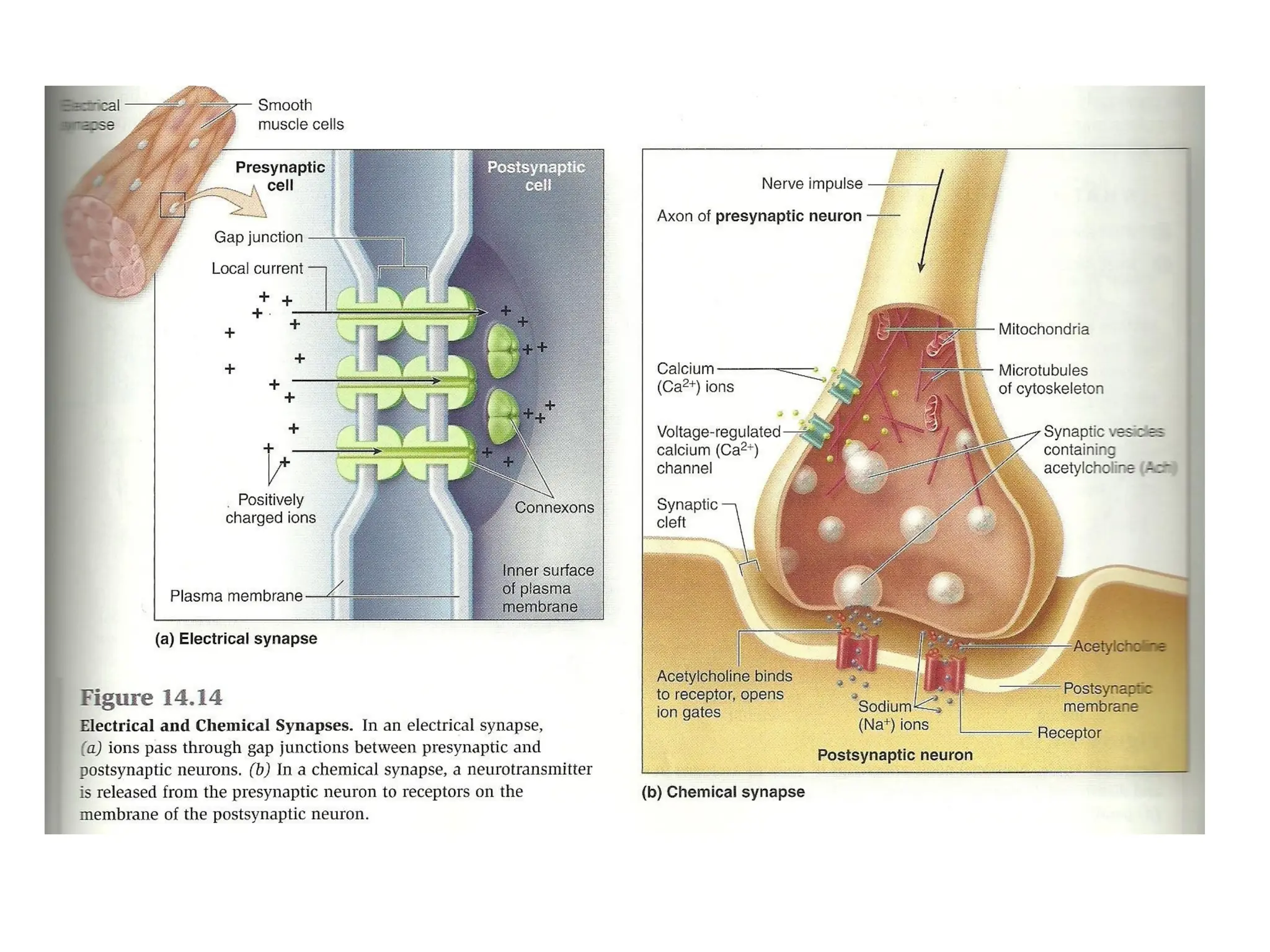
I. Structures ofNeuromuscular Junction
(Chemical Synapse and Motor End Plate)
• (Structure where axons transmit nerve impulse to
muscle fiber)
• 1. Synaptic knob (terminal)
• 2. Acetylcholine (ACh) (Synaptic Vesicles)
• 3. Synaptic cleft
• 4. Motor end plate
• 5. Sarcolemma
• 6. Acetylcholine receptor
• 7. Acetylcholinestrase
• (AChE)
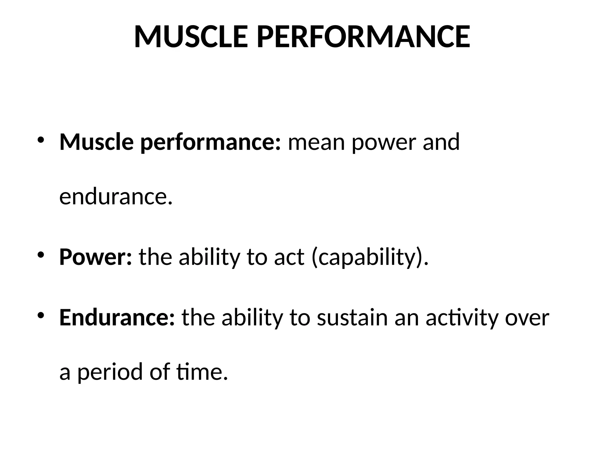
MUSCLE PERFORMANCE
• Muscleperformance: mean power and
endurance.
• Power: the ability to act (capability).
• Endurance: the ability to sustain an activity over
a period of time.
27.
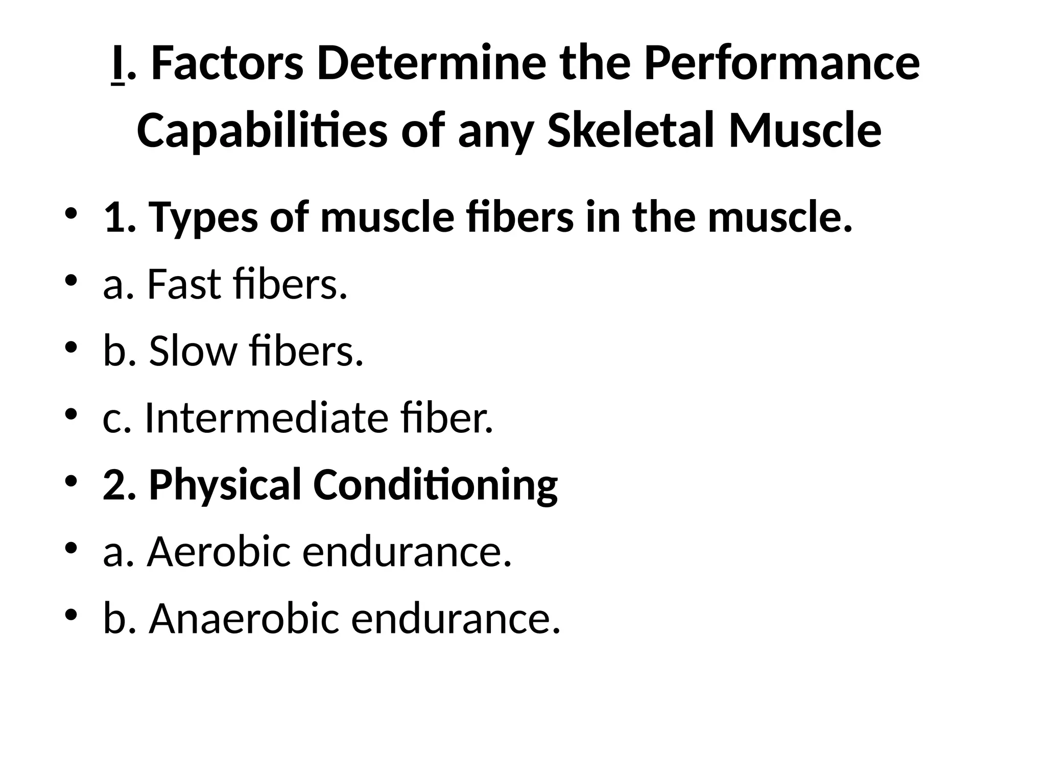
I. Factors Determinethe Performance
Capabilities of any Skeletal Muscle
• 1. Types of muscle fibers in the muscle.
• a. Fast fibers.
• b. Slow fibers.
• c. Intermediate fiber.
• 2. Physical Conditioning
• a. Aerobic endurance.
• b. Anaerobic endurance.
28.
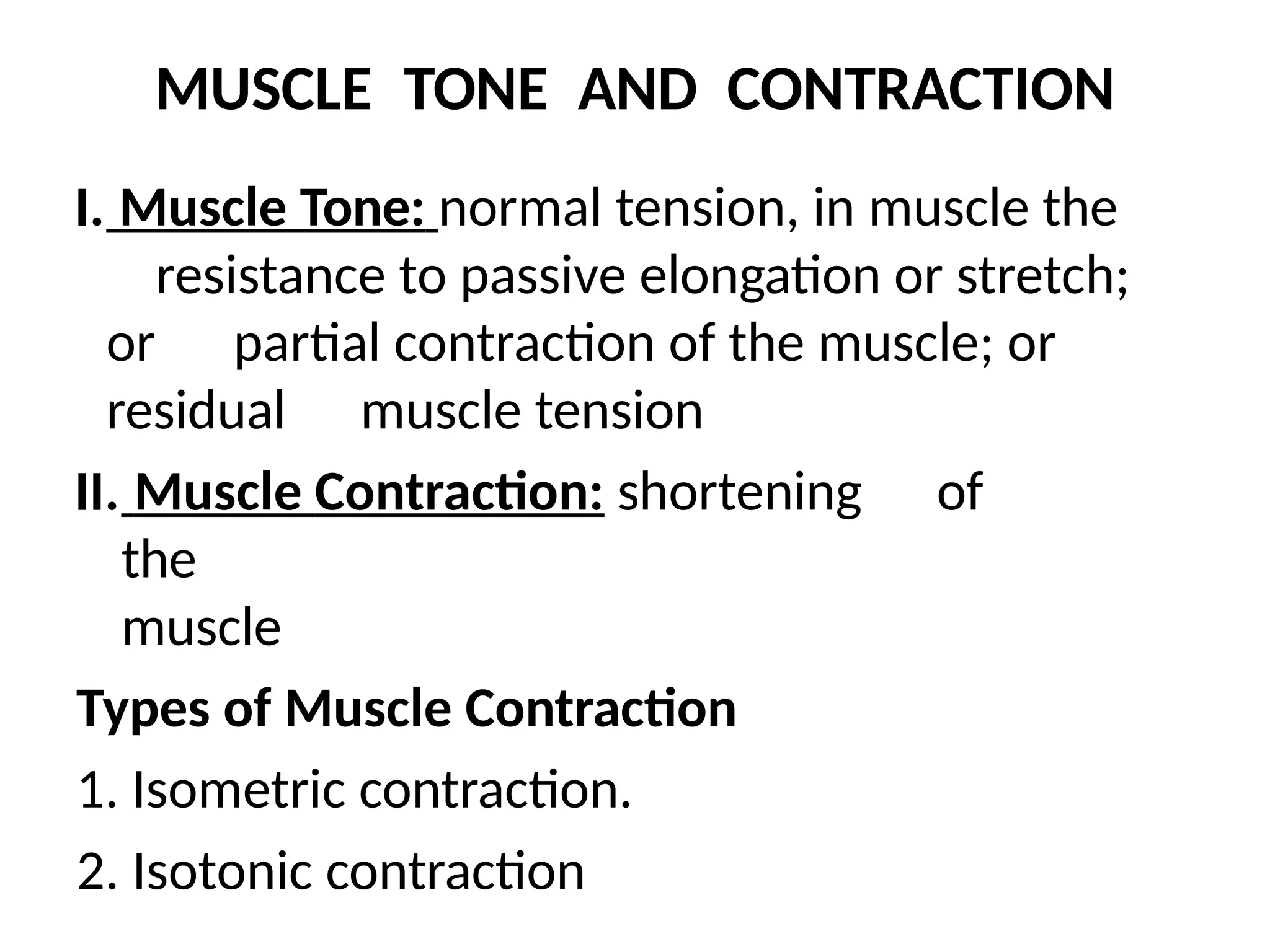
MUSCLE TONE ANDCONTRACTION
I. Muscle Tone: normal tension, in muscle the
resistance to passive elongation or stretch;
or partial contraction of the muscle; or
residual muscle tension
II. Muscle Contraction: shortening of
the
muscle
Types of Muscle Contraction
1. Isometric contraction.
2. Isotonic contraction
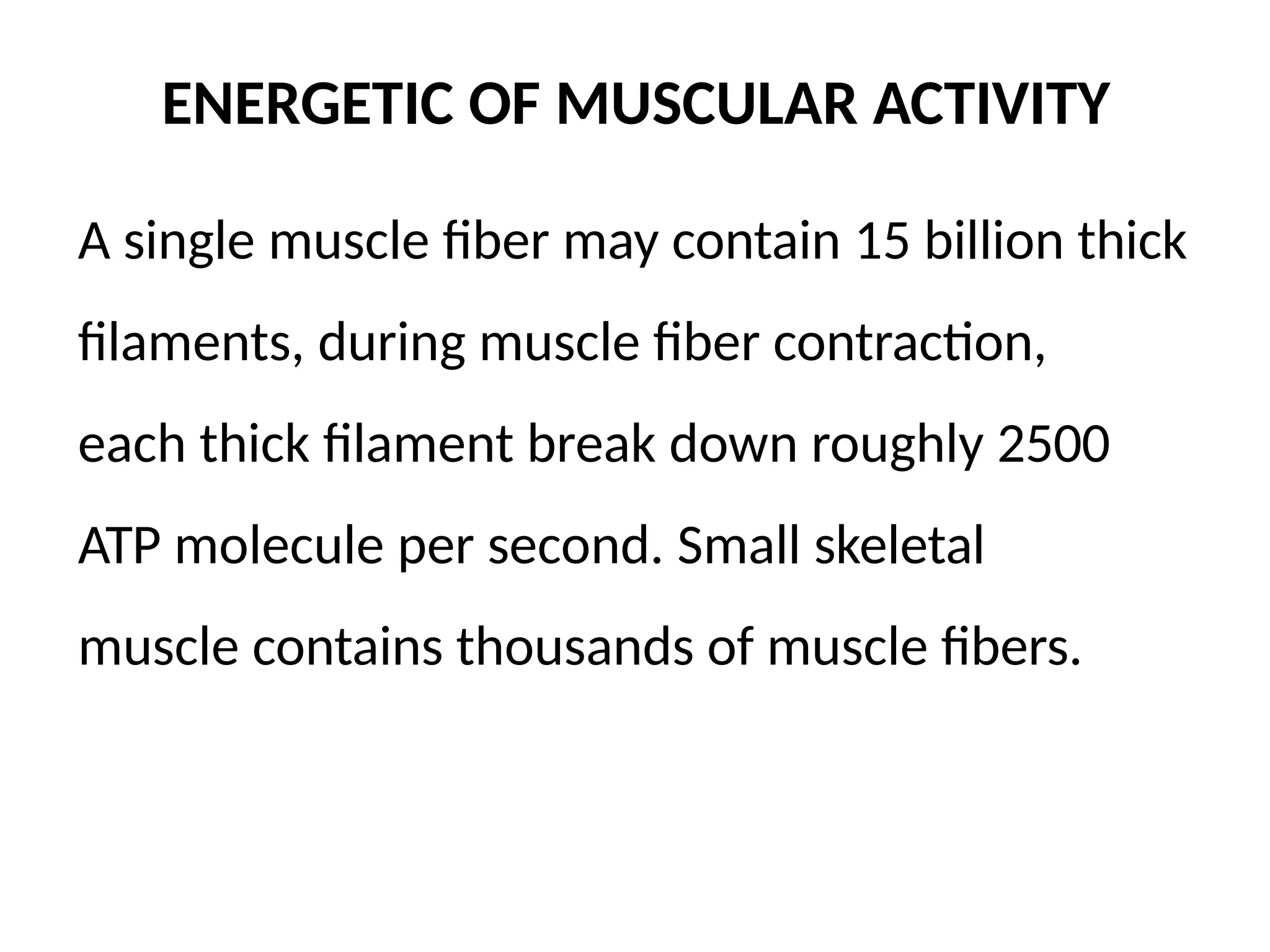
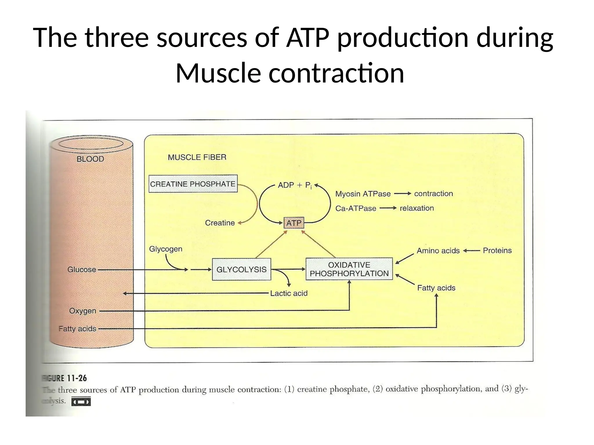
ENERGETIC OF MUSCULARACTIVITY
A single muscle fiber may contain 15 billion thick
filaments, during muscle fiber contraction,
each thick filament break down roughly 2500
ATP molecule per second. Small skeletal
muscle contains thousands of muscle fibers.
31.
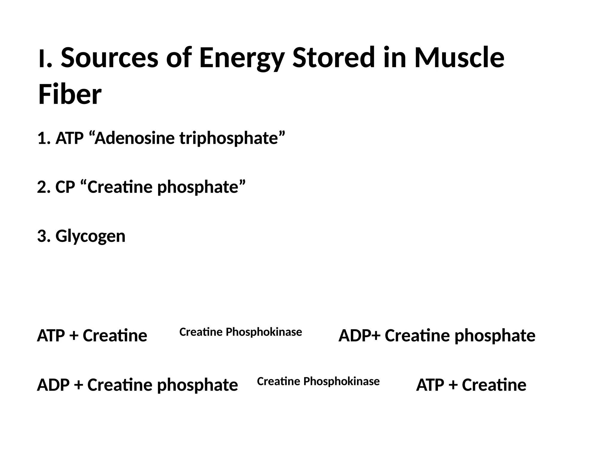
I. Sources ofEnergy Stored in Muscle
Fiber
1. ATP “Adenosine triphosphate”
2. CP “Creatine phosphate”
3. Glycogen
ATP + Creatine Creatine Phosphokinase ADP+ Creatine phosphate
ADP + Creatine phosphate Creatine Phosphokinase ATP + Creatine
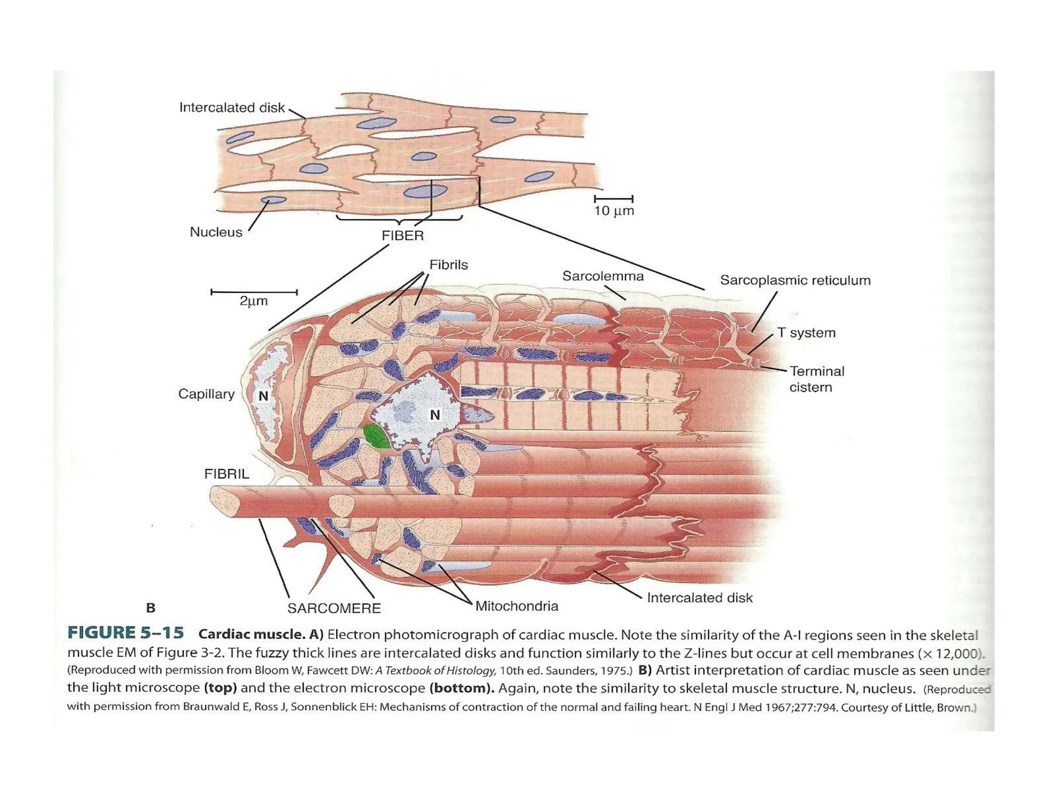
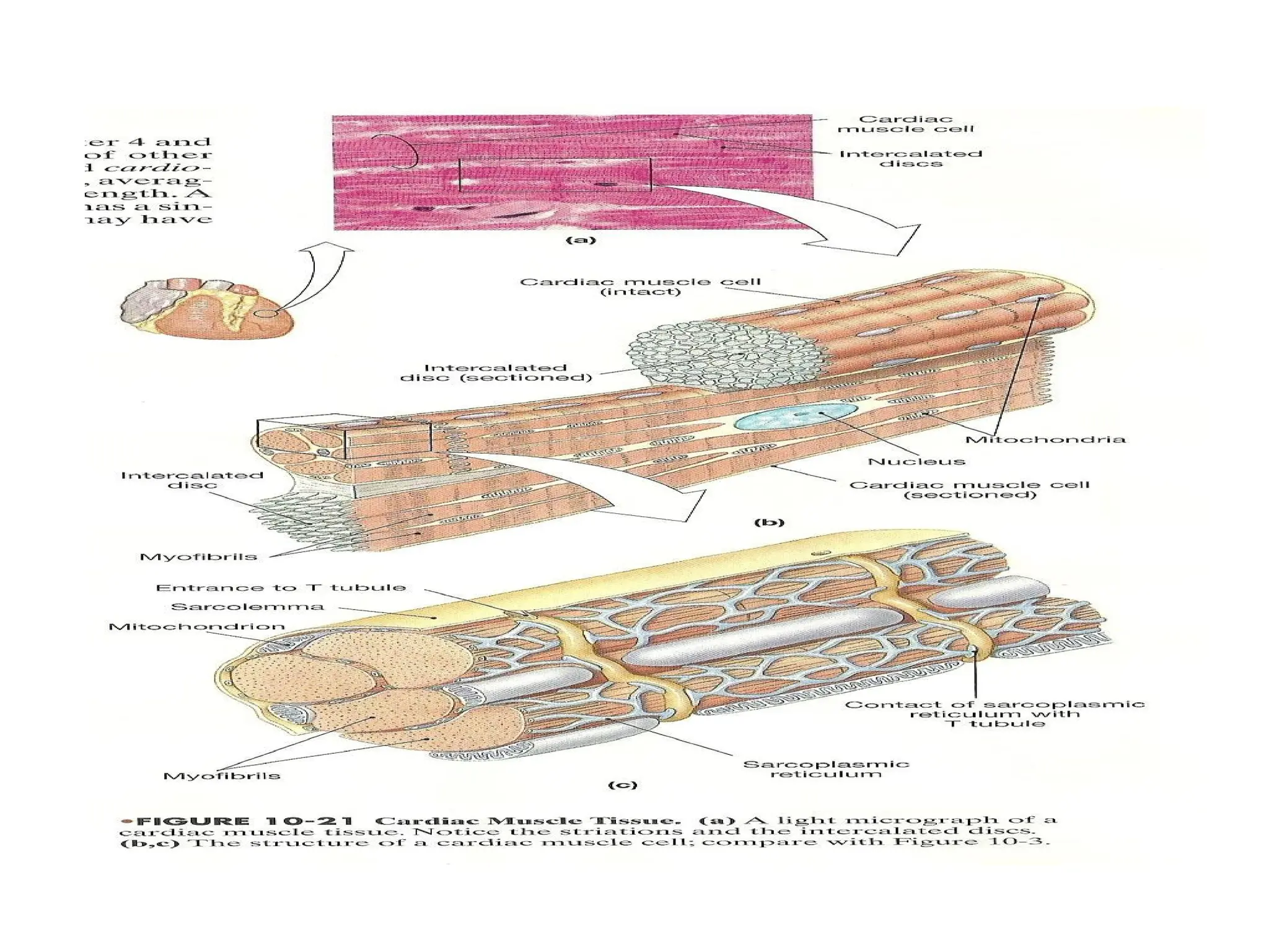
CARDIAC MUSCLE
I. CardiacMuscle Fibers are individual muscle
fibers arranged in thick bundles like skeletal
muscle fiber, but shorter and thicker, and have
one or two nuclei. Cardiac muscle fiber forms Y-
shaped branches; it is attached to adjacent muscle
fibers by junctions termed intercalated discs
40.
Specific structure ofcardiac muscle fiber
1. Intercalated disc.
2. Generally centrally located single nucleus.
3. Cardiac muscle fibers are: thinner and shorter
than skeletal muscle fiber.
4. Contractions of cardiac muscle fiber depend on
Ca++ in ECF and sarcoplasmic reticulum.
5. CMF are slower onset in contraction and
resistant to fatigue.
6. CMFs control by pacemaker cells
(Automaticity).
7. CMFs depend on aerobic metabolism (fat and
carbohydrate) to maintain energy.
41.
II. Control ofCardiac Muscle Contraction
1. Automaticity.
2. Autonomic innervations.
3. Blood born chemicals .
42.
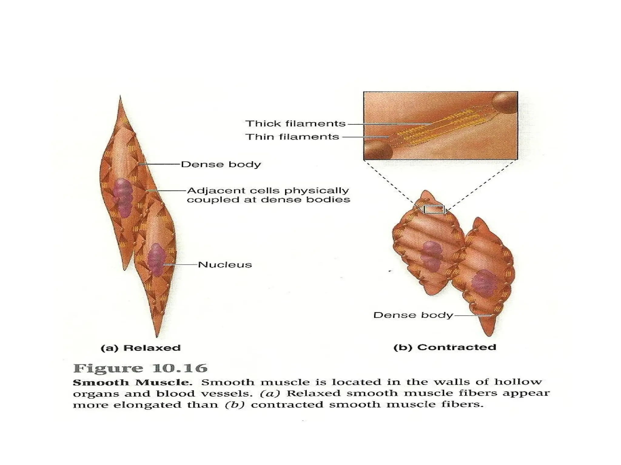
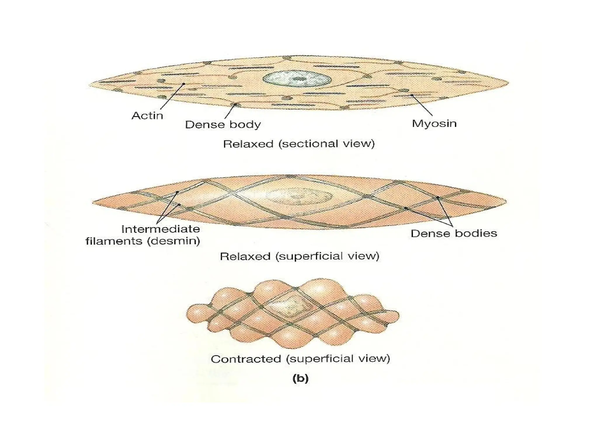
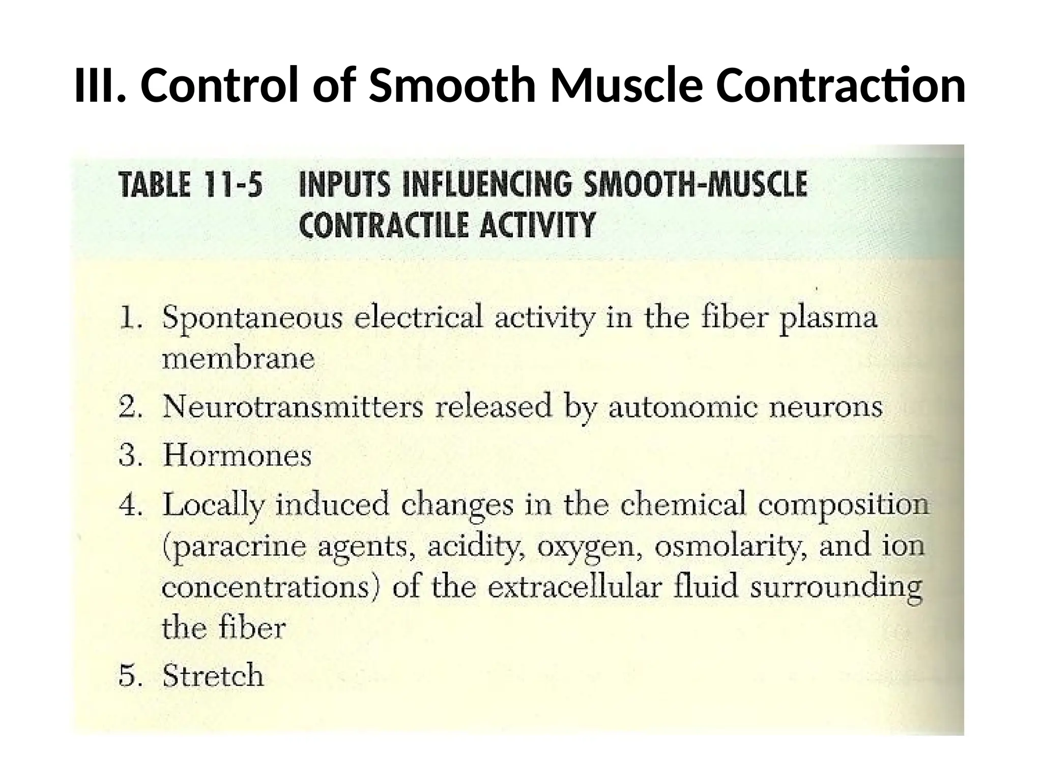
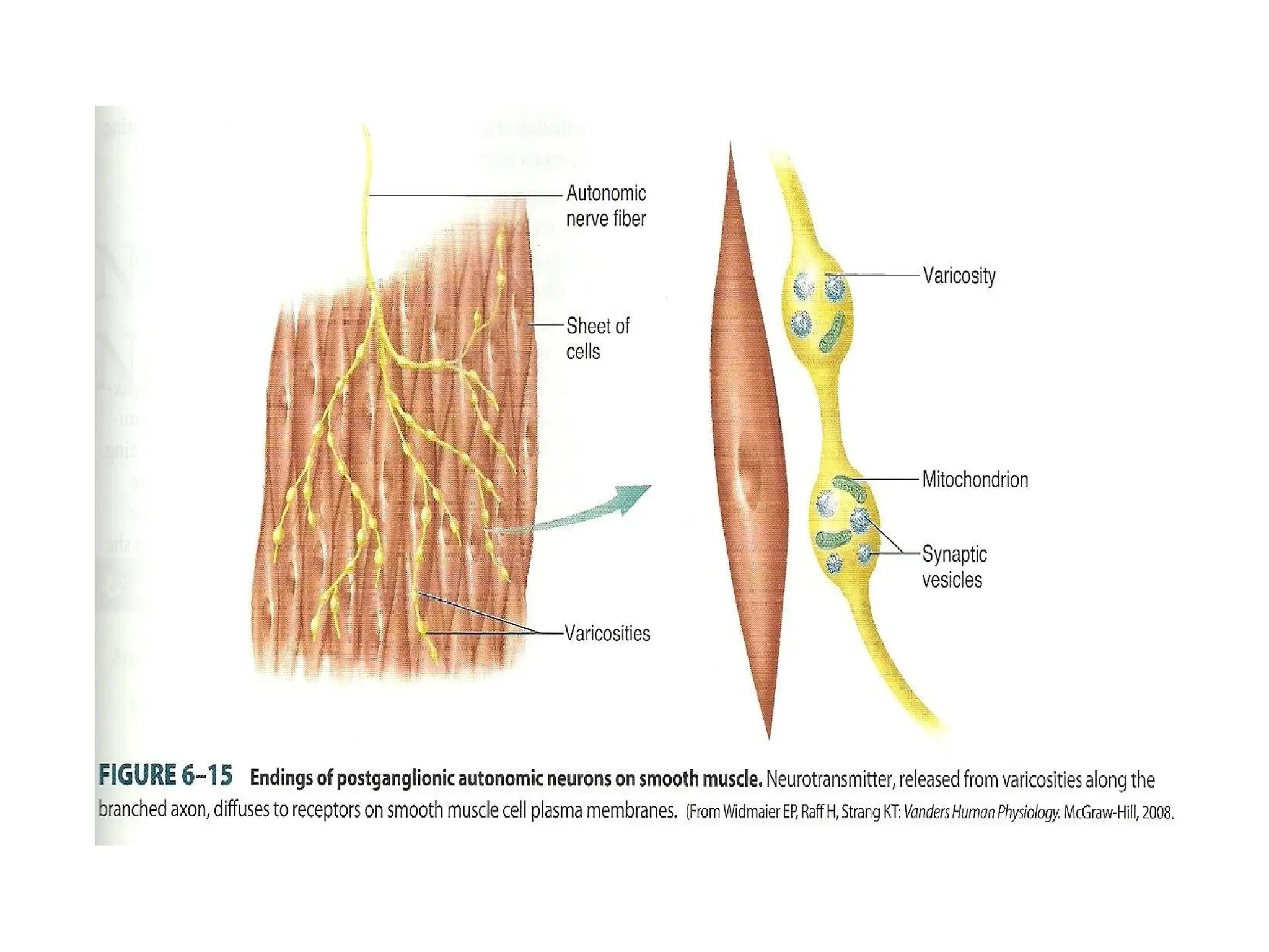
SMOOTH MUSCLEI. SmoothMuscle Fiber
Characteristic
1. Small cell.
2. Have one nucleus.
3. Capacity to divide.
4. Composed from thick and thin filaments and dense body.
5. Thick and thin filaments don’t organize into myofibrils.
6. Slow onset contraction, and may be tetanized and resistant to
fatigue.
7. Primarily aerobic metabolism.
8. Depend on ECF Ca++ to maintain contraction.
9. No T-tube and dispersed sarcoplasmic reticular throughout
sarcoplasm.
45.
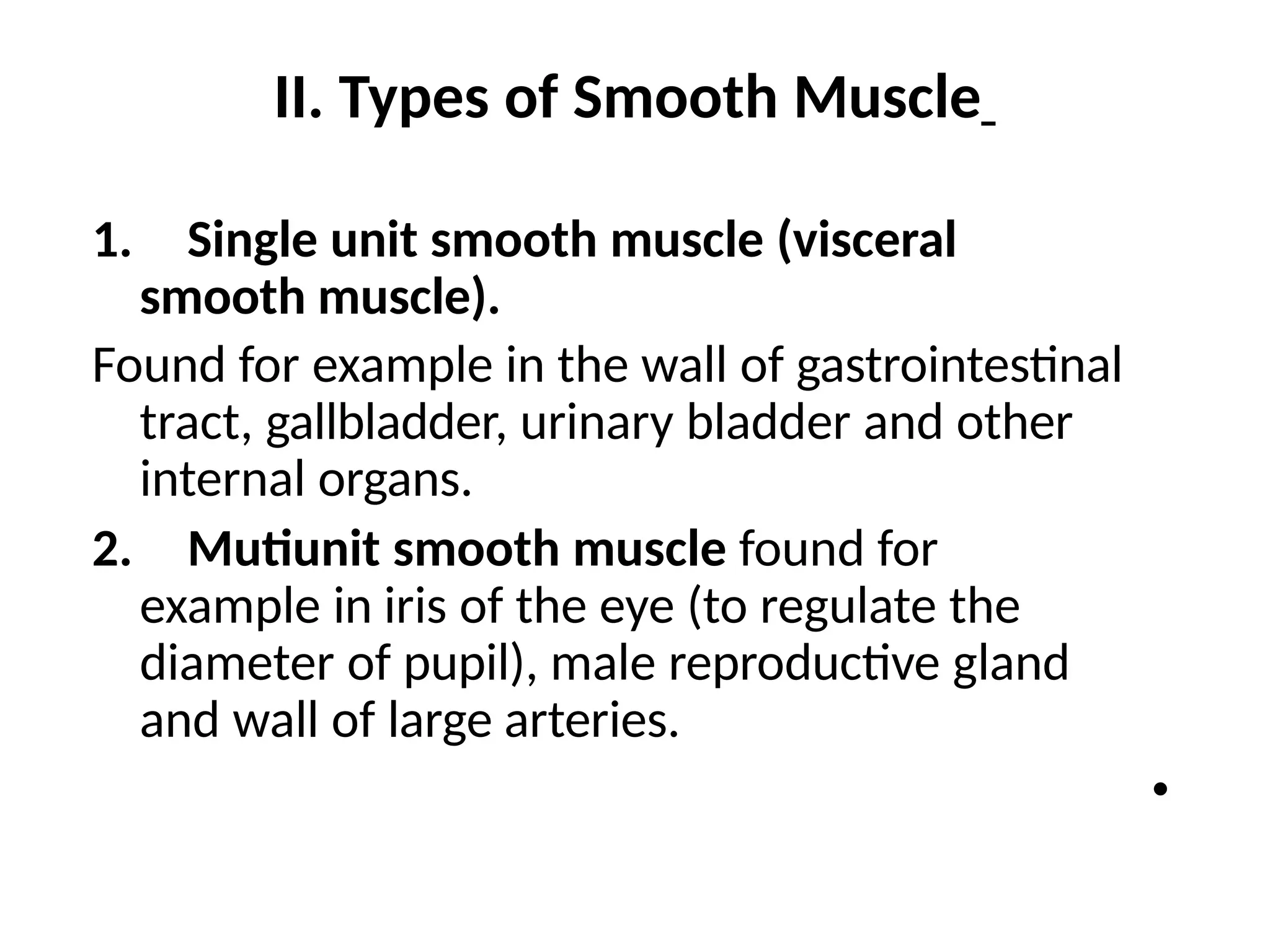
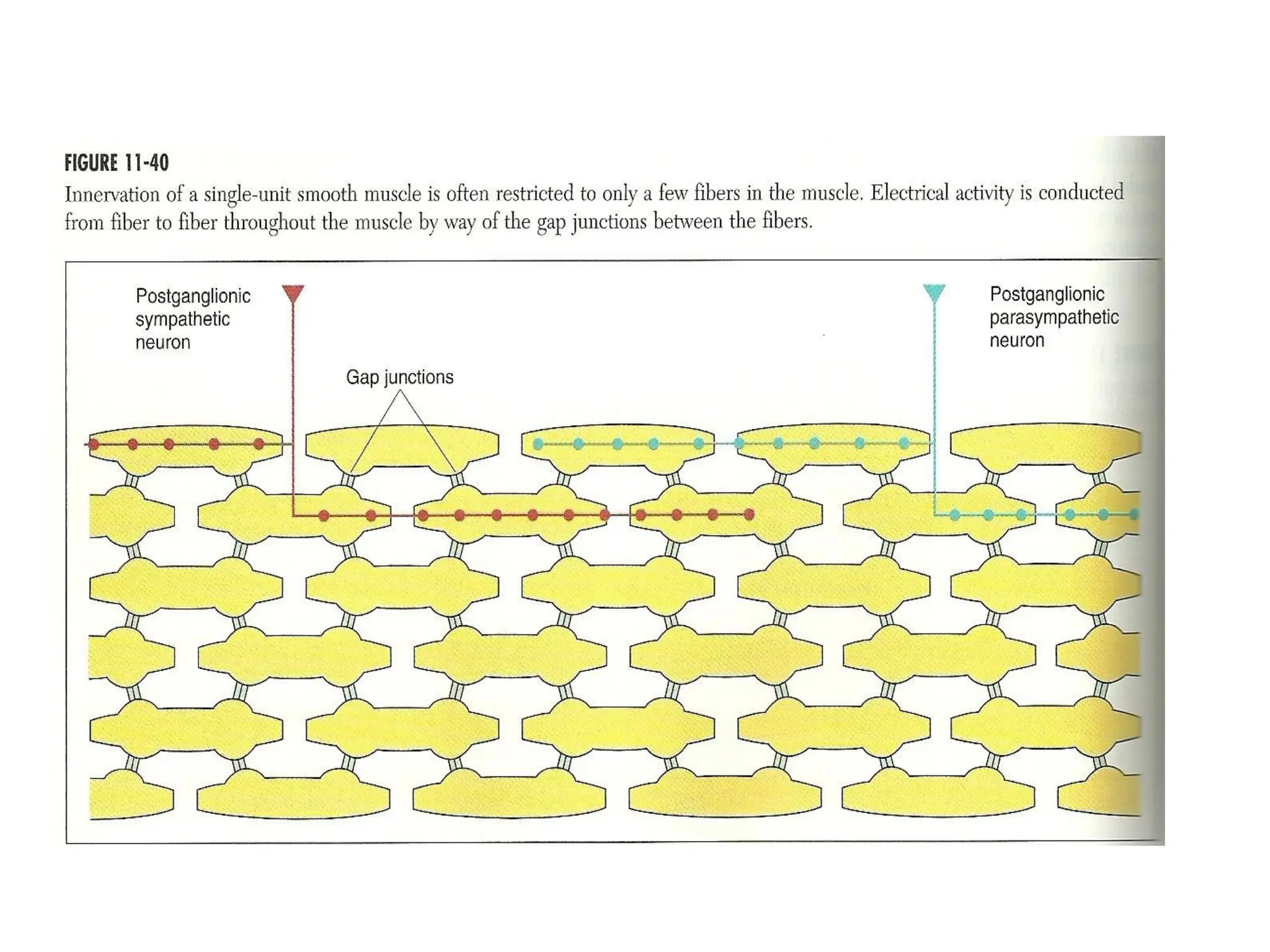
II. Types ofSmooth Muscle
1. Single unit smooth muscle (visceral
smooth muscle).
Found for example in the wall of gastrointestinal
tract, gallbladder, urinary bladder and other
internal organs.
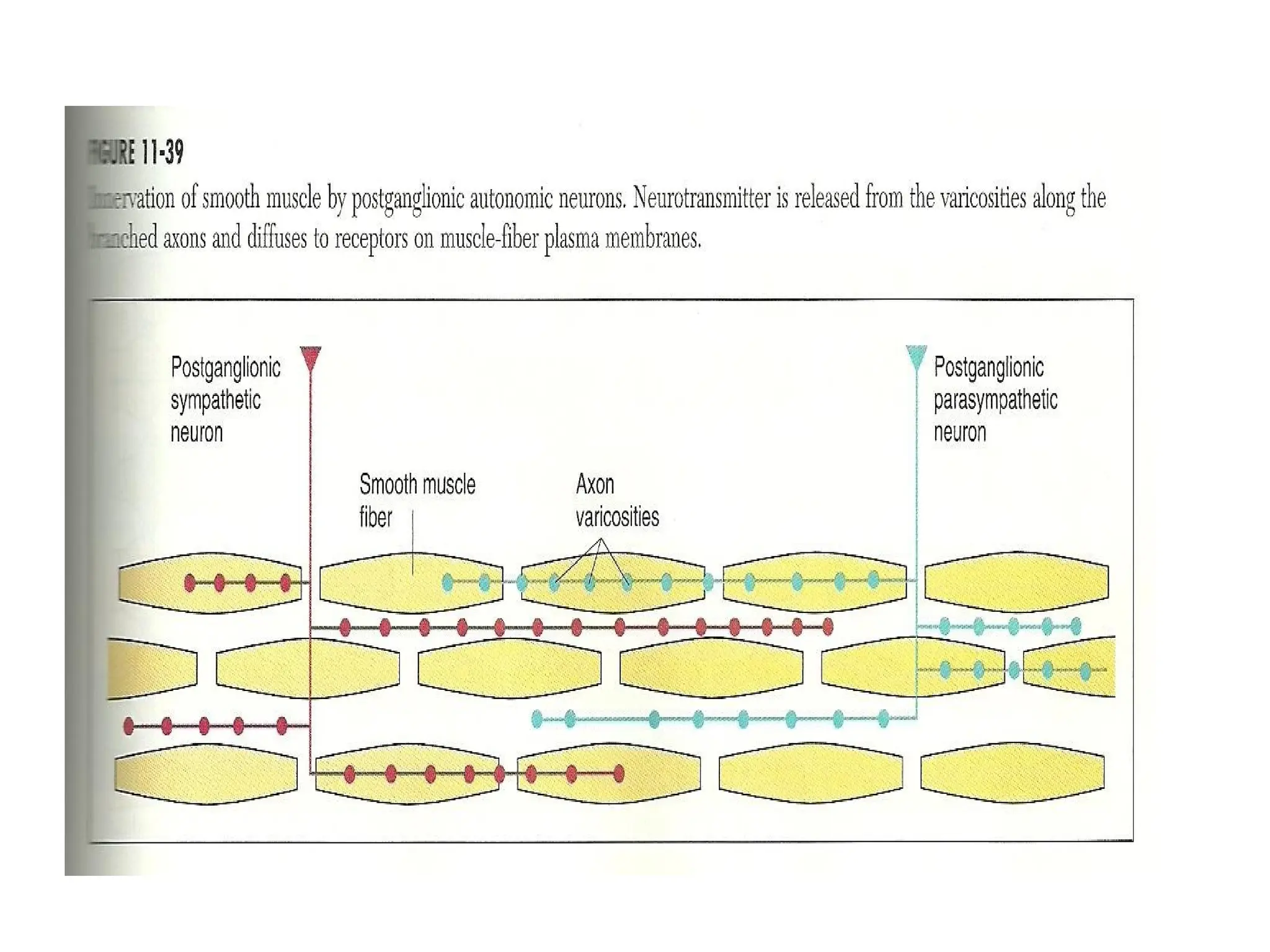
2. Mutiunit smooth muscle found for
example in iris of the eye (to regulate the
diameter of pupil), male reproductive gland
and wall of large arteries.
•
CLINICAL TERMINOLOGY OFMUSCULAR SYSTEM
Convulsion: a series of involuntary contraction of the
voluntary muscles produced hypoglycemia, hypocalcemia,
metabolic disturbances, hormonal imbalances, brain cell
injury, stroke, anoxia, hemorrhage, high fever and epilepsy.
Cramp: painful spasmodic muscular contraction.
Creatine: nonprotein substance synthesized in the body from
three amino acids: arginine, glycine and methionine
.present in the muscle to store high energy phosphate
necessary for muscle contraction.
Dystrophy : imperfect nutrition.
Endurance: the ability to sustain an activity over a period of
time.
Fibromyalgia syndrome : An inflammatory disorder
characterized by a distinctive pattern of symptoms
including tender points of body surface.
54.
Fibrosis : processin which muscle tissue is replaced
by fibrous connective tissue ,making muscle
weaker and less flexible.
Hypertonia : abnormally increased tonicity or
strength .
Muscle atrophy: skeletal muscle that is not
regularly stimulated by a motor neuron loses
muscle tone and mass. The reduction in muscle
size, tone and power is called atrophy.
Muscle fatigue: muscle can no longer contract
,because of change in pH ,due to building of lactic
acid, a lack of energy or other problem.
Muscle Contraction: shortening of the muscle.
Muscular dystrophy : degenerative myopathies that
produce muscle weakness and atrophy.
55.
Power: the abilityto act (capability).
Spasm: sudden involuntary contraction of muscle or group of
muscle accompanied by pain
Synapse: the junction between the processes of two neurons or
between a neuron and effector organ (muscle, gland or GI
neurons)
Tendonitis: inflammation of tendons and of tendon-muscle
attachments, one of the most common causes of acute pain in
the shoulder. It is frequently associated with calcium deposit
(calcium tendinitis) which may involve the bursa around the
tendon or near the joint, causing bursitis .
Tetany : continuous tonic spasm of a muscle, it is due to
abnormal calcium metabolism ,vitamin D deficiency and
alkalosis.
Tone :normal tension, in muscle the resistance to passive
elongation or stretch.
Twitch : mechanical response of skeletal muscle to single
volley
action potential