
Recommended
More Related Content
What's hot
What's hot (20)
Similar to Unit Three - Excitable Tissues (Muscle).ppt
Similar to Unit Three - Excitable Tissues (Muscle).ppt (20)
More from Wasihun Aragie
Recently uploaded
Recently uploaded (20)
Unit Three - Excitable Tissues (Muscle).ppt
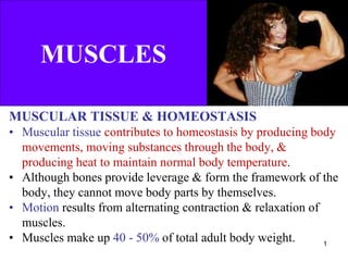
- 1. MUSCLES MUSCULAR TISSUE & HOMEOSTASIS • Muscular tissue contributes to homeostasis by producing body movements, moving substances through the body, & producing heat to maintain normal body temperature. • Although bones provide leverage & form the framework of the body, they cannot move body parts by themselves. • Motion results from alternating contraction & relaxation of muscles. • Muscles make up 40 - 50% of total adult body weight. 1
- 2. MUSCLES, cont’d • Your muscular strength reflects the primary function of muscle – transformation of chemical energy into mechanical energy to generate force, perform work & produce movement. • In addition, muscle tissues stabilize body position, regulate organ volume, generate heat, and propel fluids & food matter through various body systems. • Functions OF Muscle Tissue − Through sustained contraction or alternating contraction and relaxation, muscular tissue has four key functions: 2
- 3. Muscle Functions 1. Producing body movements – Movements of the whole body such as walking & running, and localized movements (grasping a pencil or nodding of head). 2. Stabilizing body positions – Skeletal muscle contractions stabilize joints & help maintain body positions, such as standing or sitting. 3. Storing & moving substances within the body – Food storage of in stomach or urine in urinary bladder. – Cardiac muscle contraction to pump blood & moves in blood vessels, bile storage in the gall bladder. 4. Generating heat – As muscular tissue contracts, it produces heat, a process known as thermogenesis. – This important to maintain normal body temperature. 3
- 4. Types of Muscle Tissue 4 • There are 3 types of muscle tissue
- 5. Skeletal/Voluntary muscles • Are attached to bones & moves the skeleton • Are elongated, cylindrical & multinucleated cells • Are striated (alternating light & dark bands) muscles • Are voluntary muscles, controlled by somatic NS Cardiac muscles – Are muscles of the heart - used to pump blood through circulatory system. – Are striated, uni- or binucleated – Has sarcomeres & T-tubules – Are involuntary, controlled by ANS, drugs & hormones • Have the property of autorhythmicity & syncytium – Cardiac muscle cells are joined by structures called intercalated discs. Types of Muscle Tissue 5
- 6. Types of Muscle, cont’d Smooth muscles − Are involuntary, non-striated muscle tissue, and controlled by ANS, drugs & hormones. – Located in the wall of hallow organs (GIT, blood vessels, uterus, u-bladder & iris). – Have non-striated appearance, mononucleated cells – Involuntary muscles – Have the property of autorhythmicity & syncytium. • Syncytium – is a cell made from the fusion of many others. 6
- 7. Skeletal muscle cell Cardiac muscle cell Smooth muscle cell Elongated Cells Branching Cells Spindle-Shaped Cell Multiple Peripheral Nuclei Single Central Nucleus Single Central Nucleus Visible Striations Visible Striations Lack Visible Striations Voluntary Involuntary Involuntary 7 Comparison of Skeletal, Cardiac & Smooth Muscle Cells
- 8. Properties of Muscle Tissue • Muscular tissue has four special properties that enable it to function & contribute to homeostasis: 1. Excitability • The ability to receive & respond to a stimulus 2. Contractility − The ability of muscular tissue to contract forcefully when stimulated by an action potential. 3. Extensibility − The ability of muscular tissue to stretch 4. Elasticity − The ability of muscular tissue to return to its original length & shape after contraction or extension. 8
- 9. • Each of our skeletal muscles is a separate organ composed of hundreds to thousands of cells, which are called muscle fibers b/c of their elongated shapes. • Thus, muscle cell & muscle fiber are two terms for the same structure. • Skeletal muscle also contains connective tissues surrounding the muscle fibers & the whole muscles, and blood vessels & nerves. 9 Skeletal Muscle Tissue
- 10. Whole skeletal muscle ↓ Muscle fascicles ↓ Muscle cell or Muscle fibre ↓ Myofebrile ↓ Myofilaments: - Thin myofilament (actin) - Thick myofilament (myocin) - Regulatory proteins (troponin & tropomyocin) Organization of the Skeletal Muscle 10
- 11. • Skeletal muscles are attached to bones via tendons. • Skeletal muscles are composed of connective tissue and contractile cells. Three connective tissue layers of the muscle are endomysium, perimysium & epimysium: – Bind the muscle cells together and – Provide strength & support to the entire muscle. • Connective tissue surrounding the entire muscle is epimysium. • Bundles of muscle cells are called fascicles. • Connective tissue surrounding fascicles is called perimysium. Connective tissue surrounding the individual muscle cells is endomysium: – Function to electrically insulate muscle cells from one another. Whole Skeletal Muscle 11
- 12. 12 Structure of Skeletal Muscle
- 13. • Muscle fibers: alternative name for skeletal muscle cells. Nucleus: contains the genetic material. Sarcolemma: plasma membrane of skeletal muscle cells. Sarcoplasm: muscle fiber cytoplasm. – contains a lot of mitochondria, glycogen (provide energy), myoglobin & creatine-PK, as well as myofibrils & SR. Sarcoplasmic reticulum (SR): interconnecting tubules of ER that surround each myofibril. A skeletal muscle cell has two tubular structures: a. Transverse tubules (T-tubules). Function: conduction of depolarization. b. Longitudinal tubules (sER). Function: Ca2+ storage. 13 Fine structures of the skeletal muscle
- 14. 14 Fig. Parts of a skeletal muscle fiber (cell)
- 15. Fine structures, cont’d Terminal cisternae: sac-like regions of SR that contain Ca2+ ions. T-tubules: invaginations of the sarcolemma that project deep into the cell. Triad: a group of one T-tubule lying b/n two adjacent terminal cisternae. Cytosol: intracellular fluid Mitochondria: sites of ATP synthesis Myofibril: contains contractile filaments within skeletal muscle. – Myofibril is made of myofilaments called thin filaments (actin) and thick filaments (myosin) 15
- 16. • Muscle fiber plasma membrane is known as sarcolemma • Muscle fiber cytoplasm is known as sarcoplasm • Sarcoplasm has lots of mitochondria, lots of glycogen granules (to provide glucose for energy needs) as well as myofibrils & sarcoplasmic reticuli • Sarcolemma has invaginations that penetrate through the cell called transverse tubules or T tubules 16
- 17. Sarcoplasmic Reticulum • Muscle cell version of the smooth ER. • Function: calcium storage depot in muscle cells. • Loose network of this membrane bound organelle surrounds all the myofibrils in a muscle fiber. 17
- 18. Myofibrils • Each muscle fiber contains rod-like structures called myofibrils that extend the length of the cell. • Are basically long bundles of protein structures called myofilaments & their actions give muscle the ability to contract. • Myofilaments are classified as thick filaments & thin filaments. 18
- 19. Myofilaments • 2 types of myofilaments (thick & thin) make up myofibrils. • Thick myofilaments are made the protein myosin. A single myosin protein resembles 2 golf clubs whose shafts have been twisted about one another About 300 of these myosin molecules are joined together to form a single thick filament. 19
- 20. • Each thin filament is made up of 3 different types of protein: actin, tropomyosin & troponin. • Each thin filament consists of a long helical double strand. • Actin is globular protein & on each actin subunit, there is a myosin binding site. • Loosely wrapped around actin helix & covering the myosin binding site is the filamentous protein, called tropomyosin. • Bound to both actin & tropomyosin is a triad of proteins collectively known as troponin. 20
- 21. • Each myofibril is made up 1000’s of repeating individual units known as sarcomeres. • Each sarcomere is an ordered arrangement of thick & thin filaments. Notice that it has: regions of thin filaments by themselves. a region of thick filaments by themselves. regions of thick filaments & thin filaments overlapping. Myofibrils 21
- 22. • The sarcomere is flanked by 2 protein structures known as Z lines (discs). • The figure below shows you structure of the bands in terms of major proteins, actin & myosin: In the A band 2 proteins are overlap. The I band contains only the actin protein. An H zone which contains only thick filaments. 22 Sarcomere
- 23. • The portion of the sarcomere which does not contain any thick filament is known as I band. • The I band contains only thin filament & is light under the microscope. – One I band is actually part of 2 sarcomeres. In the middle of the H zone is a structure called the M line w/c functions to hold the thick filaments to one another. Sarcomere, cont’d 23
- 24. Each muscle fiber has many T-tubules. – Typically each myofibril has a branch of a T-tubule encircling it at each A-I junction At each A-I junction, the SR will expand & form a dilated sac (terminal cisterna) Each T-tubule will be flanked by a terminal cisterna. This forms a so- called triad consisting of 2 terminal cisternae & one T-tubule branch. T-Tubules & SR 24
- 25. Place your right palm on the back of your left hand. Now slide your right palm toward your left elbow. – What happened to the distance between your elbows? It got shorter! – This is how muscle contraction occurs. – The thin filaments slide over the thick filaments. This pulls the Z discs closer together. When all the sarcomeres in a fiber do this, the entire fiber gets shorter which pulls on the endomysium, perimysium, epimysium & attached tendon & then pulls on the bone. So, that is why we have movement. Muscle Contraction: The Sliding Filament Hypothesis 25
- 26. Here is what happens as the filaments slide & the sarcomere & the muscle fiber shortens. In the process of contraction, what happens to the: 1. Distance b/n Z discs 2. Length of the A band 3. Length of the H zone 4. Length of the I band 26
- 27. • Hanson & Haxley proposed that skeletal muscle shorten during contraction because the thick & thin myofilaments slid past one another. Their model is known as the sliding filament mechanism of muscle contraction. 27 Muscle Contraction: Sliding Filament Hypothesis
- 28. All the sarcomeres in a fiber will contract together. This contracts the fiber itself. The number of fibers contracting will determine the force of contraction of the whole muscle. The process of the whole muscle contraction divided into four steps: – Excitation – Excitation-contraction coupling – Contraction – Relaxation 28 Sliding Filaments
- 29. • All cells have a voltage difference across their plasma membrane. This is the result of several things: 1. The ECF is very high in [Na+] while the ICF is very high in [K+]. The plasma membrane is impermeable to Na+ but slightly permeable to [K+]. As a result, K+ is constantly leaking out of the cell. In other words, positive charge is constantly leaking out of the cell. 29 Excitation
- 30. Excitation, cont’d 2. The Na+/K+ pump is constantly pumping 3Na+ ions out & 2K+ ions in for every ATP used. Thus, more positive charge is leaving than entering. 3. There are protein anions (i.e., negatively charged proteins) within the ICF that cannot travel through the plasma. What this adds up to is fact that the inside of the cell is negative with respect to the outside. The interior of the cell has less positive charge than the exterior. 30
- 31. Excitation, cont’d • This charge separation is known as a membrane potential (abbreviated Vm). • The value for Vm in inactive muscle cells is –90 mV. • Cells that exhibit a Vm are said to be polarized. • Vm can be changed by influx or efflux of charge. 31
- 32. Excitation, cont’d • The plasma membrane has integral proteins that act as gated ion channels. • These are channels that are normally closed, but in response to a certain signal, they will open & allow specific ions to pass through them. • Ion channels may be: – Ligand-gated the binding of an extracellular molecule (e.g., hormone, NT) causes these channels to open. – Voltage-gated Vm causes these channels to open. – Mechanically-gated stretch or mechanical pressure opens these channels. • When a channel is open, its specific ion(s) will enter or exit depending on their electrochemical gradient. 32
- 33. Excitation, cont’d In general each muscle is served by one nerve – a bundle of axons carrying signals from spinal cord to muscle. Within the muscle, each axon will go its own way & eventually branch into multiple small extensions called telodendria. Each telodendrium ends in a bulbous swelling known as the synaptic end bulb. The site of interaction b/n a neuron & any other cell is known as a synapse. The synapse b/n a neuron & a muscle is known as the neuromuscular junction (NMJ). 33
- 34. The minute space b/n the synaptic end bulb & sarcolemma is known as the synaptic cleft. There is a depression in the sarcolemma at the synaptic cleft known as the motor end plate. The synaptic end bulb is filled with vesicles that contain the neurotransmitter, acetylcholine (ACh). The motor end plate is secure full of ACh receptors (nicotinic). 34 Neuromuscular Junction (NMJ)
- 35. • Transmission is unidirectional. • There is a single NMJ per muscle fiber. • The neurotransmitter is always acetylcholine Synthesis: Choline + Acetyl-COA = ACh by the action of choline acetyl-COA transferase Storage: ACh form a complex with ATP, packed in vesicles. Release: Released by Ca-dependent exocytosis. Metabolism: Metabolized by the action of ACh-Esterase • The post junctional receptor is always nicotinic receptor. • The effect of ACh on nicotinic receptor is always excitatory producing EPSP/EPP. • It is fatigable due to depletion of ATP & NT storage. 35 Characteristics of Neuromuscular Junction
- 36. Excitation, cont’d 1. A nerve signal will arrive at the synaptic end bulb & this will cause the ACh-containing vesicles to undergo exocytosis. 2. ACh will diffuse across the synaptic cleft & bind to the ACh receptors. These receptors are actually ligand-gated Na+ channels. The binding of ACh causes them to open. 3. Na+ will rush into the cell, making the local cell interior more positive. This is known as depolarization. It is a local event! 36
- 37. Excitation, cont’d • Adjacent to the motor end plate, the sarcolemma contains voltage- gated ion channels. In order for these channels to open, the Vm must depolarize from its resting value of –90 mV to approximately –50 mV. This is the threshold. The Vm must become this positive for the voltage-gated channels to open. • The degree of depolarization depends on how much Na+ influx occurred which in turn depends on how many Na+ channels were opened by binding ACh. • If the Vm fails to depolarize to threshold, nothing will happen. The Vm will soon return to normal & no muscle contraction will occur. • If the Vm does reach the threshold, two types of voltage-gated ion channels will open: – Fast Na+ channels – Slow K+ channels 37
- 38. Excitation, cont’d • If Vm reaches threshold, fast Na+ channels open & Na+ rushes in causing the Vm to depolarize to +30mV. The depolarization stops when the Na+ channels become inactivated. • At this point, slow K+ channels have opened & K+ efflux occurs. This returns Vm back to its resting level. This is repolarization. • If we were to graph this change in Vm over time, it would look somewhat like the animation below. • This is known as an action potential. 38
- 39. Excitation, cont’d 39 • An AP can propagate itself across the surface of the plasma membrane. • The depolarization caused by the Na+ influx in one particular area of the sarcolemma causes voltage-gated channels in the adjacent membrane to open. • The resulting ionic influx then causes voltage-gated channels to open in the next patch of membrane & so on. Thus, AP propagates itself.
- 40. • When a muscle fibre membrane is depolarized, contraction of the fibre follows. The process by which depolarization initiates contraction is called excitation-contraction coupling. • It has several steps as follow: 1. AP initiated & propagated along the motor nerve fibre & arrives at the end feet. 2. Opening of voltage-gated Ca-channels & influx of Ca2+ to trigger release of Ach. 3. Ach released by Ca-dependent exocytosis & diffuse through the synaptic cleft & binds to the nicotinic receptors on postjunctional membrane. 4. Opening of ligand gated Na-channels & influx of Na+ to produce EPP. 40 Mechanism of muscle contraction (Excitation-contraction coupling)
- 41. Excitation-contraction coupling, cont’d 5. Spread of depolarization through the sarcolemma & T-tubules. 6. Depolarization of the T-tubules stimulate SR to release Ca2+ ions into the sarcoplasm. 7. Then Ca2+ binds to troponin-C. 8. Ca2+ & troponin-C combination detaches troponin-I from the active sites of actin. 9. The detachment of troponin-I from actin displaces tropomyocin, uncovering the active sites of actin filaments. 10. When the active site of actin is exposed, the heads of myosin connect to the actin, making cross-bridges. 11. The ATPase enzyme on the myosin heads hydrolysis ATP into ADP + -P plus energy. 12. The released energy causes the movement of myosin head (power stroke) towards the centre. 13. Then, head of myosin is charged with a new molecule of ATP & detached from actin leading to muscle relaxation. 41
- 42. Mechanism of Muscle Relaxation • Has the following steps: 1. Following muscle contraction, Ca2+ is re-uptake back into SR by Ca-pump requiring ATP. 2. Ca2+ in sarcoplasm → Ca2+ detaches from troponin-C → Tropomyosin covers the active sites of the actin. 3. Head of myosin charged with ATP & detached from the actin. • Muscle relaxation is an active process requiring energy. • Therefore, a large amount of energy (ATP) consumed during muscular performance for the following activities: 1. To move the head of myosin (power stroke), Myosin ATPase. 2. For active pump of Ca2+ from the sarcoplasm to SR, Ca-ATPase. 3. For Na-K pump in the membrane, Na-K ATPase. 4. For muscle relaxation 42
- 43. Rigor Mortis
- 44. Types of Skeletal Muscle Fibers • Based on the velocity of shortening, major pathways used to form ATP, & fatigability, muscle fibers are classified into two types: Fast/glycolytic fibers, have: – Myosin with high ATPase activities – Few mitochondria – High content of glycolytic enzymes – Large storage of glycogen – Little myoglobin contents Slow oxidative fibers, have: – Myosin with low ATPase activities – Numerous mitochondria – High capacity of oxidative phosphorylation – Rich in arterial blood supply, high BF – Large amount of myoglobin contents 44
- 45. Atrophy • Reduction in size of a cell, tissue, or organ Often caused by disuse. E.g. Astronauts or As result of a nerve injury. Hypertrophy • Increase in size of a cell, tissue or an organ. In muscles, hypertrophy of the organ is always due to cellular hypertrophy (increase in cell size) rather than cellular hyperplasia (increase in cell number) Muscle hypertrophy occurs due to the synthesis of more myofibrils & synthesis of larger myofibrils. As a result, there is an increase in the force & strength of contraction, decrease in fatigability. Muscle Atrophy & Hypertrophy 45