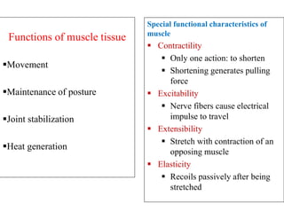
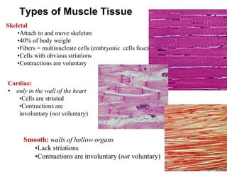
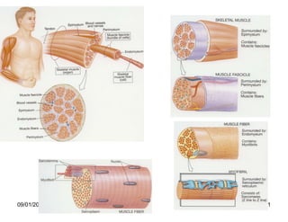
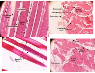
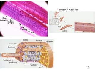
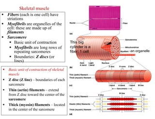
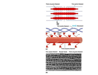
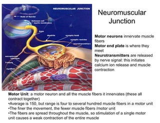
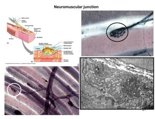
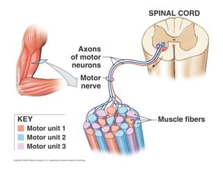
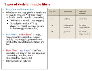
SlideShare a Scribd company logo
KILIMANJARO CHRISTIAN MMEDICAL
UNIVERSITY COLLEGE
Faculty of Rehabilitation Medicine
Department of Anatomy and Neuroscience
Anatomy Lecture for BSc.Nursing and BSc.Physiotherapy
LECTURE 7
TOPIC: MUSCULAR (CONTRACTILE) TISSUE
Date: Nov. 11th 2022, from 1:30 am -3:30pm, GYM – Physiotherapy School
Lecturer: J. S. Kauki, BSc, MSc, on PhD, Email: jskauki@gmail.com.
Office ext. 70 Block C, 3RD Floor, Anatomy dept.
Objective
 Describe the general features of muscular
tissue.
 Compare the structure, location, and
mode of control of skeletal, cardiac, and
smooth muscle tissue.
Introduction
 Muscular tissue consists of elongated cells called muscle fibers
or myocytes that can use ATP to generate force.
 As a result, muscular tissue produces body movements,
maintains posture, and generates heat.
 It also provides protection.
 Based on its location and certain structural and functional
features, muscular tissue is classified into three types: skeletal,
cardiac, and smooth
Functions of muscle tissue
Movement
Maintenance of posture
Joint stabilization
Heat generation
Special functional characteristics of
muscle
 Contractility
 Only one action: to shorten
 Shortening generates pulling
force
 Excitability
 Nerve fibers cause electrical
impulse to travel
 Extensibility
 Stretch with contraction of an
opposing muscle
 Elasticity
 Recoils passively after being
stretched
Types of Muscle Tissue
Skeletal
•Attach to and move skeleton
•40% of body weight
•Fibers = multinucleate cells (embryonic cells fuse)
•Cells with obvious striations
•Contractions are voluntary
Cardiac:
• only in the wall of the heart
•Cells are striated
•Contractions are
involuntary (not voluntary)
Smooth: walls of hollow organs
•Lack striations
•Contractions are involuntary (not voluntary)
Similarities…
 Their cells are called fibers because they are elongated
 Contraction depends on myofilaments
 Actin
 Myosin / Desmin (smooth muscles)
 Plasma membrane is called sarcolemma
 Sarcos = flesh
 Lemma = sheath
Skeletal Muscle
 Each muscle is an organ
 Consists mostly of muscle tissue
 Skeletal muscle also contains
 Connective tissue
 Blood vessels
 Nerves
 Plasma membrane is called a sarcolemma
 Cytoplasm is called sarcoplasm
Skeletal muscle
 Connective tissue and fascicles
Connective tissue sheaths
bind a skeletal muscle and its
fibers together
 Epimysium – dense regular
connective tissue surrounding
entire muscle
 Perimysium – surrounds each
fascicle
(group of muscle fiber
 Endomysium – a fine sheath
of connective tissue wrapping
each muscle cell
 Nerves and blood vessels
 Each skeletal muscle supplied by
branches of
 One nerve
 One artery
 One or more veins
Skeletal muscle
Epimysium:
surrounds
whole muscle
Perimysium
is around
fascicle
Endomysium is around each
muscle fiber
Attachments
 Many muscles span two or more joints
 Called biarticular or multijoint muscles
 Cause movements at two joints
 Direct or “fleshy” attachments
 Attachments so short that muscle
appears to attach directly to bone
 Indirect: connective tissue extends well
beyond the muscle (more common)
 Tendon: cordlike (most muscles have
tendons)
 Aponeurosis: flat sheet
 Raised bone markings where tendons
meet bones
 Tubercles, trochanters, crests, etc.
09/01/2023 11
09/01/2023 12
09/01/2023 13
Formation of Muscle fibre
Skeletal muscle
 Fibers (each is one cell) have
striations
 Myofibrils are organelles of the
cell: these are made up of
filaments
 Sarcomere
 Basic unit of contraction
 Myofibrils are long rows of
repeating sarcomeres
 Boundaries: Z discs (or
lines)
This big
cylinder is a
fiber: 1 cell -an organelle
 Basic unit of contraction of skeletal
muscle
 Z disc (Z line) – boundaries of each
sarcomere
 Thin (actin) filaments – extend
from Z disc toward the center of the
sarcomere
 Thick (myosin) filaments – located
in the center of the sarcomere
 Sarcoplasmic reticulum is smooth ER
 Tubules surround myofibrils
 Cross-channels called “terminal cisternae”
 Store Ca++ and release when muscle stimulated to contract
 To thin filaments triggering sliding filament mechanism of contraction
 T tubules are continuous with sarcolemma, therefore whole muscle (deep
parts as well) contracts simultaneously
09/01/2023 16
09/01/2023 17
Myofibrils  Made of three types of filaments
(or myofilaments):
 Thick (myosin)
 Thin (actin)
 Elastic (titin)
Sliding Filament Model
__relaxed sarcomere__ _partly contracted_
fully contracted
“A” band constant
because it is
caused by myosin,
which doesn’t
change length
Sarcomere shortens
because actin pulled
towards its middle
by myosin cross
bridges
Titin resists overstretching
Neuromuscular
Junction
Motor neurons innervate muscle
fibers
Motor end plate is where they
meet
Neurotransmitters are released
by nerve signal: this initiates
calcium ion release and muscle
contraction
Motor Unit: a motor neuron and all the muscle fibers it innervates (these all
contract together)
•Average is 150, but range is four to several hundred muscle fibers in a motor unit
•The finer the movement, the fewer muscle fibers /motor unit
•The fibers are spread throughout the muscle, so stimulation of a single motor
unit causes a weak contraction of the entire muscle
09/01/2023 21
Neuromuscular junction
Types of skeletal muscle fibers
 Fast, slow and intermediate
 Whether or not they predominantly use
oxygen to produce ATP (the energy
molecule used in muscle contraction)
 Oxidative – aerobic (use oxygen)
 Glycolytic – make ATP by
glycolysis (break down of sugars
without oxygen=anaerobic)
 Fast fibers: “white fibers” – large,
predominantly anaerobic, fatigue
rapidly (rely on glycogen reserves);
most of the skeletal muscle fibers are
fast
 Slow fibers: “red fibers” – half the
diameter, 3X slower, but can continue
contracting; aerobic, more
mitochondria, myoglobin
 Intermediate: in between
09/01/2023 24
 A) staining for
ATPase at Ph 4.2.
Type 1 fibers dark,
type 2 fibers light.
 B) Stainig to
demonstrate NADH
transferase. Type 1
dark stained, type 2
light stained.
NOTE
 A skeletal muscle contracts when its motor units are stimulated
 Amount of tension depends on
1. the frequency of stimulation
2. the number of motor units involved
 Single, momentary contraction is called a muscle twitch
 All or none principle: each muscle fiber either contracts completely or
not at all
 Amount of force: depends on how many motor units are activated
 Muscle tone
 Even at rest, some motor units are active: tense the muscle even
though not causing movement: “resting tone”
 Muscle hypertrophy
 Weight training (repeated intense workouts): increases diameter and
strength of “fast” muscle fibers by increasing production of
 Mitochondria
 Actin and myosin protein
 Myofilaments containing these contractile proteins
 The myofibril organelles these myofilaments form
 Fibers enlarge (hypertrophy) as number and size of myofibrils
increase
[Muscle fibers (=muscle cells) don’t increase in number but increase
in diameter producing large muscles]
 Endurance training (aerobic): doesn’t produce hypertrophy
 Muscle atrophy: loss of tone and mass from lack of stimulation
 Muscle becomes smaller and weaker
Note on terminology: in general, increased size is hypertrophy; increased
number of cells is hyperplasia
Cardiac muscle
 Bundles form thick
myocardium
 Cardiac muscle cells are single
cells (not called fibers)
 Cells branch
 Cells join at intercalated discs
 1-2 nuclei in center
 Here “fiber” = long row of
joined cardiac muscle cells
 Inherent rhythmicity: each
cell! (muscle cells beat
separately without any
stimulation)
Intercalated
disc__________
Smooth muscle
•Muscles are spindle-shaped cells
•One central nucleus
•Grouped into sheets: often running
perpendicular to each other
•Peristalsis
•No striations (no sarcomeres)
•Contractions are slow, sustained and
resistant to fatigue
•Does not always require a nervous signal:
can be stimulated by stretching or hormones
6 major locations:
1. inside the eye 2. walls of vessels 3. respiratory tubes
4. digestive tubes 5. urinary organs 6. reproductive organs
 Muscle tissues experience few disorders
 Heart muscle is the exception
 Skeletal muscle
 Remarkably resistant to infection
 Smooth muscle
 Problems stem from external irritants
 READ: Disorders of muscle Tissue,at least
three.
Muscle Tissue Through life
 Muscle tissue develops from myoblasts
Skeletal muscles contract by the seventh
week of development.
 Cardiac muscle
 Pumps blood from three weeks after
fertilization
 With increased age
 Amount of connective tissue increases in
muscles
 Number of muscle fibers decreases
 Loss of muscle mass with aging
 Decrease in muscular strength by 50% by
age 80
 Sarcopenia – muscle wasting
Take home Assignment
Read about the following Muscle Disorders
1. Myopathies eg Polymyositis, Dermatomyositis, Duchenne Muscular
Dystrophy, Steroid induced myopathy
2. Cramp: prolong painful involuntary contraction of skeletal muscles
3. Fibrositis: Inflammation of fibrous connective tissues in muscles.it also
effect muscles of trunk and back.
4. Myasthenia Gravis
5. Rhabdomyolysis
6. Cardiac Myopathy: Coronary Artery Disease
7. Amyotrophic Lateral Sclerosis
8. Sarcopenia: This muscle disease can be primary or secondary.
Sarcopenia causes muscle mass loss and muscle strength
Questions?
THANK YOU FOR YOUR ATTENTION

More Related Content

Similar to L.7.MUSCULAR TISSUES.pdf

muscles. explanation for kids. primary /elementary students
muscles. explanation for kids. primary /elementary studentsmuscles. explanation for kids. primary /elementary students
muscles. explanation for kids. primary /elementary students
ssuser56d277
 
muscle.ppt
muscle.pptmuscle.ppt
muscle.ppt
AidilRamadhan7
 
luctur muscular tissue physiology .pptx
luctur muscular tissue physiology  .pptxluctur muscular tissue physiology  .pptx
luctur muscular tissue physiology .pptx
SugulleXaabsey
 
muscle physiology-1.ppt
muscle physiology-1.pptmuscle physiology-1.ppt
muscle physiology-1.ppt
MonenusKedir
 
HISTOLOGY CH11 MUSCLE TISSUE
HISTOLOGY CH11 MUSCLE TISSUEHISTOLOGY CH11 MUSCLE TISSUE
HISTOLOGY CH11 MUSCLE TISSUE
Dr FARAH ABDI FARAH
 
Chapter10 muscletissuemarieb
Chapter10 muscletissuemariebChapter10 muscletissuemarieb
Chapter10 muscletissuemariebLawrence James
 
5. muscular physiology
5. muscular physiology5. muscular physiology
5. muscular physiology
Kym Anne Surmion II
 
Muscular system.pptx
Muscular system.pptxMuscular system.pptx
Muscular system.pptx
MarbahunJalaKharbhih
 
10 muscular tissue
10 muscular tissue10 muscular tissue
Muscle tissue and organisation.ppt
Muscle tissue and organisation.pptMuscle tissue and organisation.ppt
Muscle tissue and organisation.ppt
LucieNkusu
 
Muscular System
Muscular System Muscular System
Lecture 1 muscle tissue
Lecture 1   muscle tissueLecture 1   muscle tissue
Lecture 1 muscle tissuemissazyaziz
 
ANATOMY & PHYSIOLOGY OF MUSCULAR SYSTEM.ppt
ANATOMY & PHYSIOLOGY OF  MUSCULAR SYSTEM.pptANATOMY & PHYSIOLOGY OF  MUSCULAR SYSTEM.ppt
ANATOMY & PHYSIOLOGY OF MUSCULAR SYSTEM.ppt
ssuser7803c3
 
Muscle structure
Muscle structureMuscle structure
Muscle structureLloyd Dean
 
muscle-lecture.pdfbbbbbbbbbbbbbbbbbbbbbb
muscle-lecture.pdfbbbbbbbbbbbbbbbbbbbbbbmuscle-lecture.pdfbbbbbbbbbbbbbbbbbbbbbb
muscle-lecture.pdfbbbbbbbbbbbbbbbbbbbbbb
SakinaAminu
 
Muscular system
Muscular systemMuscular system
Muscular system
Komal Kp
 
Physiology of the Muscular System
Physiology of the Muscular SystemPhysiology of the Muscular System
Physiology of the Muscular System
Garry D. Lasaga
 
150 ch9 muscle
150 ch9 muscle150 ch9 muscle
150 ch9 muscle
RONALDO QUITCO
 
MUSCLE PHYSIOLOGY 1
MUSCLE PHYSIOLOGY 1MUSCLE PHYSIOLOGY 1
MUSCLE PHYSIOLOGY 1
Marilyn Soriano
 

Similar to L.7.MUSCULAR TISSUES.pdf (20)

muscles. explanation for kids. primary /elementary students
muscles. explanation for kids. primary /elementary studentsmuscles. explanation for kids. primary /elementary students
muscles. explanation for kids. primary /elementary students
 
muscle.ppt
muscle.pptmuscle.ppt
muscle.ppt
 
luctur muscular tissue physiology .pptx
luctur muscular tissue physiology  .pptxluctur muscular tissue physiology  .pptx
luctur muscular tissue physiology .pptx
 
muscle physiology-1.ppt
muscle physiology-1.pptmuscle physiology-1.ppt
muscle physiology-1.ppt
 
Muscular system
Muscular systemMuscular system
Muscular system
 
HISTOLOGY CH11 MUSCLE TISSUE
HISTOLOGY CH11 MUSCLE TISSUEHISTOLOGY CH11 MUSCLE TISSUE
HISTOLOGY CH11 MUSCLE TISSUE
 
Chapter10 muscletissuemarieb
Chapter10 muscletissuemariebChapter10 muscletissuemarieb
Chapter10 muscletissuemarieb
 
5. muscular physiology
5. muscular physiology5. muscular physiology
5. muscular physiology
 
Muscular system.pptx
Muscular system.pptxMuscular system.pptx
Muscular system.pptx
 
10 muscular tissue
10 muscular tissue10 muscular tissue
10 muscular tissue
 
Muscle tissue and organisation.ppt
Muscle tissue and organisation.pptMuscle tissue and organisation.ppt
Muscle tissue and organisation.ppt
 
Muscular System
Muscular System Muscular System
Muscular System
 
Lecture 1 muscle tissue
Lecture 1   muscle tissueLecture 1   muscle tissue
Lecture 1 muscle tissue
 
ANATOMY & PHYSIOLOGY OF MUSCULAR SYSTEM.ppt
ANATOMY & PHYSIOLOGY OF  MUSCULAR SYSTEM.pptANATOMY & PHYSIOLOGY OF  MUSCULAR SYSTEM.ppt
ANATOMY & PHYSIOLOGY OF MUSCULAR SYSTEM.ppt
 
Muscle structure
Muscle structureMuscle structure
Muscle structure
 
muscle-lecture.pdfbbbbbbbbbbbbbbbbbbbbbb
muscle-lecture.pdfbbbbbbbbbbbbbbbbbbbbbbmuscle-lecture.pdfbbbbbbbbbbbbbbbbbbbbbb
muscle-lecture.pdfbbbbbbbbbbbbbbbbbbbbbb
 
Muscular system
Muscular systemMuscular system
Muscular system
 
Physiology of the Muscular System
Physiology of the Muscular SystemPhysiology of the Muscular System
Physiology of the Muscular System
 
150 ch9 muscle
150 ch9 muscle150 ch9 muscle
150 ch9 muscle
 
MUSCLE PHYSIOLOGY 1
MUSCLE PHYSIOLOGY 1MUSCLE PHYSIOLOGY 1
MUSCLE PHYSIOLOGY 1
 

More from JuliusKauki1

L.2.L3 CELLS.pdf
L.2.L3 CELLS.pdfL.2.L3 CELLS.pdf
L.2.L3 CELLS.pdf
JuliusKauki1
 
CAT.1. ANATOMY BSc.N and BSc.Physio. 16th.Jan. 2023.pdf
CAT.1. ANATOMY BSc.N and BSc.Physio. 16th.Jan. 2023.pdfCAT.1. ANATOMY BSc.N and BSc.Physio. 16th.Jan. 2023.pdf
CAT.1. ANATOMY BSc.N and BSc.Physio. 16th.Jan. 2023.pdf
JuliusKauki1
 
L.19. JOINTS.pdf
L.19. JOINTS.pdfL.19. JOINTS.pdf
L.19. JOINTS.pdf
JuliusKauki1
 
L.9.L10.L11 EMBRYOLOGY 1. Intruduction.pdf
L.9.L10.L11 EMBRYOLOGY 1. Intruduction.pdfL.9.L10.L11 EMBRYOLOGY 1. Intruduction.pdf
L.9.L10.L11 EMBRYOLOGY 1. Intruduction.pdf
JuliusKauki1
 
L.12.L13 EMBRYOLOGY 2. FIRST 4 WEEKS.pdf
L.12.L13 EMBRYOLOGY 2. FIRST 4 WEEKS.pdfL.12.L13 EMBRYOLOGY 2. FIRST 4 WEEKS.pdf
L.12.L13 EMBRYOLOGY 2. FIRST 4 WEEKS.pdf
JuliusKauki1
 
L.8.L9 NERVOUS TISSUE.pdf
L.8.L9 NERVOUS TISSUE.pdfL.8.L9 NERVOUS TISSUE.pdf
L.8.L9 NERVOUS TISSUE.pdf
JuliusKauki1
 
L.2.L3 CELLS.pdf
L.2.L3 CELLS.pdfL.2.L3 CELLS.pdf
L.2.L3 CELLS.pdf
JuliusKauki1
 
L.6. CONNECTIVE TISSUES.pdf
L.6. CONNECTIVE TISSUES.pdfL.6. CONNECTIVE TISSUES.pdf
L.6. CONNECTIVE TISSUES.pdf
JuliusKauki1
 
NERVOUS TISSUE.pdf
NERVOUS TISSUE.pdfNERVOUS TISSUE.pdf
NERVOUS TISSUE.pdf
JuliusKauki1
 
Embryology week 1 to 4 of development.pdf
Embryology week 1 to 4 of development.pdfEmbryology week 1 to 4 of development.pdf
Embryology week 1 to 4 of development.pdf
JuliusKauki1
 
EMBRYOLOGY 2.pdf
EMBRYOLOGY 2.pdfEMBRYOLOGY 2.pdf
EMBRYOLOGY 2.pdf
JuliusKauki1
 
INTEGUMENTARY SYSTEM.pdf
INTEGUMENTARY SYSTEM.pdfINTEGUMENTARY SYSTEM.pdf
INTEGUMENTARY SYSTEM.pdf
JuliusKauki1
 
NERVOUS TISSUE.pdf
NERVOUS TISSUE.pdfNERVOUS TISSUE.pdf
NERVOUS TISSUE.pdf
JuliusKauki1
 
INTEGUMENTARY SYSTEM DEVELOPMENT.pdf
INTEGUMENTARY SYSTEM DEVELOPMENT.pdfINTEGUMENTARY SYSTEM DEVELOPMENT.pdf
INTEGUMENTARY SYSTEM DEVELOPMENT.pdf
JuliusKauki1
 
EMBRYOLOGY.pdf
EMBRYOLOGY.pdfEMBRYOLOGY.pdf
EMBRYOLOGY.pdf
JuliusKauki1
 
BSc.L2.L3 CELLS.pdf
BSc.L2.L3 CELLS.pdfBSc.L2.L3 CELLS.pdf
BSc.L2.L3 CELLS.pdf
JuliusKauki1
 
EPITHELIA TISSUES.pdf
EPITHELIA TISSUES.pdfEPITHELIA TISSUES.pdf
EPITHELIA TISSUES.pdf
JuliusKauki1
 

More from JuliusKauki1 (17)

L.2.L3 CELLS.pdf
L.2.L3 CELLS.pdfL.2.L3 CELLS.pdf
L.2.L3 CELLS.pdf
 
CAT.1. ANATOMY BSc.N and BSc.Physio. 16th.Jan. 2023.pdf
CAT.1. ANATOMY BSc.N and BSc.Physio. 16th.Jan. 2023.pdfCAT.1. ANATOMY BSc.N and BSc.Physio. 16th.Jan. 2023.pdf
CAT.1. ANATOMY BSc.N and BSc.Physio. 16th.Jan. 2023.pdf
 
L.19. JOINTS.pdf
L.19. JOINTS.pdfL.19. JOINTS.pdf
L.19. JOINTS.pdf
 
L.9.L10.L11 EMBRYOLOGY 1. Intruduction.pdf
L.9.L10.L11 EMBRYOLOGY 1. Intruduction.pdfL.9.L10.L11 EMBRYOLOGY 1. Intruduction.pdf
L.9.L10.L11 EMBRYOLOGY 1. Intruduction.pdf
 
L.12.L13 EMBRYOLOGY 2. FIRST 4 WEEKS.pdf
L.12.L13 EMBRYOLOGY 2. FIRST 4 WEEKS.pdfL.12.L13 EMBRYOLOGY 2. FIRST 4 WEEKS.pdf
L.12.L13 EMBRYOLOGY 2. FIRST 4 WEEKS.pdf
 
L.8.L9 NERVOUS TISSUE.pdf
L.8.L9 NERVOUS TISSUE.pdfL.8.L9 NERVOUS TISSUE.pdf
L.8.L9 NERVOUS TISSUE.pdf
 
L.2.L3 CELLS.pdf
L.2.L3 CELLS.pdfL.2.L3 CELLS.pdf
L.2.L3 CELLS.pdf
 
L.6. CONNECTIVE TISSUES.pdf
L.6. CONNECTIVE TISSUES.pdfL.6. CONNECTIVE TISSUES.pdf
L.6. CONNECTIVE TISSUES.pdf
 
NERVOUS TISSUE.pdf
NERVOUS TISSUE.pdfNERVOUS TISSUE.pdf
NERVOUS TISSUE.pdf
 
Embryology week 1 to 4 of development.pdf
Embryology week 1 to 4 of development.pdfEmbryology week 1 to 4 of development.pdf
Embryology week 1 to 4 of development.pdf
 
EMBRYOLOGY 2.pdf
EMBRYOLOGY 2.pdfEMBRYOLOGY 2.pdf
EMBRYOLOGY 2.pdf
 
INTEGUMENTARY SYSTEM.pdf
INTEGUMENTARY SYSTEM.pdfINTEGUMENTARY SYSTEM.pdf
INTEGUMENTARY SYSTEM.pdf
 
NERVOUS TISSUE.pdf
NERVOUS TISSUE.pdfNERVOUS TISSUE.pdf
NERVOUS TISSUE.pdf
 
INTEGUMENTARY SYSTEM DEVELOPMENT.pdf
INTEGUMENTARY SYSTEM DEVELOPMENT.pdfINTEGUMENTARY SYSTEM DEVELOPMENT.pdf
INTEGUMENTARY SYSTEM DEVELOPMENT.pdf
 
EMBRYOLOGY.pdf
EMBRYOLOGY.pdfEMBRYOLOGY.pdf
EMBRYOLOGY.pdf
 
BSc.L2.L3 CELLS.pdf
BSc.L2.L3 CELLS.pdfBSc.L2.L3 CELLS.pdf
BSc.L2.L3 CELLS.pdf
 
EPITHELIA TISSUES.pdf
EPITHELIA TISSUES.pdfEPITHELIA TISSUES.pdf
EPITHELIA TISSUES.pdf
 

Recently uploaded

Embracing GenAI - A Strategic Imperative
Embracing GenAI - A Strategic ImperativeEmbracing GenAI - A Strategic Imperative
Embracing GenAI - A Strategic Imperative
Peter Windle
 
special B.ed 2nd year old paper_20240531.pdf
special B.ed 2nd year old paper_20240531.pdfspecial B.ed 2nd year old paper_20240531.pdf
special B.ed 2nd year old paper_20240531.pdf
Special education needs
 
How libraries can support authors with open access requirements for UKRI fund...
How libraries can support authors with open access requirements for UKRI fund...How libraries can support authors with open access requirements for UKRI fund...
How libraries can support authors with open access requirements for UKRI fund...
Jisc
 
Instructions for Submissions thorugh G- Classroom.pptx
Instructions for Submissions thorugh G- Classroom.pptxInstructions for Submissions thorugh G- Classroom.pptx
Instructions for Submissions thorugh G- Classroom.pptx
Jheel Barad
 
Chapter 3 - Islamic Banking Products and Services.pptx
Chapter 3 - Islamic Banking Products and Services.pptxChapter 3 - Islamic Banking Products and Services.pptx
Chapter 3 - Islamic Banking Products and Services.pptx
Mohd Adib Abd Muin, Senior Lecturer at Universiti Utara Malaysia
 
The basics of sentences session 5pptx.pptx
The basics of sentences session 5pptx.pptxThe basics of sentences session 5pptx.pptx
The basics of sentences session 5pptx.pptx
heathfieldcps1
 
Thesis Statement for students diagnonsed withADHD.ppt
Thesis Statement for students diagnonsed withADHD.pptThesis Statement for students diagnonsed withADHD.ppt
Thesis Statement for students diagnonsed withADHD.ppt
EverAndrsGuerraGuerr
 
678020731-Sumas-y-Restas-Para-Colorear.pdf
678020731-Sumas-y-Restas-Para-Colorear.pdf678020731-Sumas-y-Restas-Para-Colorear.pdf
678020731-Sumas-y-Restas-Para-Colorear.pdf
CarlosHernanMontoyab2
 
Welcome to TechSoup New Member Orientation and Q&A (May 2024).pdf
Welcome to TechSoup   New Member Orientation and Q&A (May 2024).pdfWelcome to TechSoup   New Member Orientation and Q&A (May 2024).pdf
Welcome to TechSoup New Member Orientation and Q&A (May 2024).pdf
TechSoup
 
How to Make a Field invisible in Odoo 17
How to Make a Field invisible in Odoo 17How to Make a Field invisible in Odoo 17
How to Make a Field invisible in Odoo 17
Celine George
 
Model Attribute Check Company Auto Property
Model Attribute  Check Company Auto PropertyModel Attribute  Check Company Auto Property
Model Attribute Check Company Auto Property
Celine George
 
June 3, 2024 Anti-Semitism Letter Sent to MIT President Kornbluth and MIT Cor...
June 3, 2024 Anti-Semitism Letter Sent to MIT President Kornbluth and MIT Cor...June 3, 2024 Anti-Semitism Letter Sent to MIT President Kornbluth and MIT Cor...
June 3, 2024 Anti-Semitism Letter Sent to MIT President Kornbluth and MIT Cor...
Levi Shapiro
 
The Accursed House by Émile Gaboriau.pptx
The Accursed House by Émile Gaboriau.pptxThe Accursed House by Émile Gaboriau.pptx
The Accursed House by Émile Gaboriau.pptx
DhatriParmar
 
Supporting (UKRI) OA monographs at Salford.pptx
Supporting (UKRI) OA monographs at Salford.pptxSupporting (UKRI) OA monographs at Salford.pptx
Supporting (UKRI) OA monographs at Salford.pptx
Jisc
 
Guidance_and_Counselling.pdf B.Ed. 4th Semester
Guidance_and_Counselling.pdf B.Ed. 4th SemesterGuidance_and_Counselling.pdf B.Ed. 4th Semester
Guidance_and_Counselling.pdf B.Ed. 4th Semester
Atul Kumar Singh
 
Francesca Gottschalk - How can education support child empowerment.pptx
Francesca Gottschalk - How can education support child empowerment.pptxFrancesca Gottschalk - How can education support child empowerment.pptx
Francesca Gottschalk - How can education support child empowerment.pptx
EduSkills OECD
 
1.4 modern child centered education - mahatma gandhi-2.pptx
1.4 modern child centered education - mahatma gandhi-2.pptx1.4 modern child centered education - mahatma gandhi-2.pptx
1.4 modern child centered education - mahatma gandhi-2.pptx
JosvitaDsouza2
 
CLASS 11 CBSE B.St Project AIDS TO TRADE - INSURANCE
CLASS 11 CBSE B.St Project AIDS TO TRADE - INSURANCECLASS 11 CBSE B.St Project AIDS TO TRADE - INSURANCE
CLASS 11 CBSE B.St Project AIDS TO TRADE - INSURANCE
BhavyaRajput3
 
Digital Tools and AI for Teaching Learning and Research
Digital Tools and AI for Teaching Learning and ResearchDigital Tools and AI for Teaching Learning and Research
Digital Tools and AI for Teaching Learning and Research
Vikramjit Singh
 
CACJapan - GROUP Presentation 1- Wk 4.pdf
CACJapan - GROUP Presentation 1- Wk 4.pdfCACJapan - GROUP Presentation 1- Wk 4.pdf
CACJapan - GROUP Presentation 1- Wk 4.pdf
camakaiclarkmusic
 

Recently uploaded (20)

Embracing GenAI - A Strategic Imperative
Embracing GenAI - A Strategic ImperativeEmbracing GenAI - A Strategic Imperative
Embracing GenAI - A Strategic Imperative
 
special B.ed 2nd year old paper_20240531.pdf
special B.ed 2nd year old paper_20240531.pdfspecial B.ed 2nd year old paper_20240531.pdf
special B.ed 2nd year old paper_20240531.pdf
 
How libraries can support authors with open access requirements for UKRI fund...
How libraries can support authors with open access requirements for UKRI fund...How libraries can support authors with open access requirements for UKRI fund...
How libraries can support authors with open access requirements for UKRI fund...
 
Instructions for Submissions thorugh G- Classroom.pptx
Instructions for Submissions thorugh G- Classroom.pptxInstructions for Submissions thorugh G- Classroom.pptx
Instructions for Submissions thorugh G- Classroom.pptx
 
Chapter 3 - Islamic Banking Products and Services.pptx
Chapter 3 - Islamic Banking Products and Services.pptxChapter 3 - Islamic Banking Products and Services.pptx
Chapter 3 - Islamic Banking Products and Services.pptx
 
The basics of sentences session 5pptx.pptx
The basics of sentences session 5pptx.pptxThe basics of sentences session 5pptx.pptx
The basics of sentences session 5pptx.pptx
 
Thesis Statement for students diagnonsed withADHD.ppt
Thesis Statement for students diagnonsed withADHD.pptThesis Statement for students diagnonsed withADHD.ppt
Thesis Statement for students diagnonsed withADHD.ppt
 
678020731-Sumas-y-Restas-Para-Colorear.pdf
678020731-Sumas-y-Restas-Para-Colorear.pdf678020731-Sumas-y-Restas-Para-Colorear.pdf
678020731-Sumas-y-Restas-Para-Colorear.pdf
 
Welcome to TechSoup New Member Orientation and Q&A (May 2024).pdf
Welcome to TechSoup   New Member Orientation and Q&A (May 2024).pdfWelcome to TechSoup   New Member Orientation and Q&A (May 2024).pdf
Welcome to TechSoup New Member Orientation and Q&A (May 2024).pdf
 
How to Make a Field invisible in Odoo 17
How to Make a Field invisible in Odoo 17How to Make a Field invisible in Odoo 17
How to Make a Field invisible in Odoo 17
 
Model Attribute Check Company Auto Property
Model Attribute  Check Company Auto PropertyModel Attribute  Check Company Auto Property
Model Attribute Check Company Auto Property
 
June 3, 2024 Anti-Semitism Letter Sent to MIT President Kornbluth and MIT Cor...
June 3, 2024 Anti-Semitism Letter Sent to MIT President Kornbluth and MIT Cor...June 3, 2024 Anti-Semitism Letter Sent to MIT President Kornbluth and MIT Cor...
June 3, 2024 Anti-Semitism Letter Sent to MIT President Kornbluth and MIT Cor...
 
The Accursed House by Émile Gaboriau.pptx
The Accursed House by Émile Gaboriau.pptxThe Accursed House by Émile Gaboriau.pptx
The Accursed House by Émile Gaboriau.pptx
 
Supporting (UKRI) OA monographs at Salford.pptx
Supporting (UKRI) OA monographs at Salford.pptxSupporting (UKRI) OA monographs at Salford.pptx
Supporting (UKRI) OA monographs at Salford.pptx
 
Guidance_and_Counselling.pdf B.Ed. 4th Semester
Guidance_and_Counselling.pdf B.Ed. 4th SemesterGuidance_and_Counselling.pdf B.Ed. 4th Semester
Guidance_and_Counselling.pdf B.Ed. 4th Semester
 
Francesca Gottschalk - How can education support child empowerment.pptx
Francesca Gottschalk - How can education support child empowerment.pptxFrancesca Gottschalk - How can education support child empowerment.pptx
Francesca Gottschalk - How can education support child empowerment.pptx
 
1.4 modern child centered education - mahatma gandhi-2.pptx
1.4 modern child centered education - mahatma gandhi-2.pptx1.4 modern child centered education - mahatma gandhi-2.pptx
1.4 modern child centered education - mahatma gandhi-2.pptx
 
CLASS 11 CBSE B.St Project AIDS TO TRADE - INSURANCE
CLASS 11 CBSE B.St Project AIDS TO TRADE - INSURANCECLASS 11 CBSE B.St Project AIDS TO TRADE - INSURANCE
CLASS 11 CBSE B.St Project AIDS TO TRADE - INSURANCE
 
Digital Tools and AI for Teaching Learning and Research
Digital Tools and AI for Teaching Learning and ResearchDigital Tools and AI for Teaching Learning and Research
Digital Tools and AI for Teaching Learning and Research
 
CACJapan - GROUP Presentation 1- Wk 4.pdf
CACJapan - GROUP Presentation 1- Wk 4.pdfCACJapan - GROUP Presentation 1- Wk 4.pdf
CACJapan - GROUP Presentation 1- Wk 4.pdf
 

L.7.MUSCULAR TISSUES.pdf

  • 1. KILIMANJARO CHRISTIAN MMEDICAL UNIVERSITY COLLEGE Faculty of Rehabilitation Medicine Department of Anatomy and Neuroscience Anatomy Lecture for BSc.Nursing and BSc.Physiotherapy LECTURE 7 TOPIC: MUSCULAR (CONTRACTILE) TISSUE Date: Nov. 11th 2022, from 1:30 am -3:30pm, GYM – Physiotherapy School Lecturer: J. S. Kauki, BSc, MSc, on PhD, Email: jskauki@gmail.com. Office ext. 70 Block C, 3RD Floor, Anatomy dept.
  • 2. Objective  Describe the general features of muscular tissue.  Compare the structure, location, and mode of control of skeletal, cardiac, and smooth muscle tissue.
  • 3. Introduction  Muscular tissue consists of elongated cells called muscle fibers or myocytes that can use ATP to generate force.  As a result, muscular tissue produces body movements, maintains posture, and generates heat.  It also provides protection.  Based on its location and certain structural and functional features, muscular tissue is classified into three types: skeletal, cardiac, and smooth
  • 4. Functions of muscle tissue Movement Maintenance of posture Joint stabilization Heat generation Special functional characteristics of muscle  Contractility  Only one action: to shorten  Shortening generates pulling force  Excitability  Nerve fibers cause electrical impulse to travel  Extensibility  Stretch with contraction of an opposing muscle  Elasticity  Recoils passively after being stretched
  • 5. Types of Muscle Tissue Skeletal •Attach to and move skeleton •40% of body weight •Fibers = multinucleate cells (embryonic cells fuse) •Cells with obvious striations •Contractions are voluntary Cardiac: • only in the wall of the heart •Cells are striated •Contractions are involuntary (not voluntary) Smooth: walls of hollow organs •Lack striations •Contractions are involuntary (not voluntary)
  • 6. Similarities…  Their cells are called fibers because they are elongated  Contraction depends on myofilaments  Actin  Myosin / Desmin (smooth muscles)  Plasma membrane is called sarcolemma  Sarcos = flesh  Lemma = sheath
  • 7. Skeletal Muscle  Each muscle is an organ  Consists mostly of muscle tissue  Skeletal muscle also contains  Connective tissue  Blood vessels  Nerves  Plasma membrane is called a sarcolemma  Cytoplasm is called sarcoplasm
  • 8. Skeletal muscle  Connective tissue and fascicles Connective tissue sheaths bind a skeletal muscle and its fibers together  Epimysium – dense regular connective tissue surrounding entire muscle  Perimysium – surrounds each fascicle (group of muscle fiber  Endomysium – a fine sheath of connective tissue wrapping each muscle cell  Nerves and blood vessels  Each skeletal muscle supplied by branches of  One nerve  One artery  One or more veins
  • 9. Skeletal muscle Epimysium: surrounds whole muscle Perimysium is around fascicle Endomysium is around each muscle fiber
  • 10. Attachments  Many muscles span two or more joints  Called biarticular or multijoint muscles  Cause movements at two joints  Direct or “fleshy” attachments  Attachments so short that muscle appears to attach directly to bone  Indirect: connective tissue extends well beyond the muscle (more common)  Tendon: cordlike (most muscles have tendons)  Aponeurosis: flat sheet  Raised bone markings where tendons meet bones  Tubercles, trochanters, crests, etc.
  • 14. Skeletal muscle  Fibers (each is one cell) have striations  Myofibrils are organelles of the cell: these are made up of filaments  Sarcomere  Basic unit of contraction  Myofibrils are long rows of repeating sarcomeres  Boundaries: Z discs (or lines) This big cylinder is a fiber: 1 cell -an organelle  Basic unit of contraction of skeletal muscle  Z disc (Z line) – boundaries of each sarcomere  Thin (actin) filaments – extend from Z disc toward the center of the sarcomere  Thick (myosin) filaments – located in the center of the sarcomere
  • 15.  Sarcoplasmic reticulum is smooth ER  Tubules surround myofibrils  Cross-channels called “terminal cisternae”  Store Ca++ and release when muscle stimulated to contract  To thin filaments triggering sliding filament mechanism of contraction  T tubules are continuous with sarcolemma, therefore whole muscle (deep parts as well) contracts simultaneously
  • 17. 09/01/2023 17 Myofibrils  Made of three types of filaments (or myofilaments):  Thick (myosin)  Thin (actin)  Elastic (titin)
  • 18. Sliding Filament Model __relaxed sarcomere__ _partly contracted_ fully contracted “A” band constant because it is caused by myosin, which doesn’t change length Sarcomere shortens because actin pulled towards its middle by myosin cross bridges Titin resists overstretching
  • 19.
  • 20. Neuromuscular Junction Motor neurons innervate muscle fibers Motor end plate is where they meet Neurotransmitters are released by nerve signal: this initiates calcium ion release and muscle contraction Motor Unit: a motor neuron and all the muscle fibers it innervates (these all contract together) •Average is 150, but range is four to several hundred muscle fibers in a motor unit •The finer the movement, the fewer muscle fibers /motor unit •The fibers are spread throughout the muscle, so stimulation of a single motor unit causes a weak contraction of the entire muscle
  • 22.
  • 23. Types of skeletal muscle fibers  Fast, slow and intermediate  Whether or not they predominantly use oxygen to produce ATP (the energy molecule used in muscle contraction)  Oxidative – aerobic (use oxygen)  Glycolytic – make ATP by glycolysis (break down of sugars without oxygen=anaerobic)  Fast fibers: “white fibers” – large, predominantly anaerobic, fatigue rapidly (rely on glycogen reserves); most of the skeletal muscle fibers are fast  Slow fibers: “red fibers” – half the diameter, 3X slower, but can continue contracting; aerobic, more mitochondria, myoglobin  Intermediate: in between
  • 24. 09/01/2023 24  A) staining for ATPase at Ph 4.2. Type 1 fibers dark, type 2 fibers light.  B) Stainig to demonstrate NADH transferase. Type 1 dark stained, type 2 light stained.
  • 25. NOTE  A skeletal muscle contracts when its motor units are stimulated  Amount of tension depends on 1. the frequency of stimulation 2. the number of motor units involved  Single, momentary contraction is called a muscle twitch  All or none principle: each muscle fiber either contracts completely or not at all  Amount of force: depends on how many motor units are activated  Muscle tone  Even at rest, some motor units are active: tense the muscle even though not causing movement: “resting tone”
  • 26.  Muscle hypertrophy  Weight training (repeated intense workouts): increases diameter and strength of “fast” muscle fibers by increasing production of  Mitochondria  Actin and myosin protein  Myofilaments containing these contractile proteins  The myofibril organelles these myofilaments form  Fibers enlarge (hypertrophy) as number and size of myofibrils increase [Muscle fibers (=muscle cells) don’t increase in number but increase in diameter producing large muscles]  Endurance training (aerobic): doesn’t produce hypertrophy  Muscle atrophy: loss of tone and mass from lack of stimulation  Muscle becomes smaller and weaker Note on terminology: in general, increased size is hypertrophy; increased number of cells is hyperplasia
  • 27. Cardiac muscle  Bundles form thick myocardium  Cardiac muscle cells are single cells (not called fibers)  Cells branch  Cells join at intercalated discs  1-2 nuclei in center  Here “fiber” = long row of joined cardiac muscle cells  Inherent rhythmicity: each cell! (muscle cells beat separately without any stimulation) Intercalated disc__________
  • 28. Smooth muscle •Muscles are spindle-shaped cells •One central nucleus •Grouped into sheets: often running perpendicular to each other •Peristalsis •No striations (no sarcomeres) •Contractions are slow, sustained and resistant to fatigue •Does not always require a nervous signal: can be stimulated by stretching or hormones 6 major locations: 1. inside the eye 2. walls of vessels 3. respiratory tubes 4. digestive tubes 5. urinary organs 6. reproductive organs
  • 29.
  • 30.
  • 31.
  • 32.  Muscle tissues experience few disorders  Heart muscle is the exception  Skeletal muscle  Remarkably resistant to infection  Smooth muscle  Problems stem from external irritants  READ: Disorders of muscle Tissue,at least three.
  • 33. Muscle Tissue Through life  Muscle tissue develops from myoblasts Skeletal muscles contract by the seventh week of development.  Cardiac muscle  Pumps blood from three weeks after fertilization  With increased age  Amount of connective tissue increases in muscles  Number of muscle fibers decreases  Loss of muscle mass with aging  Decrease in muscular strength by 50% by age 80  Sarcopenia – muscle wasting
  • 34. Take home Assignment Read about the following Muscle Disorders 1. Myopathies eg Polymyositis, Dermatomyositis, Duchenne Muscular Dystrophy, Steroid induced myopathy 2. Cramp: prolong painful involuntary contraction of skeletal muscles 3. Fibrositis: Inflammation of fibrous connective tissues in muscles.it also effect muscles of trunk and back. 4. Myasthenia Gravis 5. Rhabdomyolysis 6. Cardiac Myopathy: Coronary Artery Disease 7. Amyotrophic Lateral Sclerosis 8. Sarcopenia: This muscle disease can be primary or secondary. Sarcopenia causes muscle mass loss and muscle strength
  • 36. THANK YOU FOR YOUR ATTENTION