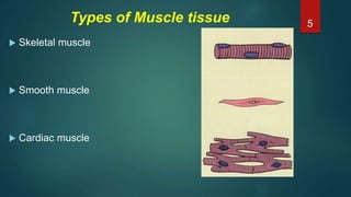
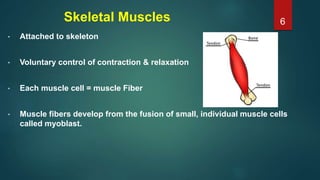
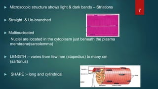
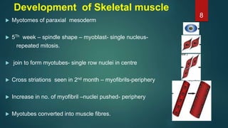
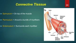
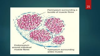
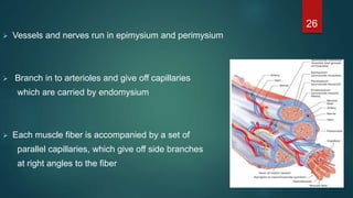
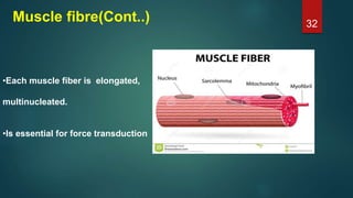
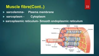
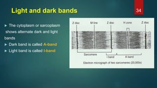
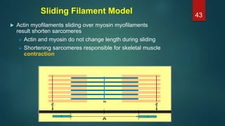
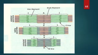
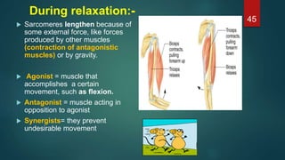
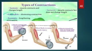
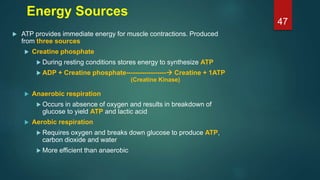
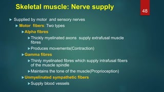
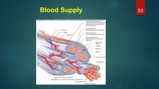
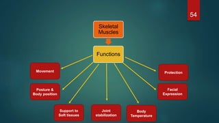
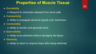
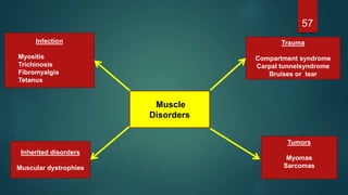
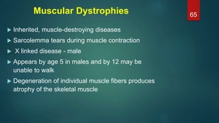
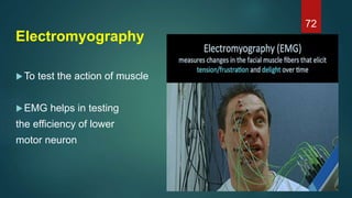
SKELETAL
MUSCLE
Smoking is injurious to health
Rakesh kumar
1
 Introduction
 Classification
 Microscopic structure
 Physiology
 Applied Anatomy
 References
Protocol
2
Introduction
 MUSCULUS = LITTLE MOUSE
 Muscle tissue is the one of the basic tissues of the body.
 Responsible for movement .
 Composed of differentiated cells containing contractile
proteins(actin, myosin etc.)
3
 Muscles – machines - chemical energy - mechanical energy.
 40-50% of the body mass.
 Attaches to bone, skin or fascia
 Developed from MESODERM except
a)Arrector pili
b)Muscles of iris
c)Myoepithelial cells of sweat ,salivary and lacrimal glands - ECTODERM
4
Types of Muscle tissue
 Skeletal muscle
 Smooth muscle
 Cardiac muscle
5
Skeletal Muscles
• Attached to skeleton
• Voluntary control of contraction & relaxation
• Each muscle cell = muscle Fiber
• Muscle fibers develop from the fusion of small, individual muscle cells
called myoblast.
6
 Microscopic structure shows light & dark bands – Striations
 Straight & Un-branched
 Multinucleated
Nuclei are located in the cytoplasm just beneath the plasma
membrane(sarcolemma)
 LENGTH :- varies from few mm (stapedius) to many cm
(sartorius)
 SHAPE :- long and cylindrical
7
Development of Skeletal muscle
 Myotomes of paraxial mesoderm
 5Th week – spindle shape – myoblast- single nucleus-
repeated mitosis.
 join to form myotubes- single row nuclei in centre
 Cross striations seen in 2nd month – myofibrils-periphery
 Increase in no. of myofibril –nuclei pushed- periphery
 Myotubes converted into muscle fibres.
8
Parts of Voluntary Muscle 9
Two parts of a muscle
1. Belly part:- Contractile part
2. Fibrous part:- Non contractile and non
elastic
a) Tendon
b) Aponeurosis
10
Classification
 According to colour
 According to Insertion of muscle
 According to direction of muscle fibre
- Parallel
- Pennate
- Spiral
- Cruciate
11
1. Slow (or Red) Oxidative Fibers Type I
 Slow but continuous contraction for extended periods
 Smaller diameter
 contain myoglobin
 more capillaries
 more mitochondria
Does not fatigue as fast due to aerobic production of ATP
Eg: Soleus.
12
2. Fast (Red) Oxidative Fibers Type IIa
Have attributes in between fast and slow types
13
3. Fast Glycolytic (white)Fibers Type IIb
 Fast contraction after nervous stimulation
 Large diameter
 Large glycogen reserve
 Fewer mitochondria
 Densely packed myofibrils
Fatigue fast due to mainly anaerobic respiration
Eg. Biceps brachii, Gastrocnemius.
14
15
According to insertion of muscle
 Insertion near the proximal end of bone
Eg – biceps brachii, psoas major
 Insertion towards the distal end of bone,
away from the joint
Eg- brachioradialis
 Insertion at the middle of shaft of bone
Eg- Coracobrachialis, pronator teres
16
According to direction of muscle fiber
a) Parallel muscles
 The muscle fibers are parallel to line of
pull
 The fibers are long, but their numbers are
few
Functions:
 Range of movement .
 ↓ total force of contraction
17
Sub divisions of parallel muscles
 Strap muscle- Sartorius, Rectus abdominis
 Quadrate muscle- Quadratus lumborum
 Fusiform muscle- Biceps brachii
18
b) Pennate muscles
 Fibers are oblique to line of pull
19
 Circumpennate
 The muscle is cylindrical
 Oblique fibers converge into central tendon from all
sides
Eg- Tibialis anterior
c) Spiral muscle
 Muscles are twisted in arrangement
close to their insertion
Eg- Pectoralis major & latissimus dorsi
20
d) Cruciate muscle
Fasciculi in the muscle are crossed
Eg: Sternocliedomastoid, Masseter
21
22
Circular: surrounds a body
orifice, constricting it when
contracted
Eg: Orbicularis occuli
(closes the eyelids)
23
Connective Tisssue
 Epimysium = On top of the muscle
 Perimysium = Around a bundle of myofibers
 Endomysium = Surrounds each myofiber
24
25
 Vessels and nerves run in epimysium and perimysium
 Branch in to arterioles and give off capillaries
which are carried by endomysium
 Each muscle fiber is accompanied by a set of
parallel capillaries, which give off side branches
at right angles to the fiber
26
• Epimysium consists mainly type I collagen fibers
• Perimysium contain type I and III collagen fibers
• Endomysium contain type III and IV collagen fibers
• Collagen IV is associated with the basal lamina that invests each
muscle fiber
• All these connective tissue layers extend beyond the muscle
belly to form the tendons, aponeurosis and fascia.
27
Attachment of tendon to skeletal muscle 28
Fascicles
Myofibrils
Myofilaments
Actin Myosin
It is important to remember the Heirarchy
29
Microscopic Structure
Some vocabulary:
Skeletal muscle cell = fiber or myofiber
Sarcolemma
Sarcoplasm
Sarcoplasmic reticulum
Myofibril
Myofilaments
T-tubules
Muscle
Epimysium
Myofibrils
Sarcomere
Myofilament
Myofibrils
30
Muscle and Muscle fibre
• Each muscle divides into several
muscle bundles or fasciculi
by perimysium
• Each fasciculus is composed of
variable number of muscle fibers
• Individual muscles are separated by
fascia, which also forms tendons
and aponeurosis
31
Muscle fibre(Cont..)
•Each muscle fiber is elongated,
multinucleated.
•Is essential for force transduction
32
Muscle fibre(Cont..)
 sarcolemma- Plasma membrane
 sarcoplasm - Cytoplasm
 sarcoplasmic reticulum- Smooth endoplasmic reticulum
33
Light and dark bands
 The cytoplasm or sarcoplasm
shows alternate dark and light
bands
 Dark band is called A-band
 Light band is called I-band
34
Light and dark bands 35
Sarcomere
 The portion of myofibrils between every two Z-lines
 Z disc: filamentous network of protein. Serves as attachment for
actin myofilaments
 Titin filaments: elastic chains of amino acids; make muscles
extensible and elastic
36
Myofibril
 Muscle fibers are filled with threads called
myofibrils, separated by SR (sarcoplasmic
reticulum)
 Myofilaments (thick & thin filaments) are the
contractile proteins of muscle
37
Transverse Tubules
 T (transverse) tubules are invaginations of the
sarcolemma into the center of the cell
 filled with extracellular fluid
 carry muscle action potentials down into cell
 Mitochondria lie in rows throughout the cell
 near the muscle proteins that use ATP during contraction
Sarcoplasmic Reticulum (SR)
 System of tubular sacs similar to smooth ER
 Placed horizontally between T-tubules
 Stores Ca2+ in a relaxed muscle
 Release of Ca2+ triggers muscle contraction
39
Proteins of Muscle
 Myofibrils are built of 3 kinds of protein
 Contractile proteins
 myosin and actin
 Regulatory proteins which turn contraction on & off
 troponin and tropomyosin
 Structural proteins which provide proper alignment, elasticity and
extensibility
 titin, myomesin, nebulin and dystrophin
40
Neuromuscular Junction
SYNAPSE: axon terminal
resting in an invagination
of the sarcolemma
Neuromuscular
junction (NMJ):
 Presynaptic terminal: axon
terminal with synaptic
vesicles
 Synaptic cleft: space
 Postsynaptic membrane or
motor end-plate
41
42
Sliding Filament Model
 Actin myofilaments sliding over myosin myofilaments
result shorten sarcomeres
 Actin and myosin do not change length during sliding
 Shortening sarcomeres responsible for skeletal muscle
contraction
43
44
During relaxation:-
 Sarcomeres lengthen because of
some external force, like forces
produced by other muscles
(contraction of antagonistic
muscles) or by gravity.
 Agonist = muscle that
accomplishes a certain
movement, such as flexion.
 Antagonist = muscle acting in
opposition to agonist
 Synergists= they prevent
undesirable movement
45
46
Energy Sources
 ATP provides immediate energy for muscle contractions. Produced
from three sources
 Creatine phosphate
 During resting conditions stores energy to synthesize ATP
 ADP + Creatine phosphate------------------ Creatine + 1ATP
 Anaerobic respiration
 Occurs in absence of oxygen and results in breakdown of
glucose to yield ATP and lactic acid
 Aerobic respiration
 Requires oxygen and breaks down glucose to produce ATP,
carbon dioxide and water
 More efficient than anaerobic
(Creatine Kinase)
47
Skeletal muscle: Nerve supply
 Supplied by motor and sensory nerves
 Motor fibers: Two types
Alpha fibres
Thickly myelinated axons supply extrafusal muscle
fibres
Produces movements(Contraction)
Gamma fibres
Thinly myelinated fibres which supply intrafusal fibers
of the muscle spindle
Maintains the tone of the muscle(Proprioception)
Unmyelinated sympathetic fibers
Supply blood vessels
48
Skeletal muscle: Nerve supply
 Motor end plate : Motor neuron (efferent) axons
originating in the spinal cord contact
muscle fibers via a structure known as the
motor endplate.
 Motor point:
 The site where the motor nerve
enters the muscle
49
Skeletal muscle: Nerve supply
 Motor unit/functional unit
 A single motor neuron
together with the muscle
fibers supplied by it.
 The size of the unit
depends on the
precision of the
movement
 Large & Small
50
Sensory fibers
 Arises from specialized endings
 Muscle spindle/tendon spindle
 Essential for muscle tone and body posture
 Essential for coordinated voluntary movements
51
Deep Tendon Reflex 52
Blood Supply 53
54
Movement
Posture &
Body position
Support to
Soft tissues
Joint
stabilization
Body
Temperature
Skeletal
Muscles
Functions
Facial
Expression
Protection
Properties of Muscle Tissue
 Excitability
 Respond to chemicals released from nerve cells
 Conductivity
 Ability to propagate electrical signals over membrane
 Contractility
 Ability to shorten and generate force
 Extensibility
 Ability to be stretched without damaging the tissue
 Elasticity
 Ability to return to original shape after being stretched
55
APPLIED ANATOMY
56
57
Muscle
Disorders
Trauma
Compartment syndrome
Carpal tunnelsyndrome
Bruises or tear
Infection
Myositis
Trichinosis
Fibromyalgia
Tetanus
Inherited disorders
Muscular dystrophies
Tumors
Myomas
Sarcomas
58
Secondary
Disorders
Immune problems:
Myasthenia gravis
Guilliain Barre syndrome
Nervous system:
Botulism
Poliomyelitis
Cardiovascular system:
Anemia
Heart failure
Metabolic problems:
Hypercalcemia
Hypocalcemia
Fatigue
Decreased capacity to work and reduced
efficiency of performance
Cause -
 Exhaustion of Ach in motor endplate
 Accumulation of metabolites like lactic acid and
phosphoric acid
 Lack of nutrients like glucose
 Lack of oxygen
59
Spasticity -Tone is increased
(Eg- stroke)
Flaccidity -Tone is decreased
(Eg -spinal shock)
Atrophy- (wasting of muscles)
(Eg- Poliomyelitis)
60
 Hypertrophy - New muscle fibers are not
formed, Enlargement due to
production of more myofibrils and
cell organelles
 Hyperplasia – increase
in number
 Damage of skeletal
muscle – replaced by
fibrous scar tissue
61
Myasthenia gravis
 Autoimmune disorder - blocks the ACh receptors- NMJ
 The more receptors are damaged the weaker the muscle.
 More common in women 20 to 40 with possible line to
thymus gland tumors
 Symptoms
 Treatment includes steroids that reduce antibodies that bind
to ACh receptors and inhibitors of acetylcholinesterase
62
63
64
Muscular Dystrophies
 Inherited, muscle-destroying diseases
 Sarcolemma tears during muscle contraction
 X linked disease - male
 Appears by age 5 in males and by 12 may be
unable to walk
 Degeneration of individual muscle fibers produces
atrophy of the skeletal muscle
65
66
Poliomyelitis
 Poliovirus
 Under the age of 3
 Poliovirus destroys motor neuron in the AHC.
 Muscle of leg more affected than arm but
paralysis can spread to the muscle of the thorax
and abdomen
67
Sports Injuries
 5 year study of college football players
indicate:
• Mild – 73.5%
• Moderate – 21.5%
• Severe – 11.6%
 A study of 1650 joggers running 27 miles
a week reported 1819 injuries in a single
year.
68
Sports related conditions
 Bone bruise: bleeding within the periosteum
 Bursitis: an inflammation of bursae
 Muscle cramps: prolonged,involuntary,and painful muscular
contraction
 Sprains: tear or break in ligaments or tendons
 Strains: tears in muscles
 Tendinitis: an inflammation of connective tissue surrounding a
tendon
69
Muscle Memory
 Muscle memory is not a memory stored in your muscles,but memories
stored in your brain.
 Motor Learning.
 Muscle Encoding.
 Muscle Re-education or Rehabilitaion.
70
71
Electromyography
To test the action of muscle
EMG helps in testing
the efficiency of lower
motor neuron
72
REFERENCES
1. Gray’s Anatomy- Susan Standring. 40th Edition.
2. Inderbir singh. Textbook of human histology with color atlas, 2006, 5th
edition
3. DiFiore’s , Atlas of histology, 12th edition
4. Vander’s Human physiology, 12th edition
5. Manipal manual of physiology,1st edition
6. Principal of general anatomy A.k.datta,7th edition
7. General anatomy,B D Chaurasia,4th edition
8. Principles of Human Anatomy,Gerard J.Tortora/Mark T.Nielsen, 13th
edition
9. Gross Anatomy,Kyung Won Chung/Harold M. chung, 6th edition
10. A&P ,Applications Manual ,Martini &Welch
73
74

Skeletal muscle must watch

  • 1.
    SKELETAL MUSCLE Smoking is injuriousto health Rakesh kumar 1
  • 2.
     Introduction  Classification Microscopic structure  Physiology  Applied Anatomy  References Protocol 2
  • 3.
    Introduction  MUSCULUS =LITTLE MOUSE  Muscle tissue is the one of the basic tissues of the body.  Responsible for movement .  Composed of differentiated cells containing contractile proteins(actin, myosin etc.) 3
  • 4.
     Muscles –machines - chemical energy - mechanical energy.  40-50% of the body mass.  Attaches to bone, skin or fascia  Developed from MESODERM except a)Arrector pili b)Muscles of iris c)Myoepithelial cells of sweat ,salivary and lacrimal glands - ECTODERM 4
  • 5.
    Types of Muscletissue  Skeletal muscle  Smooth muscle  Cardiac muscle 5
  • 6.
    Skeletal Muscles • Attachedto skeleton • Voluntary control of contraction & relaxation • Each muscle cell = muscle Fiber • Muscle fibers develop from the fusion of small, individual muscle cells called myoblast. 6
  • 7.
     Microscopic structureshows light & dark bands – Striations  Straight & Un-branched  Multinucleated Nuclei are located in the cytoplasm just beneath the plasma membrane(sarcolemma)  LENGTH :- varies from few mm (stapedius) to many cm (sartorius)  SHAPE :- long and cylindrical 7
  • 8.
    Development of Skeletalmuscle  Myotomes of paraxial mesoderm  5Th week – spindle shape – myoblast- single nucleus- repeated mitosis.  join to form myotubes- single row nuclei in centre  Cross striations seen in 2nd month – myofibrils-periphery  Increase in no. of myofibril –nuclei pushed- periphery  Myotubes converted into muscle fibres. 8
  • 9.
  • 10.
    Two parts ofa muscle 1. Belly part:- Contractile part 2. Fibrous part:- Non contractile and non elastic a) Tendon b) Aponeurosis 10
  • 11.
    Classification  According tocolour  According to Insertion of muscle  According to direction of muscle fibre - Parallel - Pennate - Spiral - Cruciate 11
  • 12.
    1. Slow (orRed) Oxidative Fibers Type I  Slow but continuous contraction for extended periods  Smaller diameter  contain myoglobin  more capillaries  more mitochondria Does not fatigue as fast due to aerobic production of ATP Eg: Soleus. 12
  • 13.
    2. Fast (Red)Oxidative Fibers Type IIa Have attributes in between fast and slow types 13
  • 14.
    3. Fast Glycolytic(white)Fibers Type IIb  Fast contraction after nervous stimulation  Large diameter  Large glycogen reserve  Fewer mitochondria  Densely packed myofibrils Fatigue fast due to mainly anaerobic respiration Eg. Biceps brachii, Gastrocnemius. 14
  • 15.
  • 16.
    According to insertionof muscle  Insertion near the proximal end of bone Eg – biceps brachii, psoas major  Insertion towards the distal end of bone, away from the joint Eg- brachioradialis  Insertion at the middle of shaft of bone Eg- Coracobrachialis, pronator teres 16
  • 17.
    According to directionof muscle fiber a) Parallel muscles  The muscle fibers are parallel to line of pull  The fibers are long, but their numbers are few Functions:  Range of movement .  ↓ total force of contraction 17
  • 18.
    Sub divisions ofparallel muscles  Strap muscle- Sartorius, Rectus abdominis  Quadrate muscle- Quadratus lumborum  Fusiform muscle- Biceps brachii 18
  • 19.
    b) Pennate muscles Fibers are oblique to line of pull 19
  • 20.
     Circumpennate  Themuscle is cylindrical  Oblique fibers converge into central tendon from all sides Eg- Tibialis anterior c) Spiral muscle  Muscles are twisted in arrangement close to their insertion Eg- Pectoralis major & latissimus dorsi 20
  • 21.
    d) Cruciate muscle Fasciculiin the muscle are crossed Eg: Sternocliedomastoid, Masseter 21
  • 22.
    22 Circular: surrounds abody orifice, constricting it when contracted Eg: Orbicularis occuli (closes the eyelids)
  • 23.
  • 24.
    Connective Tisssue  Epimysium= On top of the muscle  Perimysium = Around a bundle of myofibers  Endomysium = Surrounds each myofiber 24
  • 25.
  • 26.
     Vessels andnerves run in epimysium and perimysium  Branch in to arterioles and give off capillaries which are carried by endomysium  Each muscle fiber is accompanied by a set of parallel capillaries, which give off side branches at right angles to the fiber 26
  • 27.
    • Epimysium consistsmainly type I collagen fibers • Perimysium contain type I and III collagen fibers • Endomysium contain type III and IV collagen fibers • Collagen IV is associated with the basal lamina that invests each muscle fiber • All these connective tissue layers extend beyond the muscle belly to form the tendons, aponeurosis and fascia. 27
  • 28.
    Attachment of tendonto skeletal muscle 28
  • 29.
    Fascicles Myofibrils Myofilaments Actin Myosin It isimportant to remember the Heirarchy 29
  • 30.
    Microscopic Structure Some vocabulary: Skeletalmuscle cell = fiber or myofiber Sarcolemma Sarcoplasm Sarcoplasmic reticulum Myofibril Myofilaments T-tubules Muscle Epimysium Myofibrils Sarcomere Myofilament Myofibrils 30
  • 31.
    Muscle and Musclefibre • Each muscle divides into several muscle bundles or fasciculi by perimysium • Each fasciculus is composed of variable number of muscle fibers • Individual muscles are separated by fascia, which also forms tendons and aponeurosis 31
  • 32.
    Muscle fibre(Cont..) •Each musclefiber is elongated, multinucleated. •Is essential for force transduction 32
  • 33.
    Muscle fibre(Cont..)  sarcolemma-Plasma membrane  sarcoplasm - Cytoplasm  sarcoplasmic reticulum- Smooth endoplasmic reticulum 33
  • 34.
    Light and darkbands  The cytoplasm or sarcoplasm shows alternate dark and light bands  Dark band is called A-band  Light band is called I-band 34
  • 35.
  • 36.
    Sarcomere  The portionof myofibrils between every two Z-lines  Z disc: filamentous network of protein. Serves as attachment for actin myofilaments  Titin filaments: elastic chains of amino acids; make muscles extensible and elastic 36
  • 37.
    Myofibril  Muscle fibersare filled with threads called myofibrils, separated by SR (sarcoplasmic reticulum)  Myofilaments (thick & thin filaments) are the contractile proteins of muscle 37
  • 38.
    Transverse Tubules  T(transverse) tubules are invaginations of the sarcolemma into the center of the cell  filled with extracellular fluid  carry muscle action potentials down into cell  Mitochondria lie in rows throughout the cell  near the muscle proteins that use ATP during contraction
  • 39.
    Sarcoplasmic Reticulum (SR) System of tubular sacs similar to smooth ER  Placed horizontally between T-tubules  Stores Ca2+ in a relaxed muscle  Release of Ca2+ triggers muscle contraction 39
  • 40.
    Proteins of Muscle Myofibrils are built of 3 kinds of protein  Contractile proteins  myosin and actin  Regulatory proteins which turn contraction on & off  troponin and tropomyosin  Structural proteins which provide proper alignment, elasticity and extensibility  titin, myomesin, nebulin and dystrophin 40
  • 41.
    Neuromuscular Junction SYNAPSE: axonterminal resting in an invagination of the sarcolemma Neuromuscular junction (NMJ):  Presynaptic terminal: axon terminal with synaptic vesicles  Synaptic cleft: space  Postsynaptic membrane or motor end-plate 41
  • 42.
  • 43.
    Sliding Filament Model Actin myofilaments sliding over myosin myofilaments result shorten sarcomeres  Actin and myosin do not change length during sliding  Shortening sarcomeres responsible for skeletal muscle contraction 43
  • 44.
  • 45.
    During relaxation:-  Sarcomereslengthen because of some external force, like forces produced by other muscles (contraction of antagonistic muscles) or by gravity.  Agonist = muscle that accomplishes a certain movement, such as flexion.  Antagonist = muscle acting in opposition to agonist  Synergists= they prevent undesirable movement 45
  • 46.
  • 47.
    Energy Sources  ATPprovides immediate energy for muscle contractions. Produced from three sources  Creatine phosphate  During resting conditions stores energy to synthesize ATP  ADP + Creatine phosphate------------------ Creatine + 1ATP  Anaerobic respiration  Occurs in absence of oxygen and results in breakdown of glucose to yield ATP and lactic acid  Aerobic respiration  Requires oxygen and breaks down glucose to produce ATP, carbon dioxide and water  More efficient than anaerobic (Creatine Kinase) 47
  • 48.
    Skeletal muscle: Nervesupply  Supplied by motor and sensory nerves  Motor fibers: Two types Alpha fibres Thickly myelinated axons supply extrafusal muscle fibres Produces movements(Contraction) Gamma fibres Thinly myelinated fibres which supply intrafusal fibers of the muscle spindle Maintains the tone of the muscle(Proprioception) Unmyelinated sympathetic fibers Supply blood vessels 48
  • 49.
    Skeletal muscle: Nervesupply  Motor end plate : Motor neuron (efferent) axons originating in the spinal cord contact muscle fibers via a structure known as the motor endplate.  Motor point:  The site where the motor nerve enters the muscle 49
  • 50.
    Skeletal muscle: Nervesupply  Motor unit/functional unit  A single motor neuron together with the muscle fibers supplied by it.  The size of the unit depends on the precision of the movement  Large & Small 50
  • 51.
    Sensory fibers  Arisesfrom specialized endings  Muscle spindle/tendon spindle  Essential for muscle tone and body posture  Essential for coordinated voluntary movements 51
  • 52.
  • 53.
  • 54.
    54 Movement Posture & Body position Supportto Soft tissues Joint stabilization Body Temperature Skeletal Muscles Functions Facial Expression Protection
  • 55.
    Properties of MuscleTissue  Excitability  Respond to chemicals released from nerve cells  Conductivity  Ability to propagate electrical signals over membrane  Contractility  Ability to shorten and generate force  Extensibility  Ability to be stretched without damaging the tissue  Elasticity  Ability to return to original shape after being stretched 55
  • 56.
  • 57.
    57 Muscle Disorders Trauma Compartment syndrome Carpal tunnelsyndrome Bruisesor tear Infection Myositis Trichinosis Fibromyalgia Tetanus Inherited disorders Muscular dystrophies Tumors Myomas Sarcomas
  • 58.
    58 Secondary Disorders Immune problems: Myasthenia gravis GuilliainBarre syndrome Nervous system: Botulism Poliomyelitis Cardiovascular system: Anemia Heart failure Metabolic problems: Hypercalcemia Hypocalcemia
  • 59.
    Fatigue Decreased capacity towork and reduced efficiency of performance Cause -  Exhaustion of Ach in motor endplate  Accumulation of metabolites like lactic acid and phosphoric acid  Lack of nutrients like glucose  Lack of oxygen 59
  • 60.
    Spasticity -Tone isincreased (Eg- stroke) Flaccidity -Tone is decreased (Eg -spinal shock) Atrophy- (wasting of muscles) (Eg- Poliomyelitis) 60
  • 61.
     Hypertrophy -New muscle fibers are not formed, Enlargement due to production of more myofibrils and cell organelles  Hyperplasia – increase in number  Damage of skeletal muscle – replaced by fibrous scar tissue 61
  • 62.
    Myasthenia gravis  Autoimmunedisorder - blocks the ACh receptors- NMJ  The more receptors are damaged the weaker the muscle.  More common in women 20 to 40 with possible line to thymus gland tumors  Symptoms  Treatment includes steroids that reduce antibodies that bind to ACh receptors and inhibitors of acetylcholinesterase 62
  • 63.
  • 64.
  • 65.
    Muscular Dystrophies  Inherited,muscle-destroying diseases  Sarcolemma tears during muscle contraction  X linked disease - male  Appears by age 5 in males and by 12 may be unable to walk  Degeneration of individual muscle fibers produces atrophy of the skeletal muscle 65
  • 66.
  • 67.
    Poliomyelitis  Poliovirus  Underthe age of 3  Poliovirus destroys motor neuron in the AHC.  Muscle of leg more affected than arm but paralysis can spread to the muscle of the thorax and abdomen 67
  • 68.
    Sports Injuries  5year study of college football players indicate: • Mild – 73.5% • Moderate – 21.5% • Severe – 11.6%  A study of 1650 joggers running 27 miles a week reported 1819 injuries in a single year. 68
  • 69.
    Sports related conditions Bone bruise: bleeding within the periosteum  Bursitis: an inflammation of bursae  Muscle cramps: prolonged,involuntary,and painful muscular contraction  Sprains: tear or break in ligaments or tendons  Strains: tears in muscles  Tendinitis: an inflammation of connective tissue surrounding a tendon 69
  • 70.
    Muscle Memory  Musclememory is not a memory stored in your muscles,but memories stored in your brain.  Motor Learning.  Muscle Encoding.  Muscle Re-education or Rehabilitaion. 70
  • 71.
  • 72.
    Electromyography To test theaction of muscle EMG helps in testing the efficiency of lower motor neuron 72
  • 73.
    REFERENCES 1. Gray’s Anatomy-Susan Standring. 40th Edition. 2. Inderbir singh. Textbook of human histology with color atlas, 2006, 5th edition 3. DiFiore’s , Atlas of histology, 12th edition 4. Vander’s Human physiology, 12th edition 5. Manipal manual of physiology,1st edition 6. Principal of general anatomy A.k.datta,7th edition 7. General anatomy,B D Chaurasia,4th edition 8. Principles of Human Anatomy,Gerard J.Tortora/Mark T.Nielsen, 13th edition 9. Gross Anatomy,Kyung Won Chung/Harold M. chung, 6th edition 10. A&P ,Applications Manual ,Martini &Welch 73
  • 74.