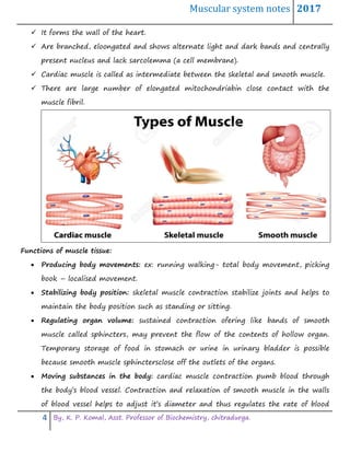
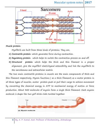
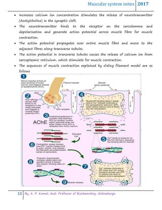
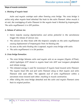
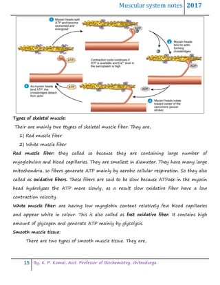
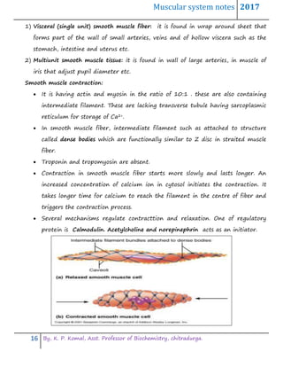
Muscular system notes 2017
1 By, K. P. Komal, Asst. Professor of Biochemistry, chitradurga.
Lecture Notes
On
Muscular system
By,
K. P. KOMAL
ASSISTANT PROFESSOR
DEPARTMENT OF BIOCHEMISTRY
GOVERNMENT SCIENCE COLLEGE, CHITRADURGA. 577501
KARNATAKA STATE.
Muscular system notes 2017
2 By, K. P. Komal, Asst. Professor of Biochemistry, chitradurga.
Bone forms the framework of the body, they can not move the body parts by
themselves. Motion results from the alternate contraction and relaxation of
muscle, which constitute 40-50%of the total body weight.
The specific study of muscle is known as myology.
Muscles,
 Changes chemical energy into mechanical energy to generate the force,
perform work and produce movement.
 Stabilizes the body positions.
 Regulate organ volume
 Generate heat
 Propel fluids and food matter through various body system.
Types of muscle tissue: there are three types of muscles,
 Skeletal muscle
 Cardiac muscle
 Smooth muscle
These three types of muscle tissues share same properties such as function but
differ in microscopic anatomy locations. They are controlled by nervous and endocrine
system.
Skeletal muscle tissue:
 is named so because the function of most skeletal muscle is to move the bones of
the skeleton (a few skeletal muscle attaches to the tissues other than bone). Most
attached by tendons to bones.
 It is termed straited because alternating light and dark bands( straitions) are
visible when examined under the microscope.
 Located at the peripheral region of the organism
 Can be easily contracted but can not be remained contracted for long time.
Muscular system notes 2017
3 By, K. P. Komal, Asst. Professor of Biochemistry, chitradurga.
 It is long elongated cell with multinuclei.
 Voluntary –subject to consious control
 Cells are surrounded by connective tissues.
 Connective tissue wrappings of skeletal muscle:
 Endomysium- around single muscle fiber
 Perimysium- around a fascicle(bundle) of fobers
 Epimysium – covers the entire skeletal muscle
 Fascia – on the outside of the epimysium
 Skeletal muscle atttachments:
 Epimysium blends into a connective tissue attachments
 Tendon – cord like structure
 Aponeurose- sheet like structure
 Sites of muscle attachment
 Bones
 Cartilage
 Connective tissue coverings
Smooth muscle tissue:
 Is located in the walls of hallow internal structures such as blood vessels also found
in skin attached to hair follicle.
 Under microscope it look like a smooth or nonstraited
 Spindle or fused in structure
 Nucleus is centrally presentarranged in layers.
 Contraction is slow and can be remained in contracted form for longer time so
called involuntary muscle
 It is controlled by autonomic nervous system.
Cardiac muscle tissue:
 Only the heart contains the cardiac muscle.
Muscular system notes 2017
4 By, K. P. Komal, Asst. Professor of Biochemistry, chitradurga.
 It forms the wall of the heart.
 Are branched, eloongated and shows alternate light and dark bands and centrally
present nucleus and lack sarcolemma (a cell membrane).
 Cardiac muscle is called as intermediate between the skeletal and smooth muscle.
 There are large number of elongated mitochondriabin close contact with the
muscle fibril.
Functions of muscle tissue:
 Producing body movements: ex: running walking- total body movement, picking
book – localised movement.
 Stabilizing body position: skeletal muscle contraction stabilize joints and helps to
maintain the body position such as standing or sitting.
 Regulating organ volume: sustained contraction ofering like bands of smooth
muscle called sphincters, may prevent the flow of the contents of hollow organ.
Temporary storage of food in stomach or urine in urinary bladder is possible
because smooth muscle sphinctersclose off the outlets of the organs.
 Moving substances in the body: cardiac muscle contraction pumb blood through
the body’s blood vessel. Contraction and relaxation of smooth muscle in the walls
of blood vessel helps to adjust it’s diameter and thus regulates the rate of blood
Muscular system notes 2017
5 By, K. P. Komal, Asst. Professor of Biochemistry, chitradurga.
flow. Smooth muscle contraction also moves food and substances such as juices,
like bile, and enzymes through the gastrointestinal tract etc..
 Producing heat: muscle contraction produces the heat which is used to maintain
the normal body temperature. Involuntary contraction of skeletal muscle known
as shivering can increase the rate of heat production several folds.
Propertise of muscle tissue:
Muscle tissue has four special properties that enable it to function and contribute
to homeostasis (condition of a body to maintain temperature and blood pressure at a
constant level). All muscle cells share several properties: contractility, excitability,
extensibility, and elasticity:
1. Contractility is the ability of muscle cells to forcefully shorten. For instance, in
order to flex (decrease the angle of a joint) your elbow you need to contract
(shorten) the biceps brachii and other elbow flexor muscles in the anterior arm.
Notice that in order to extend your elbow, the posterior arm extensor muscles
need to contract. Thus, muscles can only pull, never push.
2. Excitability is the ability to respond to a stimulus, which may be delivered from a
motor neuron or a hormone.
3. Extensibility is the ability of a muscle to be stretched. For instance, let's reconsider
our elbow flexing motion we discussed earlier. In order to be able to flex the
elbow, the elbow extensor muscles must extend in order to allow flexion to occur.
Lack of extensibility is known as spasticity.
4. Elasticity is the ability to recoil or bounce back to the muscle's original length
after being stretched.
Skeletal muscle tissue:
Connective tissue is present in all muscles as fascia. Enclosing each muscle is a
layer of connective tissue known as the epimysium; enclosing each fascicle is a layer
called the perimysium, and enclosing each muscle fiber is a layer of connective tissue
Muscular system notes 2017
6 By, K. P. Komal, Asst. Professor of Biochemistry, chitradurga.
called the
endomysium.
Muscle fibers
Muscular system notes 2017
7 By, K. P. Komal, Asst. Professor of Biochemistry, chitradurga.
Muscle fibres are the individual contractile units within muscle. A single muscle
such as the biceps brachii contains many muscle fibres.
Another group of cells, the myosatellite cells are found between the basement
membrane and the sarcolemma of muscle fibers. These cells are normally quiescent but
can be activated by exercise or pathology to provide additional myonuclei for muscle
growth or repair.
Development:
Individual muscle fibers are formed during development from the fusion of several
undifferentiated immature cells known as myoblasts into long, cylindrical, multi-
nucleated cells. Differentiation into this state is primarily completed before birth with
the cells continuing to grow in size thereafter.
Microanatomy:
Skeletal muscle exhibits a distinctive banding pattern when viewed under the
microscope due to the arrangement of cytoskeletal elements in the cytoplasm of the
muscle fibers. The principal cytoplasmic proteins are myosin and actin (also known as
"thick" and "thin" filaments, respectively) which are arranged in a repeating unit called
a sarcomere. The interaction of myosin and actin is responsible for muscle contraction.
Every single organelle and macromolecule of a muscle fiber is arranged to ensure
form meets function. The cell membrane is called the sarcolemma with the cytoplasm
known as the sarcoplasm. In the sarcoplasm are the myofibrils. The myofibrils are long
protein bundles about 1 micrometer in diameter each containing myofilaments. Pressed
against the inside of the sarcolemma are the unusual flattened myonuclei. Between the
myofibrils are the mitochondria.
While the muscle fiber does not have a smooth endoplasmic reticulum, it contains
a sarcoplasmic reticulum. The sarcoplasmic reticulum surrounds the myofibrils and
holds a reserve of the calcium ions needed to cause a muscle contraction. Periodically, it
has dilated end sacs known as terminal cisternae. These cross the muscle fiber from one
Muscular system notes 2017
8 By, K. P. Komal, Asst. Professor of Biochemistry, chitradurga.
side to the other. In between two terminal cisternae is a tubular infolding called a
transverse tubule (T tubule). T tubules are the pathways for action potentials to signal
the sarcoplasmic reticulum to release calcium, causing a muscle contraction. Together,
two terminal cisternae and a transverse tubule form a triad. A sarcomere is the basic
unit of muscle tissue in both cardiac and skeletal muscle. Sarcomeres appear under the
microscope as striations, with alternating dark and light bands. Sarcomeres are
connected to a plasma membrane, called a sarcolemma, by T-tubules, which speed up
the rate of depolarization within the sarcomere.
Individual sarcomeres are composed of long, fibrous proteins that slide past each
other when the muscles contract and relax. The two most important proteins within
sarcomeres are myosin, which forms a thick, flexible filament, and actin, which forms
the thin, more rigid filament. Myosin has a long, fibrous tail and a globular head which
binds to actin. The myosin head also binds to ATP, the source of energy for muscle
movement. Actin molecules are bound to the Z-disc (Zwischeibe disc), which forms the
borders of the sarcomere, separetes one sarcomere from other. Together, myosin and
actin form myofibrils, the repeating molecular structure of sarcomeres. The darker
middle portion of sarcomere is A band (Anisotropic), which extends the entire length of
the thich filament. There are two thin filaments for every thick filament . the I band
(Isotropic) is a ligghter, less dense area. A z disc passes through the centre of each I
band, a narrow h zone (Henson zone), in the centre of each A band contains thick but
no thin filament. Supporting protein that holds thick filament together at the centre
of H zone form M line (Merkel’s line) so named because of sarcomere.
Myofibril activity is required for muscle contraction on the molecular level. When
ATP binds to myosin, it separates from the actin of the myofibril, which causes a
contraction. Muscle contraction is a complex process regulated by calcium influx and the
stimulus of electrical impulses.
Muscular system notes 2017
9 By, K. P. Komal, Asst. Professor of Biochemistry, chitradurga.
Muscle protein:
Myofibrils are built from three kinds of proteins. They are,
1) Contractile protein: which generates force during contraction.
2) Regulatory protein: which helps to switch the contraction process on and off.
3) Structural protein: which helps the thick and thin filament in a proper
alignment, give the myofibril elasticityand extensibility and link the myofibrils to
the sarcolemma and extracellular matrix.
The two main contractile proteins in muscle are the main components of thick and
thin filament respectively. Myosin functions ( as a thick filament) as a motor protein in
all three types of muscles, motor protein push or pull their cargo to achieve movement
by converting the chemical energy in ATP to mechanical energy of motion or force
production. About 300 molecules of myosin form a single thick filasment. Each myosin
molecule is shape like two golf sticks clubs twisted together.
Muscular system notes 2017
10 By, K. P. Komal, Asst. Professor of Biochemistry, chitradurga.
The myosin tail points towards the M line of the sarcomere. Tails of neighbouring
myosin molecule lie parallel to one another, forming the shaft of thick filament. The
two projections of each myosin molecule are called head and are crossbridges.
The tail section of myosin is the carboxyl end of the chain. It is coiled around itself in
an α helical arrangement. Treatment with trypsin results in cleavage of tail about one
third of the way down from the head to produce heavy meromyosin (the head group
with short tail) and light meromyosin (the remained part of the tail).
The heavy meromyosin can be further separated by treating with papain. The “C”
protein is involved in their assembly. The M protein is also involved, presumably in
holding the tail section together as well as anchoring them to the centre M line of the H
zone.
The globular head section of the myosin is the working unit. It contains the
ATPase activity that provides the energy for the contractile process and it contains
actin binding site. ATP does not provide the energy directly for contraction but, its
binding to myosin leads to its hydrolysis by myosin ATPase induce the reversible
conformational changes of the head group that allows its binding to and dissociation
from actin during the process of pulling the Z lines towards the centre of the
sarcomere.
Muscular system notes 2017
11 By, K. P. Komal, Asst. Professor of Biochemistry, chitradurga.
Actin: thin filament extended from anchoring point with in the Z discs. Their main
component is the protein , actin. Individual actin molecule joins to form an actin
filament that is twisted into a helix. On each actin molecule is a binding site for myosin.
It makes up about 20-25% of muscle protein. It is a globular proteinwith molecular
weight of about 42000 dal.
Regulatory proteins:
Tropomyosin & troponin are also part of thin filament. In relaxed muscle myosin is
blocked from binding to actin because tropomyosin covers the myosin binding site on
actin. The tropomyosin strand inturn is held in place by troponin. Troponin also results
in conformational change which results in the exposure of actin-myosin binding site
and initiating the contraction process by permitting the free myosin to bind to the
actin.
Beside contractile and regulatory proteins, muscle contains about a dozen of
structural proteins which contribute to the alignment, stability, elasticitty and
extensibility of myofibrils. Several key structural proteins are Titin, myomesin, nebulin
and dystrophin. Titin, is the third most plentiful protein in skeletal muscle (after actin
and myosin). Titin anchors the thick filament to both a Z disc to an M line. Thereby
helping to stabilize the position of the thick filament. Titin probably helps the sarcomere
return to its resting length after a muscle has contracted or been stretched.
Nebulin: large but inelastic protein, lies along side the thin filament and also attaches to
Z disc. It helps to maintain the alignment of the thion filament in the sarcomere.
Dystrophin: it is a cytoskeletal protein that links thin filament of the sarcomere to the
integral membrane protein of the sarcolemma. Inturn the membrane protein attaches
to the connective tissue matrix that surrounds muscle tissue fibers. Hence dystrophin
and its associated proteinare thougght to reinforce the sarcolemma and helps to
transmit the tension generated in sarcolemma to tendons.
Muscular system notes 2017
12 By, K. P. Komal, Asst. Professor of Biochemistry, chitradurga.
Sliding filament mechanism:
The model describes the contraction of muscle is known as sliding filament theory.
Muscle contraction
Contraction mechanism:
 When the nerve impulse from brain and spinal cord are carried along motor
neuron to muscle fibre Ca++ ions are released in the terminal axon.
Muscular system notes 2017
13 By, K. P. Komal, Asst. Professor of Biochemistry, chitradurga.
 Increases calcium ion concentration stimulates the release of neurotransmitter
(Acetylcholine) in the synaptic cleft.
 The neurotransmitter binds to the receptor on the sarcolemma and
depolarization and generate action potential across muscle fibre for muscle
contraction.
 The action potential propagates over entire muscle fibre and move to the
adjacent fibres along transverse tubules.
 The action potential in transverse tubules causes the release of calcium ion from
sarcoplasmic reticulum, which stimulate for muscle contraction.
 The sequences of muscle contraction explained by sliding filament model are as
follows
Muscular system notes 2017
14 By, K. P. Komal, Asst. Professor of Biochemistry, chitradurga.
Steps of muscle contraction
1. Blocking of myosin head:
Actin and myosin overlaps each other forming cross bridge. The cross bridge is
active only when myosin head attached like hook to the actin filament. When muscle is
at rest, the overlapping of actin filament to the myosin head is blocked by tropomyosin.
The actin myofilament is in OFF position.
2. Release of calcium ion:
 Nerve impulse causing depolarization and action potential in the sarcolemma
trigger the release of calcium ions.
 The calcium ion then binds with the troponin complex on the actin myofilament
causing displacement of tropomyosin from its blocking site.
 As soon as the actin binding site is exposed, myosin cross bridge with actin.
 The actin myofilament is in ON position.
3. Cross bridge formation:
 The cross bridge between actin and myosin acts as an enzyme (Myosin ATPase),
which hydrolyses ATP stored in myosin head into ADP and inorganic phosphate
and release energy.
 This released energy is used for movement of myosin head toward actin filament.
The myosin head tilts and pull actin filament along so that myosin and actin
filament slide each other. The opposite end of actin myofilament within a
sarcomere move toward each other, resulting in muscle contraction.
 After sliding the cross bridge detached and the actin and myosin filament come
back to original position.
Muscular system notes 2017
15 By, K. P. Komal, Asst. Professor of Biochemistry, chitradurga.
Types of skeletal muscle:
Their are mainly two ttypes of skeletal muscle fiber. They are,
1) Red muscle fiber
2) White muscle fiber
Red muscle fiber: they called so because they are containing large number of
myoglobulins and blood capillaries. They are smallest in diameter. They have many large
mitochondria, so fibers generate ATP mainly by aerobic cellular respiration. So they also
called as oxidative fibers. These fibers are said to be slow because ATPase in the myosin
head hydrolyzes the ATP more slowly, as a result slow oxidative fiber have a low
contraction velocity.
White muscle fiber: are having low myoglobin content relatively few blood capillaries
and appear white in colour. This is also called as fast oxidative fiber. It contains high
amount of glycogen and generate ATP mainly by glycolysis.
Smooth muscle tissue:
There are two types of smooth muscle tissue. They are,
Muscular system notes 2017
16 By, K. P. Komal, Asst. Professor of Biochemistry, chitradurga.
1) Visceral (single unit) smooth muscle fiber: it is found in wrap around sheet that
forms part of the wall of small arteries, veins and of hollow viscera such as the
stomach, intestine and uterus etc.
2) Multiunit smooth muscle tissue: it is found in wall of large arteries, in muscle of
iris that adjust pupil diameter etc.
Smooth muscle contraction:
 It is having actin and myosin in the ratio of 10:1 . these are also containing
intermediate filament. These are lacking transverse tubule having sarcoplasmic
reticulum for storage of Ca2+.
 In smooth muscle fiber, intermediate filament such as attached to structure
called dense bodies which are functionally similar to Z disc in straited muscle
fiber.
 Troponin and tropomyosin are absent.
 Contraction in smooth muscle fiber starts more slowly and lasts longer. An
increased concentration of calcium ion in cytosol initiates the contraction. It
takes longer time for calcium to reach the filament in the centre of fiber and
triggers the contraction process.
 Several mechanisms regulate contracttion and relaxation. One of regulatory
protein is Calmodulin. Acetylcholine and norepinephrin acts as an initiator.
Muscular system notes 2017
17 By, K. P. Komal, Asst. Professor of Biochemistry, chitradurga.
Role of calmodulin in smooth muscle contraction:
Calmodulin (CaM) (an abbreviation for calcium-modulated protein) is a multifunctional
intermediate calcium-binding messenger protein expressed in alleukaryotic cells. It is an
intracellular target of the secondary messenger Ca2+, and the binding of Ca2+ is required
for the activation of calmodulin. Once bound to Ca2+, calmodulin acts as part of a
calcium signal transduction pathway by modifying its interactions with various target
proteins such as kinases or phosphatases.
Calmodulin plays an important role in excitation contraction (EC) coupling and
the initiation of the cross-bridge cycling in smooth muscle, ultimately causing smooth
muscle contraction. In order to activate contraction of smooth muscle, the head of
the myosin light chain must be phosphorylated. This phosphorylation is done by Myosin
Light Chain (MLC) Kinase. This MLC Kinase is activated by a calmodulin when it is
bound by calcium, thus making smooth muscle contraction dependent on the presence
of calcium, through the binding of calmodulin and activation of MLC kinase.
Another way that calmodulin affects muscle contraction is by controlling the
movement of Ca2+ across both the cell and sarcoplasmic reticulum membranes. The
Ca2+ channels, such as the ryanodine receptor of the sarcoplasmic reticulum, can be
inhibited by calmodulin bound to calcium, thus affecting the overall levels of calcium in
the cell. Calcium pumps take calcium out of the cytoplasm and/or store it in the
endoplasmic reticulum and this control helps regulate many downstream processes.
This is a very important function of calmodulin because it indirectly plays a role
in every physiological process that is affected by smooth muscle contraction such as
digestion and contraction of arteries (which helps distribute blood and regulate blood
pressure).
Muscular system notes 2017
18 By, K. P. Komal, Asst. Professor of Biochemistry, chitradurga.
Role of creatine phosphate in muscle contraction:
Phosphocreatine, also known as creatine phosphate (CP) or PCr (Pcr), is a
phosphorylated creatine molecule that serves as a rapidly mobilizable reserve of high-
energy phosphates in skeletal muscle and the brain to recycleadenosine triphosphate,
the energy currency of the cell.
Muscular system notes 2017
19 By, K. P. Komal, Asst. Professor of Biochemistry, chitradurga.
Definitions:
1) the latent period: The latent period is the time from when the stimulus is delivered
to the first indications of contraction in the muscle.
2) the contraction period: The contraction period, or contraction time, is the time it
takes the muscle to reach its peak contraction after the latent period.
3) the relaxation period : The relaxation period is the time the muscle takes to return
to resting tension after reaching its peak contraction.
Muscular system notes 2017
20 By, K. P. Komal, Asst. Professor of Biochemistry, chitradurga.

Muscular system

  • 1.
    Muscular system notes2017 1 By, K. P. Komal, Asst. Professor of Biochemistry, chitradurga. Lecture Notes On Muscular system By, K. P. KOMAL ASSISTANT PROFESSOR DEPARTMENT OF BIOCHEMISTRY GOVERNMENT SCIENCE COLLEGE, CHITRADURGA. 577501 KARNATAKA STATE.
  • 2.
    Muscular system notes2017 2 By, K. P. Komal, Asst. Professor of Biochemistry, chitradurga. Bone forms the framework of the body, they can not move the body parts by themselves. Motion results from the alternate contraction and relaxation of muscle, which constitute 40-50%of the total body weight. The specific study of muscle is known as myology. Muscles,  Changes chemical energy into mechanical energy to generate the force, perform work and produce movement.  Stabilizes the body positions.  Regulate organ volume  Generate heat  Propel fluids and food matter through various body system. Types of muscle tissue: there are three types of muscles,  Skeletal muscle  Cardiac muscle  Smooth muscle These three types of muscle tissues share same properties such as function but differ in microscopic anatomy locations. They are controlled by nervous and endocrine system. Skeletal muscle tissue:  is named so because the function of most skeletal muscle is to move the bones of the skeleton (a few skeletal muscle attaches to the tissues other than bone). Most attached by tendons to bones.  It is termed straited because alternating light and dark bands( straitions) are visible when examined under the microscope.  Located at the peripheral region of the organism  Can be easily contracted but can not be remained contracted for long time.
  • 3.
    Muscular system notes2017 3 By, K. P. Komal, Asst. Professor of Biochemistry, chitradurga.  It is long elongated cell with multinuclei.  Voluntary –subject to consious control  Cells are surrounded by connective tissues.  Connective tissue wrappings of skeletal muscle:  Endomysium- around single muscle fiber  Perimysium- around a fascicle(bundle) of fobers  Epimysium – covers the entire skeletal muscle  Fascia – on the outside of the epimysium  Skeletal muscle atttachments:  Epimysium blends into a connective tissue attachments  Tendon – cord like structure  Aponeurose- sheet like structure  Sites of muscle attachment  Bones  Cartilage  Connective tissue coverings Smooth muscle tissue:  Is located in the walls of hallow internal structures such as blood vessels also found in skin attached to hair follicle.  Under microscope it look like a smooth or nonstraited  Spindle or fused in structure  Nucleus is centrally presentarranged in layers.  Contraction is slow and can be remained in contracted form for longer time so called involuntary muscle  It is controlled by autonomic nervous system. Cardiac muscle tissue:  Only the heart contains the cardiac muscle.
  • 4.
    Muscular system notes2017 4 By, K. P. Komal, Asst. Professor of Biochemistry, chitradurga.  It forms the wall of the heart.  Are branched, eloongated and shows alternate light and dark bands and centrally present nucleus and lack sarcolemma (a cell membrane).  Cardiac muscle is called as intermediate between the skeletal and smooth muscle.  There are large number of elongated mitochondriabin close contact with the muscle fibril. Functions of muscle tissue:  Producing body movements: ex: running walking- total body movement, picking book – localised movement.  Stabilizing body position: skeletal muscle contraction stabilize joints and helps to maintain the body position such as standing or sitting.  Regulating organ volume: sustained contraction ofering like bands of smooth muscle called sphincters, may prevent the flow of the contents of hollow organ. Temporary storage of food in stomach or urine in urinary bladder is possible because smooth muscle sphinctersclose off the outlets of the organs.  Moving substances in the body: cardiac muscle contraction pumb blood through the body’s blood vessel. Contraction and relaxation of smooth muscle in the walls of blood vessel helps to adjust it’s diameter and thus regulates the rate of blood
  • 5.
    Muscular system notes2017 5 By, K. P. Komal, Asst. Professor of Biochemistry, chitradurga. flow. Smooth muscle contraction also moves food and substances such as juices, like bile, and enzymes through the gastrointestinal tract etc..  Producing heat: muscle contraction produces the heat which is used to maintain the normal body temperature. Involuntary contraction of skeletal muscle known as shivering can increase the rate of heat production several folds. Propertise of muscle tissue: Muscle tissue has four special properties that enable it to function and contribute to homeostasis (condition of a body to maintain temperature and blood pressure at a constant level). All muscle cells share several properties: contractility, excitability, extensibility, and elasticity: 1. Contractility is the ability of muscle cells to forcefully shorten. For instance, in order to flex (decrease the angle of a joint) your elbow you need to contract (shorten) the biceps brachii and other elbow flexor muscles in the anterior arm. Notice that in order to extend your elbow, the posterior arm extensor muscles need to contract. Thus, muscles can only pull, never push. 2. Excitability is the ability to respond to a stimulus, which may be delivered from a motor neuron or a hormone. 3. Extensibility is the ability of a muscle to be stretched. For instance, let's reconsider our elbow flexing motion we discussed earlier. In order to be able to flex the elbow, the elbow extensor muscles must extend in order to allow flexion to occur. Lack of extensibility is known as spasticity. 4. Elasticity is the ability to recoil or bounce back to the muscle's original length after being stretched. Skeletal muscle tissue: Connective tissue is present in all muscles as fascia. Enclosing each muscle is a layer of connective tissue known as the epimysium; enclosing each fascicle is a layer called the perimysium, and enclosing each muscle fiber is a layer of connective tissue
  • 6.
    Muscular system notes2017 6 By, K. P. Komal, Asst. Professor of Biochemistry, chitradurga. called the endomysium. Muscle fibers
  • 7.
    Muscular system notes2017 7 By, K. P. Komal, Asst. Professor of Biochemistry, chitradurga. Muscle fibres are the individual contractile units within muscle. A single muscle such as the biceps brachii contains many muscle fibres. Another group of cells, the myosatellite cells are found between the basement membrane and the sarcolemma of muscle fibers. These cells are normally quiescent but can be activated by exercise or pathology to provide additional myonuclei for muscle growth or repair. Development: Individual muscle fibers are formed during development from the fusion of several undifferentiated immature cells known as myoblasts into long, cylindrical, multi- nucleated cells. Differentiation into this state is primarily completed before birth with the cells continuing to grow in size thereafter. Microanatomy: Skeletal muscle exhibits a distinctive banding pattern when viewed under the microscope due to the arrangement of cytoskeletal elements in the cytoplasm of the muscle fibers. The principal cytoplasmic proteins are myosin and actin (also known as "thick" and "thin" filaments, respectively) which are arranged in a repeating unit called a sarcomere. The interaction of myosin and actin is responsible for muscle contraction. Every single organelle and macromolecule of a muscle fiber is arranged to ensure form meets function. The cell membrane is called the sarcolemma with the cytoplasm known as the sarcoplasm. In the sarcoplasm are the myofibrils. The myofibrils are long protein bundles about 1 micrometer in diameter each containing myofilaments. Pressed against the inside of the sarcolemma are the unusual flattened myonuclei. Between the myofibrils are the mitochondria. While the muscle fiber does not have a smooth endoplasmic reticulum, it contains a sarcoplasmic reticulum. The sarcoplasmic reticulum surrounds the myofibrils and holds a reserve of the calcium ions needed to cause a muscle contraction. Periodically, it has dilated end sacs known as terminal cisternae. These cross the muscle fiber from one
  • 8.
    Muscular system notes2017 8 By, K. P. Komal, Asst. Professor of Biochemistry, chitradurga. side to the other. In between two terminal cisternae is a tubular infolding called a transverse tubule (T tubule). T tubules are the pathways for action potentials to signal the sarcoplasmic reticulum to release calcium, causing a muscle contraction. Together, two terminal cisternae and a transverse tubule form a triad. A sarcomere is the basic unit of muscle tissue in both cardiac and skeletal muscle. Sarcomeres appear under the microscope as striations, with alternating dark and light bands. Sarcomeres are connected to a plasma membrane, called a sarcolemma, by T-tubules, which speed up the rate of depolarization within the sarcomere. Individual sarcomeres are composed of long, fibrous proteins that slide past each other when the muscles contract and relax. The two most important proteins within sarcomeres are myosin, which forms a thick, flexible filament, and actin, which forms the thin, more rigid filament. Myosin has a long, fibrous tail and a globular head which binds to actin. The myosin head also binds to ATP, the source of energy for muscle movement. Actin molecules are bound to the Z-disc (Zwischeibe disc), which forms the borders of the sarcomere, separetes one sarcomere from other. Together, myosin and actin form myofibrils, the repeating molecular structure of sarcomeres. The darker middle portion of sarcomere is A band (Anisotropic), which extends the entire length of the thich filament. There are two thin filaments for every thick filament . the I band (Isotropic) is a ligghter, less dense area. A z disc passes through the centre of each I band, a narrow h zone (Henson zone), in the centre of each A band contains thick but no thin filament. Supporting protein that holds thick filament together at the centre of H zone form M line (Merkel’s line) so named because of sarcomere. Myofibril activity is required for muscle contraction on the molecular level. When ATP binds to myosin, it separates from the actin of the myofibril, which causes a contraction. Muscle contraction is a complex process regulated by calcium influx and the stimulus of electrical impulses.
  • 9.
    Muscular system notes2017 9 By, K. P. Komal, Asst. Professor of Biochemistry, chitradurga. Muscle protein: Myofibrils are built from three kinds of proteins. They are, 1) Contractile protein: which generates force during contraction. 2) Regulatory protein: which helps to switch the contraction process on and off. 3) Structural protein: which helps the thick and thin filament in a proper alignment, give the myofibril elasticityand extensibility and link the myofibrils to the sarcolemma and extracellular matrix. The two main contractile proteins in muscle are the main components of thick and thin filament respectively. Myosin functions ( as a thick filament) as a motor protein in all three types of muscles, motor protein push or pull their cargo to achieve movement by converting the chemical energy in ATP to mechanical energy of motion or force production. About 300 molecules of myosin form a single thick filasment. Each myosin molecule is shape like two golf sticks clubs twisted together.
  • 10.
    Muscular system notes2017 10 By, K. P. Komal, Asst. Professor of Biochemistry, chitradurga. The myosin tail points towards the M line of the sarcomere. Tails of neighbouring myosin molecule lie parallel to one another, forming the shaft of thick filament. The two projections of each myosin molecule are called head and are crossbridges. The tail section of myosin is the carboxyl end of the chain. It is coiled around itself in an α helical arrangement. Treatment with trypsin results in cleavage of tail about one third of the way down from the head to produce heavy meromyosin (the head group with short tail) and light meromyosin (the remained part of the tail). The heavy meromyosin can be further separated by treating with papain. The “C” protein is involved in their assembly. The M protein is also involved, presumably in holding the tail section together as well as anchoring them to the centre M line of the H zone. The globular head section of the myosin is the working unit. It contains the ATPase activity that provides the energy for the contractile process and it contains actin binding site. ATP does not provide the energy directly for contraction but, its binding to myosin leads to its hydrolysis by myosin ATPase induce the reversible conformational changes of the head group that allows its binding to and dissociation from actin during the process of pulling the Z lines towards the centre of the sarcomere.
  • 11.
    Muscular system notes2017 11 By, K. P. Komal, Asst. Professor of Biochemistry, chitradurga. Actin: thin filament extended from anchoring point with in the Z discs. Their main component is the protein , actin. Individual actin molecule joins to form an actin filament that is twisted into a helix. On each actin molecule is a binding site for myosin. It makes up about 20-25% of muscle protein. It is a globular proteinwith molecular weight of about 42000 dal. Regulatory proteins: Tropomyosin & troponin are also part of thin filament. In relaxed muscle myosin is blocked from binding to actin because tropomyosin covers the myosin binding site on actin. The tropomyosin strand inturn is held in place by troponin. Troponin also results in conformational change which results in the exposure of actin-myosin binding site and initiating the contraction process by permitting the free myosin to bind to the actin. Beside contractile and regulatory proteins, muscle contains about a dozen of structural proteins which contribute to the alignment, stability, elasticitty and extensibility of myofibrils. Several key structural proteins are Titin, myomesin, nebulin and dystrophin. Titin, is the third most plentiful protein in skeletal muscle (after actin and myosin). Titin anchors the thick filament to both a Z disc to an M line. Thereby helping to stabilize the position of the thick filament. Titin probably helps the sarcomere return to its resting length after a muscle has contracted or been stretched. Nebulin: large but inelastic protein, lies along side the thin filament and also attaches to Z disc. It helps to maintain the alignment of the thion filament in the sarcomere. Dystrophin: it is a cytoskeletal protein that links thin filament of the sarcomere to the integral membrane protein of the sarcolemma. Inturn the membrane protein attaches to the connective tissue matrix that surrounds muscle tissue fibers. Hence dystrophin and its associated proteinare thougght to reinforce the sarcolemma and helps to transmit the tension generated in sarcolemma to tendons.
  • 12.
    Muscular system notes2017 12 By, K. P. Komal, Asst. Professor of Biochemistry, chitradurga. Sliding filament mechanism: The model describes the contraction of muscle is known as sliding filament theory. Muscle contraction Contraction mechanism:  When the nerve impulse from brain and spinal cord are carried along motor neuron to muscle fibre Ca++ ions are released in the terminal axon.
  • 13.
    Muscular system notes2017 13 By, K. P. Komal, Asst. Professor of Biochemistry, chitradurga.  Increases calcium ion concentration stimulates the release of neurotransmitter (Acetylcholine) in the synaptic cleft.  The neurotransmitter binds to the receptor on the sarcolemma and depolarization and generate action potential across muscle fibre for muscle contraction.  The action potential propagates over entire muscle fibre and move to the adjacent fibres along transverse tubules.  The action potential in transverse tubules causes the release of calcium ion from sarcoplasmic reticulum, which stimulate for muscle contraction.  The sequences of muscle contraction explained by sliding filament model are as follows
  • 14.
    Muscular system notes2017 14 By, K. P. Komal, Asst. Professor of Biochemistry, chitradurga. Steps of muscle contraction 1. Blocking of myosin head: Actin and myosin overlaps each other forming cross bridge. The cross bridge is active only when myosin head attached like hook to the actin filament. When muscle is at rest, the overlapping of actin filament to the myosin head is blocked by tropomyosin. The actin myofilament is in OFF position. 2. Release of calcium ion:  Nerve impulse causing depolarization and action potential in the sarcolemma trigger the release of calcium ions.  The calcium ion then binds with the troponin complex on the actin myofilament causing displacement of tropomyosin from its blocking site.  As soon as the actin binding site is exposed, myosin cross bridge with actin.  The actin myofilament is in ON position. 3. Cross bridge formation:  The cross bridge between actin and myosin acts as an enzyme (Myosin ATPase), which hydrolyses ATP stored in myosin head into ADP and inorganic phosphate and release energy.  This released energy is used for movement of myosin head toward actin filament. The myosin head tilts and pull actin filament along so that myosin and actin filament slide each other. The opposite end of actin myofilament within a sarcomere move toward each other, resulting in muscle contraction.  After sliding the cross bridge detached and the actin and myosin filament come back to original position.
  • 15.
    Muscular system notes2017 15 By, K. P. Komal, Asst. Professor of Biochemistry, chitradurga. Types of skeletal muscle: Their are mainly two ttypes of skeletal muscle fiber. They are, 1) Red muscle fiber 2) White muscle fiber Red muscle fiber: they called so because they are containing large number of myoglobulins and blood capillaries. They are smallest in diameter. They have many large mitochondria, so fibers generate ATP mainly by aerobic cellular respiration. So they also called as oxidative fibers. These fibers are said to be slow because ATPase in the myosin head hydrolyzes the ATP more slowly, as a result slow oxidative fiber have a low contraction velocity. White muscle fiber: are having low myoglobin content relatively few blood capillaries and appear white in colour. This is also called as fast oxidative fiber. It contains high amount of glycogen and generate ATP mainly by glycolysis. Smooth muscle tissue: There are two types of smooth muscle tissue. They are,
  • 16.
    Muscular system notes2017 16 By, K. P. Komal, Asst. Professor of Biochemistry, chitradurga. 1) Visceral (single unit) smooth muscle fiber: it is found in wrap around sheet that forms part of the wall of small arteries, veins and of hollow viscera such as the stomach, intestine and uterus etc. 2) Multiunit smooth muscle tissue: it is found in wall of large arteries, in muscle of iris that adjust pupil diameter etc. Smooth muscle contraction:  It is having actin and myosin in the ratio of 10:1 . these are also containing intermediate filament. These are lacking transverse tubule having sarcoplasmic reticulum for storage of Ca2+.  In smooth muscle fiber, intermediate filament such as attached to structure called dense bodies which are functionally similar to Z disc in straited muscle fiber.  Troponin and tropomyosin are absent.  Contraction in smooth muscle fiber starts more slowly and lasts longer. An increased concentration of calcium ion in cytosol initiates the contraction. It takes longer time for calcium to reach the filament in the centre of fiber and triggers the contraction process.  Several mechanisms regulate contracttion and relaxation. One of regulatory protein is Calmodulin. Acetylcholine and norepinephrin acts as an initiator.
  • 17.
    Muscular system notes2017 17 By, K. P. Komal, Asst. Professor of Biochemistry, chitradurga. Role of calmodulin in smooth muscle contraction: Calmodulin (CaM) (an abbreviation for calcium-modulated protein) is a multifunctional intermediate calcium-binding messenger protein expressed in alleukaryotic cells. It is an intracellular target of the secondary messenger Ca2+, and the binding of Ca2+ is required for the activation of calmodulin. Once bound to Ca2+, calmodulin acts as part of a calcium signal transduction pathway by modifying its interactions with various target proteins such as kinases or phosphatases. Calmodulin plays an important role in excitation contraction (EC) coupling and the initiation of the cross-bridge cycling in smooth muscle, ultimately causing smooth muscle contraction. In order to activate contraction of smooth muscle, the head of the myosin light chain must be phosphorylated. This phosphorylation is done by Myosin Light Chain (MLC) Kinase. This MLC Kinase is activated by a calmodulin when it is bound by calcium, thus making smooth muscle contraction dependent on the presence of calcium, through the binding of calmodulin and activation of MLC kinase. Another way that calmodulin affects muscle contraction is by controlling the movement of Ca2+ across both the cell and sarcoplasmic reticulum membranes. The Ca2+ channels, such as the ryanodine receptor of the sarcoplasmic reticulum, can be inhibited by calmodulin bound to calcium, thus affecting the overall levels of calcium in the cell. Calcium pumps take calcium out of the cytoplasm and/or store it in the endoplasmic reticulum and this control helps regulate many downstream processes. This is a very important function of calmodulin because it indirectly plays a role in every physiological process that is affected by smooth muscle contraction such as digestion and contraction of arteries (which helps distribute blood and regulate blood pressure).
  • 18.
    Muscular system notes2017 18 By, K. P. Komal, Asst. Professor of Biochemistry, chitradurga. Role of creatine phosphate in muscle contraction: Phosphocreatine, also known as creatine phosphate (CP) or PCr (Pcr), is a phosphorylated creatine molecule that serves as a rapidly mobilizable reserve of high- energy phosphates in skeletal muscle and the brain to recycleadenosine triphosphate, the energy currency of the cell.
  • 19.
    Muscular system notes2017 19 By, K. P. Komal, Asst. Professor of Biochemistry, chitradurga. Definitions: 1) the latent period: The latent period is the time from when the stimulus is delivered to the first indications of contraction in the muscle. 2) the contraction period: The contraction period, or contraction time, is the time it takes the muscle to reach its peak contraction after the latent period. 3) the relaxation period : The relaxation period is the time the muscle takes to return to resting tension after reaching its peak contraction.
  • 20.
    Muscular system notes2017 20 By, K. P. Komal, Asst. Professor of Biochemistry, chitradurga.