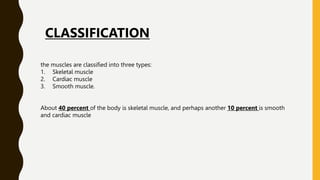
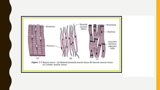
1. There are three types of muscle in the body: skeletal, cardiac, and smooth muscle. Skeletal muscle makes up about 40% of body mass.
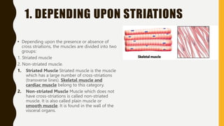
2. Muscles are classified based on their structure and control. Skeletal and cardiac muscles are striated due to cross-striations and voluntary, while smooth muscle is non-striated and involuntary.
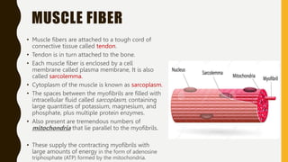
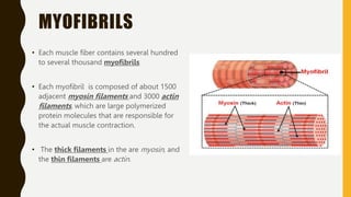
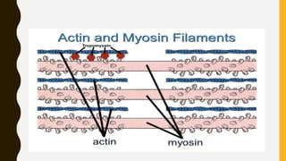
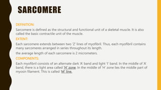
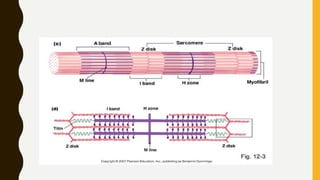
3. Each muscle fiber contains myofibrils which are made up of repeating sarcomere units consisting of actin and myosin filaments that overlap to enable contraction.
1. MUSCLES
P R E PA R E D B Y M I S S FAT I M A S U N D U S
2. the muscles are classified into three types:
1. Skeletal muscle
2. Cardiac muscle
3. Smooth muscle.
About 40 percent of the body is skeletal muscle, and perhaps another 10 percent is smooth
and cardiac muscle
CLASSIFICATION
3. 1. DEPENDING UPON STRIATIONS
• Depending upon the presence or absence of
cross striations, the muscles are divided into two
groups:
1. Striated muscle
2. Non-striated muscle.
1. Striated Muscle Striated muscle is the muscle
which has a large number of cross-striations
(transverse lines). Skeletal muscle and
cardiac muscle belong to this category.
2. Non-striated Muscle Muscle which does not
have cross-striations is called non-striated
muscle. It is also called plain muscle or
smooth muscle. It is found in the wall of the
visceral organs.
4.
5. 2. DEPENDING UPON CONTROL
• Depending upon control, the muscles are classified into two types:
1.Voluntary muscle
2. Involuntary muscle.
1) Voluntary Muscle
Voluntary muscle is the muscle that is controlled by the will. Skeletal muscles are the
voluntary muscles. These muscles are innervated by somatic nerves.
2) Involuntary Muscle
Muscle that cannot be controlled by the will is called involuntary muscle. Cardiac
muscle and smooth muscle are involuntary muscles. These muscles are innervated by
autonomic nerves.
6. STRUCTURE OF MUCSLE
• muscle tissue is made up of a large number of individual
muscle cells or myocytes.
• The muscle cells are commonly called muscle fibers
because these cells are long and slender in appearance.
• Skeletal muscle fibers are multinucleated and are arranged
parallel to one another with some connective tissue in
between.
• Muscle mass is separated from the neighboring tissues by a
thick fibrous tissue layer known as fascia.
• Beneath the fascia, muscle is covered by a connective tissue
sheath called epimysium.
• In the muscle, the muscle fibers are arranged in various
groups called bundles or fasciculi.
• Connective tissue sheath that covers each fasciculus is called
perimysium.
• Each muscle fiber is covered by a connective tissue layer
7. MUSCLE FIBER
• Muscle fibers are attached to a tough cord of
connective tissue called tendon.
• Tendon is in turn attached to the bone.
• Each muscle fiber is enclosed by a cell
membrane called plasma membrane, It is also
called sarcolemma.
• Cytoplasm of the muscle is known as sarcoplasm.
• The spaces between the myofibrils are filled with
intracellular fluid called sarcoplasm, containing
large quantities of potassium, magnesium, and
phosphate, plus multiple protein enzymes.
• Also present are tremendous numbers of
mitochondria that lie parallel to the myofibrils.
• These supply the contracting myofibrils with
large amounts of energy in the form of adenosine
triphosphate (ATP) formed by the mitochondria.
8. • in the sarcoplasm surrounding the myofibrils of each muscle fiber is an extensive reticulum, called
the sarcoplasmic reticulum.
• This reticulum has a special organization that is extremely important in regulating calcium, storage,
release and uptake and therefore muscle contraction
• Each muscle fiber has got one or more nuclei. In long muscle fibers, many nuclei are seen. Nuclei
are oval or elongated and situated just beneath the sarcolemma.
9.
10. MYOFIBRILS
• Each muscle fiber contains several hundred
to several thousand myofibrils
• Each myofibril is composed of about 1500
adjacent myosin filaments and 3000 actin
filaments, which are large polymerized
protein molecules that are responsible for
the actual muscle contraction.
• The thick filaments in the are myosin, and
the thin filaments are actin.
11.
12. each myofibril consists of a number of two alternating bands
These bands are formed by muscle proteins. The two bands are:
1. Light band or ‘I’ band.
2. Dark band or ‘A’ band
Light Band or ‘I’ Band
• Light band is called ‘I’ (isotropic) band because it is isotropic to polarized light.
• Light band contain only actin filaments.
Dark Band or ‘A’ Band
• Dark band is called ‘A’ (anisotropic) band because it is anisotropic to polarized light.
• The dark bands contain myosin filaments
These bands give skeletal and cardiac muscle their striated appearance.
13.
14. • Between I band a narrow and dark line called ‘Z’ line or ‘Z’ disk .
• The portion of myofibril in between two ‘Z’ lines is called sarcomere.
15. SARCOMERE
DEFINITION:
Sarcomere is defined as the structural and functional unit of a skeletal muscle. It is also
called the basic contractile unit of the muscle.
EXTENT:
Each sarcomere extends between two ‘Z’ lines of myofibril. Thus, each myofibril contains
many sarcomeres arranged in series throughout its length.
the average length of each sarcomere is 2 micrometers.
COMPONENTS:
Each myofibril consists of an alternate dark ‘A’ band and light ‘I’ band. In the middle of ‘A’
band, there is a light area called ‘H’ zone. In the middle of ‘H’ zone lies the middle part of
myosin filament. This is called ‘M’ line.
16.
17. Titin Filamentous Molecules Keep the Myosin and Actin Filaments in
Place.
• Each titin molecule has a molecular weight of about 3 million, which makes it one of the largest
protein molecules in the body.
• it is filamentous, it is very springy.
• One end of the titin molecule is elastic and is attached to the Z disk, acting as a spring and
changing length as the sarcomere contracts and relaxes.
• The other part of the titin molecule tethers it to the myosin thick filament.
19. MYOSIN FILAMENTS
• Myosin Filaments Are Composed of Multiple Myosin Molecules.
• Each of the myosin molecule has a molecular weight of about 480,000.
• The myosin molecule is composed of six polypeptide chains—two heavy chains, each
with a
molecular weight of about 200,000, and four light chains with molecular weights of
about 20,000 each.
• The two heavy chains wrap spirally around each other to form a double helix, which is
called the tail of the myosin molecule. One end of each of these chains is folded
bilaterally into a globular polypeptide structure called a myosin head.
20. • The myosin filament is made up of 200 or more individual myosin molecules.
• The protruding arms and heads together are called cross-bridges.
• Each cross-bridge is flexible at two points called hinges—one where the arm leaves the body of
the myosin filament, and the other where the head attaches to the arm.
• The total length of each myosin filament is uniform, almost exactly 1.6 micrometers.
21. the myosin head that is essential for muscle contraction is that it functions as an ATPase enzyme
22.
23. ACTIN FILAMENT
• Actin Filaments Are Composed of Actin, Tropomyosin, and Troponin
• Actin molecules are the major constituents of the thin actin filaments.
• Each actin molecule is called F-actin and it is the polymer of a small protein known as
G-actin.
• The molecular weight of each molecule is 42,000.
• The actin molecules in the actin filament are also arranged in the form of a double
helix.
• Each F-actin molecule has an active site to which the myosin head is attached.
• Each actin filament is about 1 micrometer long.
24.
25. TROPOMYOSIN
• tropomyosin molecules are situated along the double helix strand of actin filament.
• Each tropomyosin molecule has the molecular weight of 70,000 and a length of 40
nanometers.
• In relaxed condition of the muscle, the tropomyosin molecules cover all the active sites of F-
F-actin molecules.
TROPONIN
It is formed by three subunits:
1. Troponin I, which is attached to F-actin
2. Troponin T, which is attached to tropomyosin
3. Troponin C, which is attached to calcium ions.