Downloaded 866 times

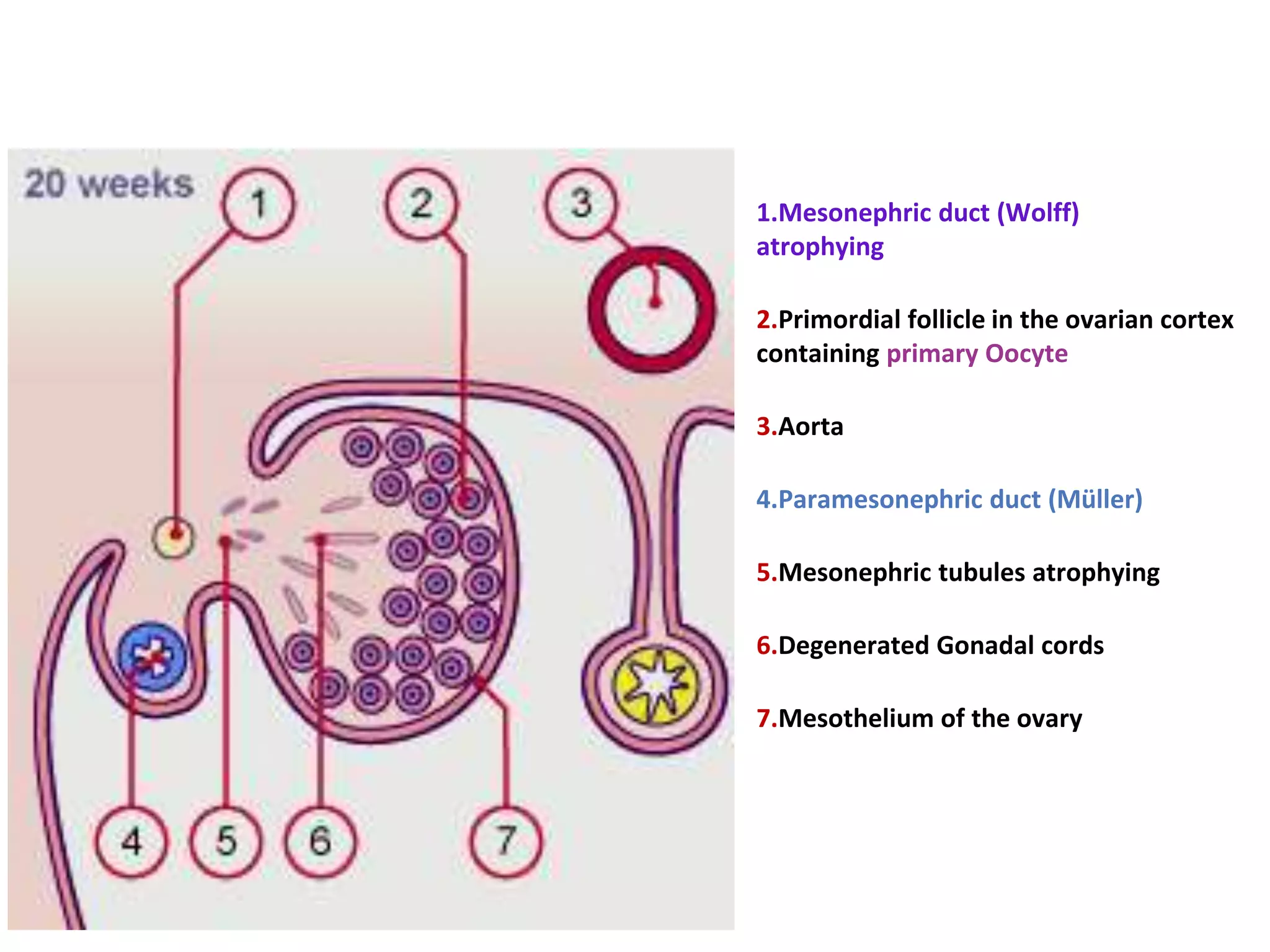
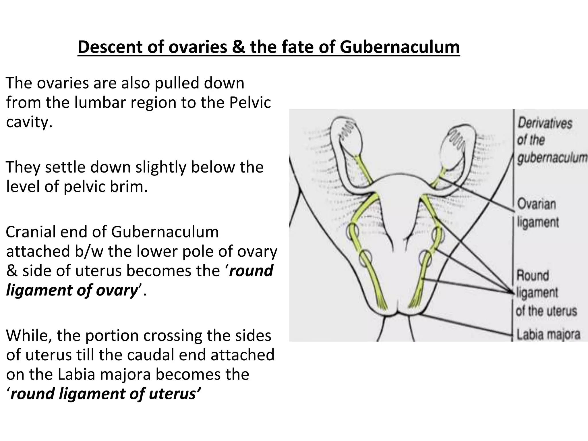
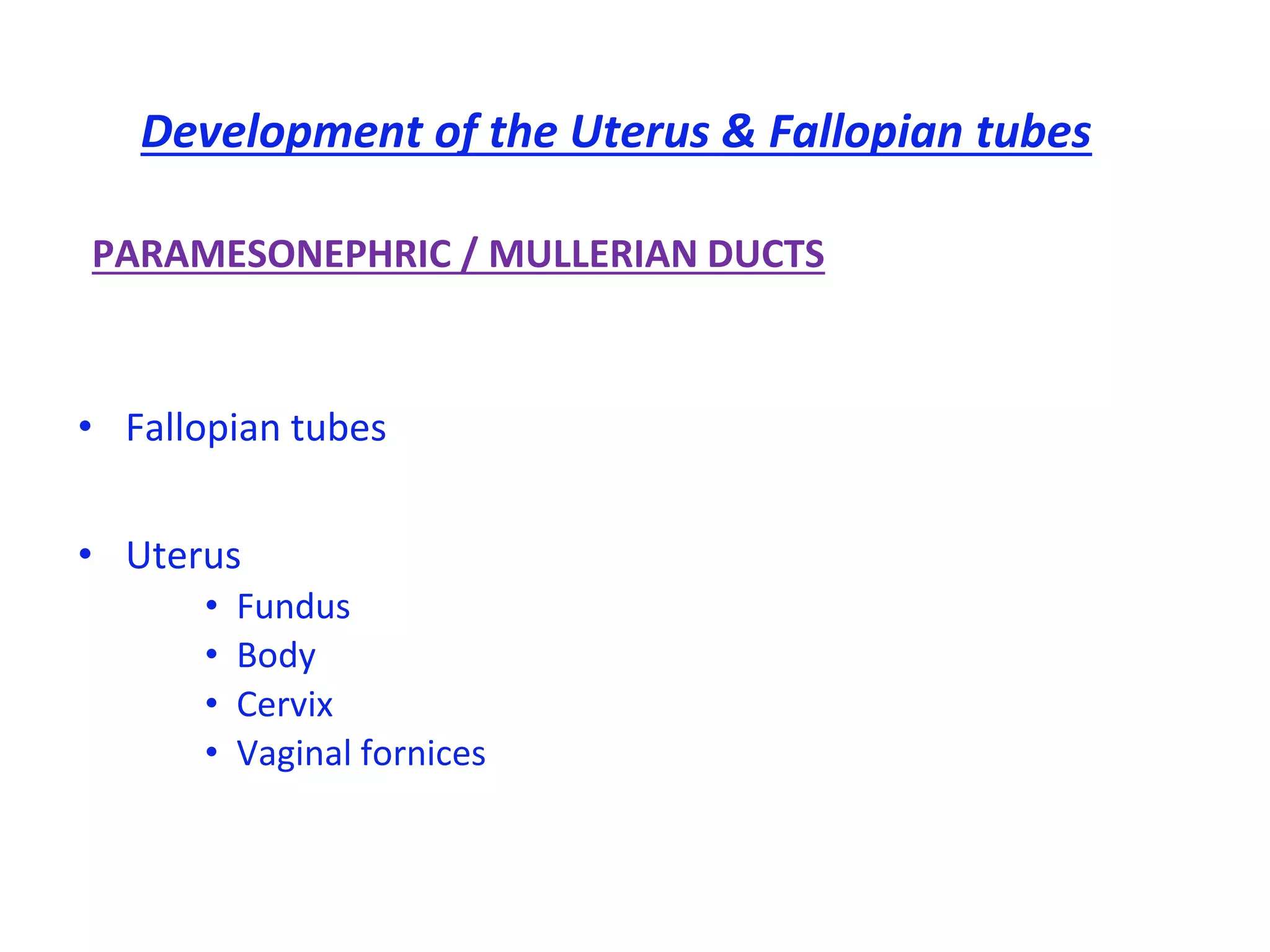
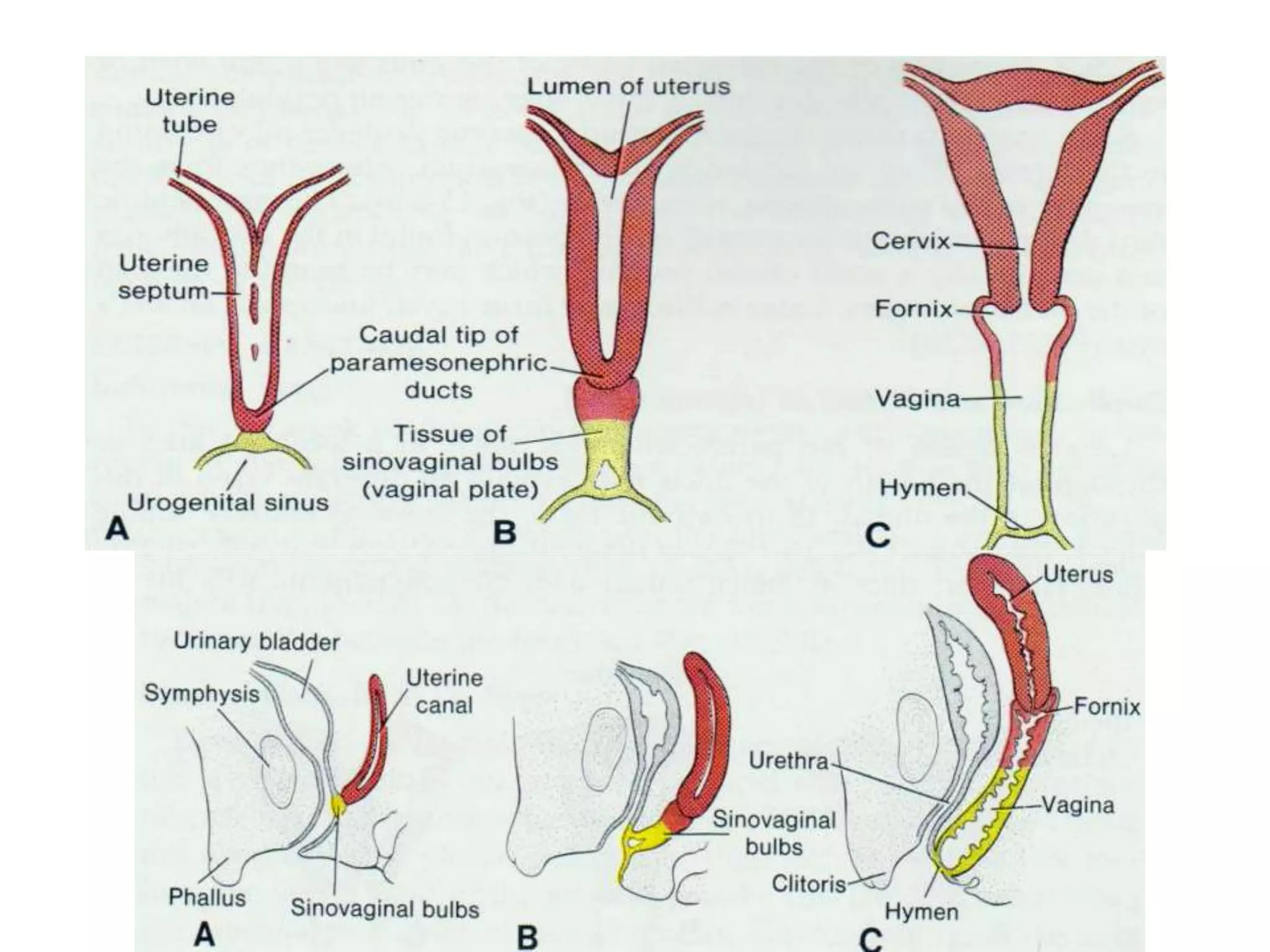
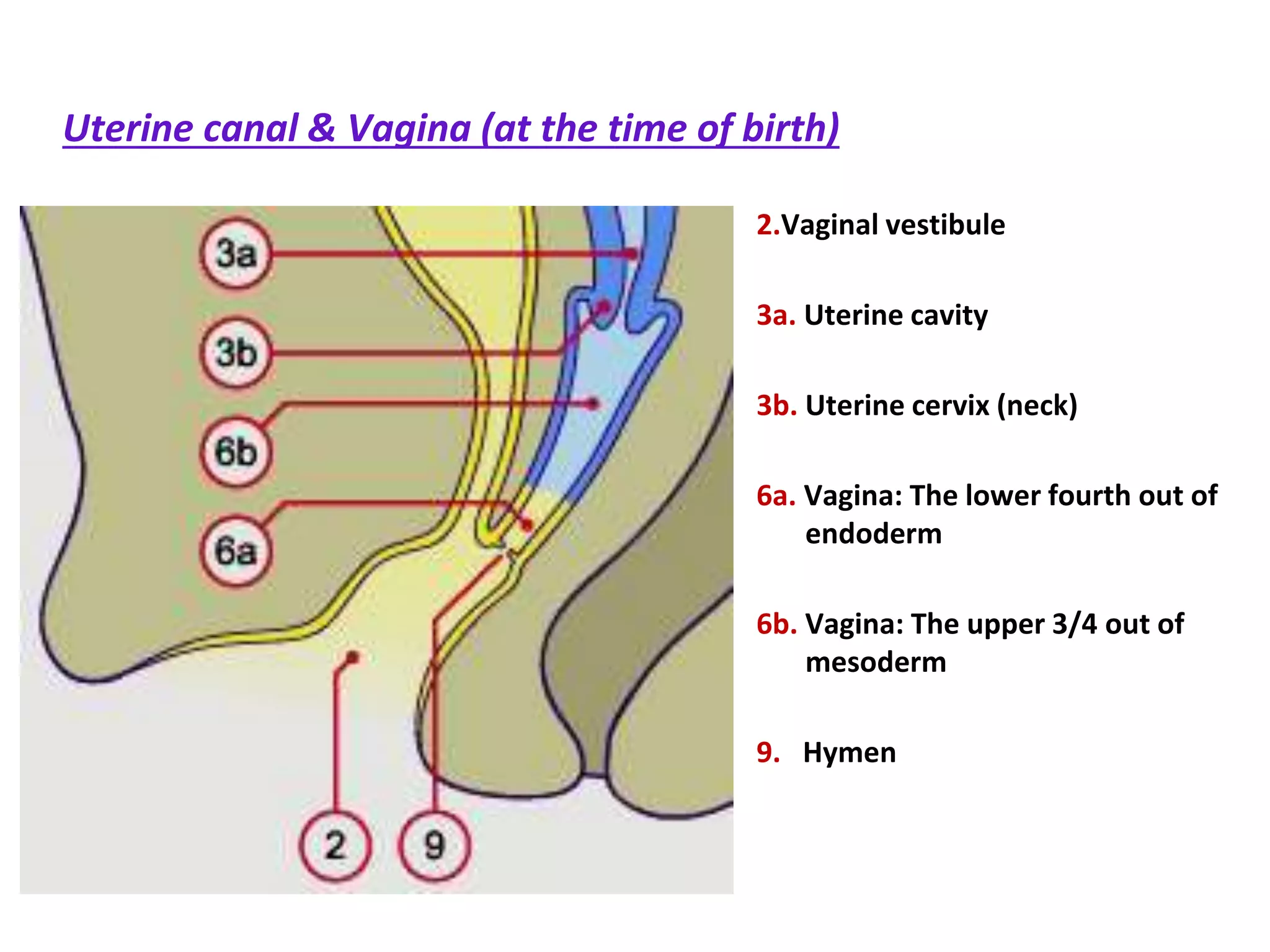
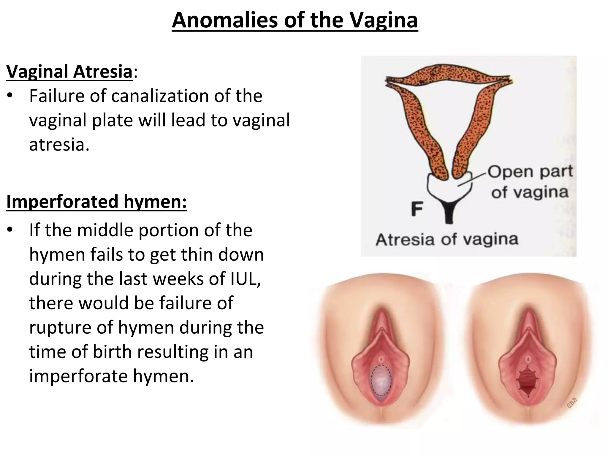
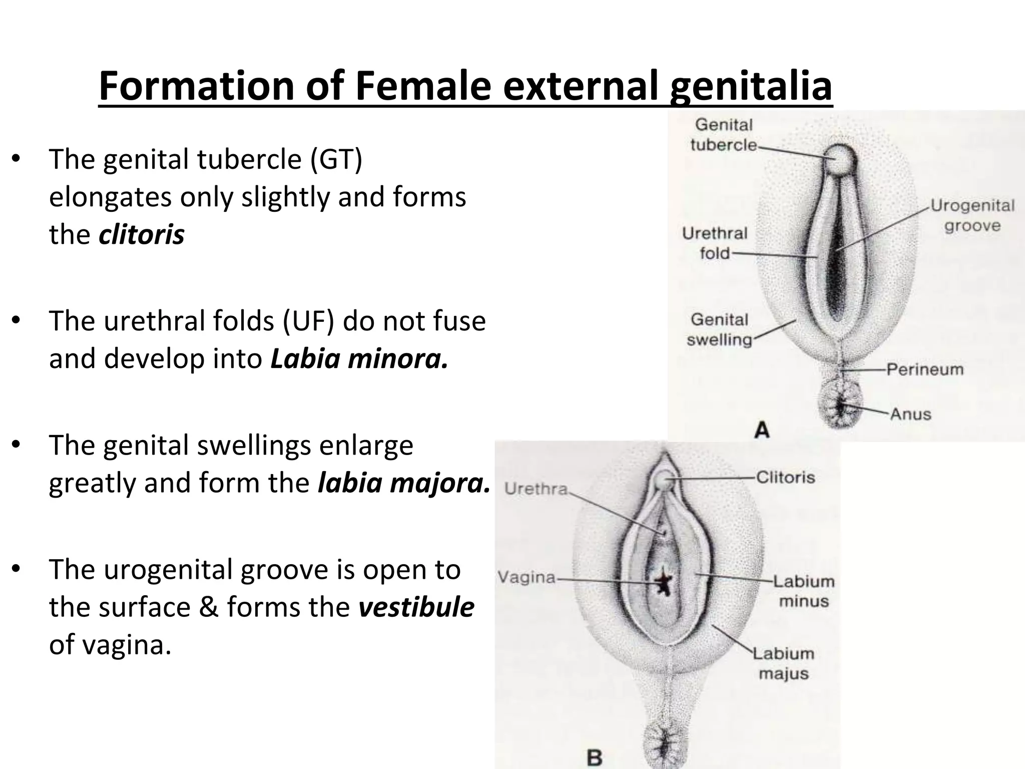
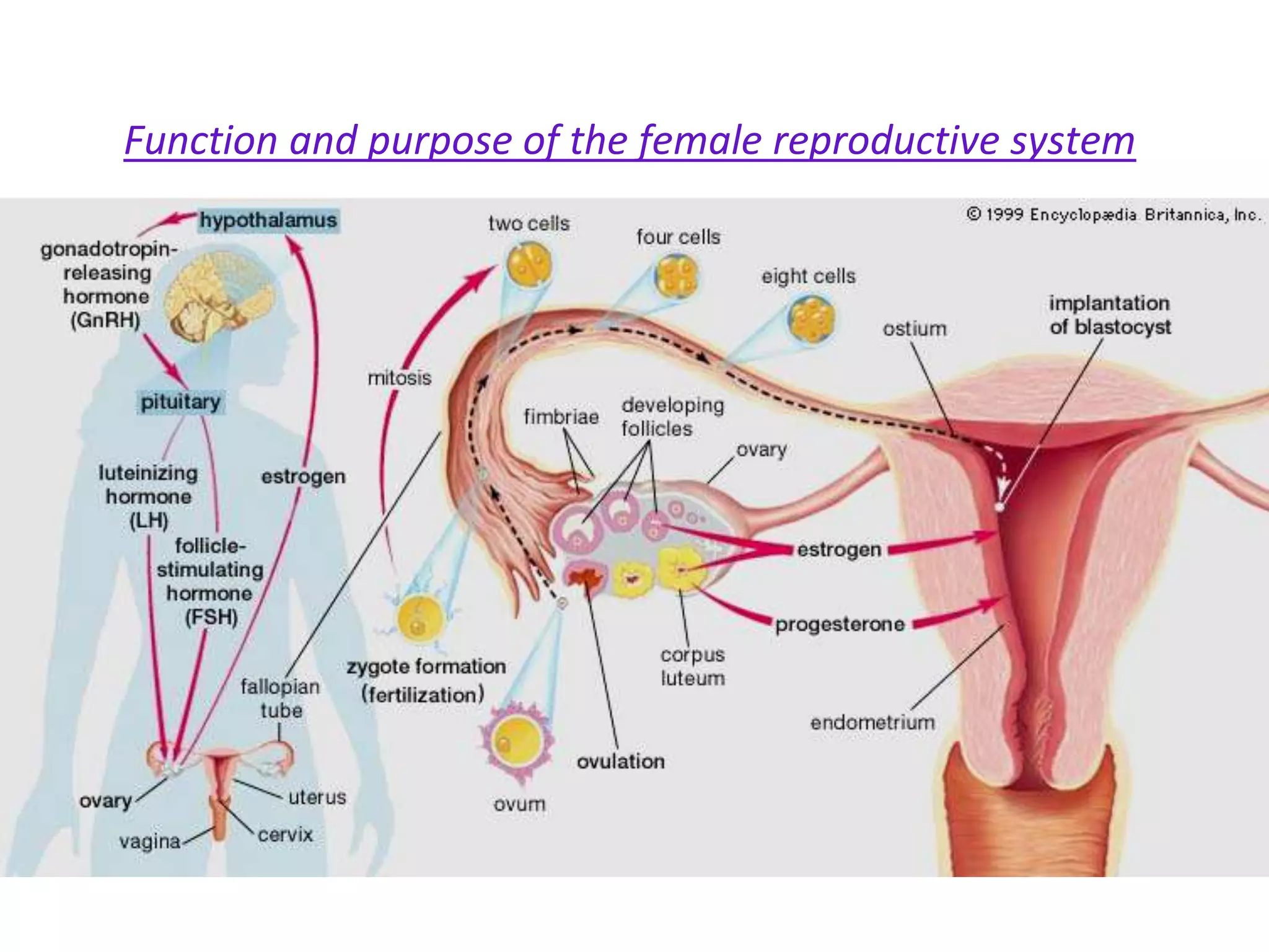
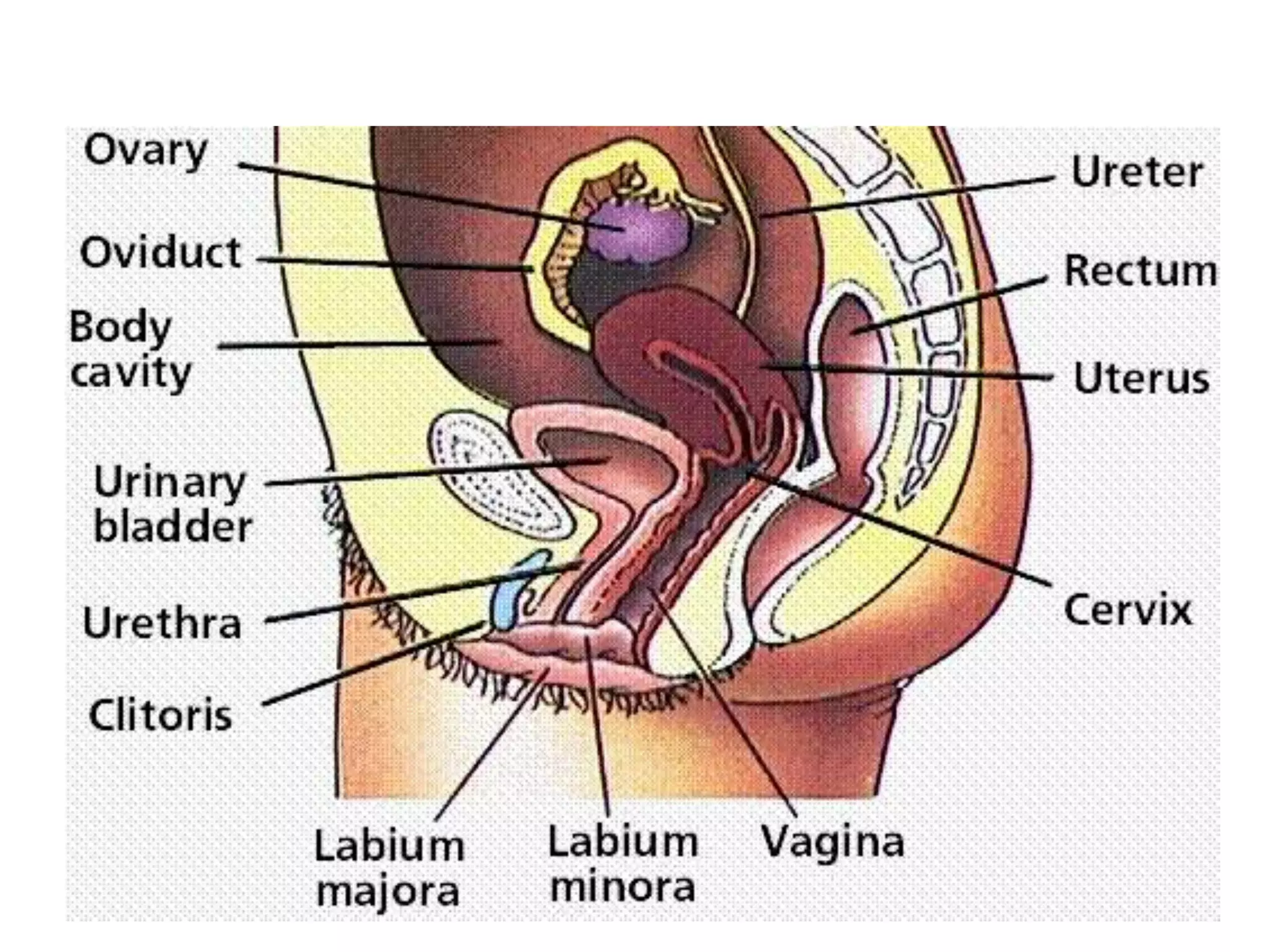

The document discusses the development of the female reproductive system, detailing the differentiation of gonads into ovaries, the descent of ovaries, and the formation of the uterus and fallopian tubes from paramesonephric ducts. It also covers the development and anomalies of the vagina and female external genitalia, including congenital malformations such as uterus didelphys and vaginal atresia. The lecture aims to provide students with an understanding of these processes and their implications in human anatomy and reproductive health.


