Downloaded 803 times
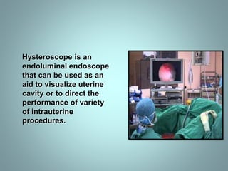
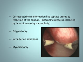
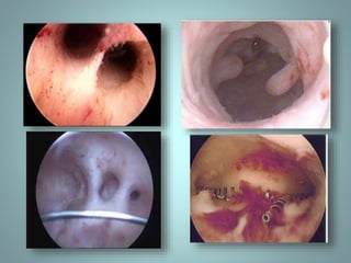


This document provides information about hysteroscopy, including: - A hysteroscope is an endoscope used to visualize the uterine cavity and perform procedures. - It describes the historical development of hysteroscopy from the 19th century to modern techniques. - The types of hysteroscopes and instrumentation used are outlined, including distention media, electrodes, sheaths, and cameras. - The document discusses the procedures, indications, contraindications and complications of diagnostic and operative hysteroscopy.


