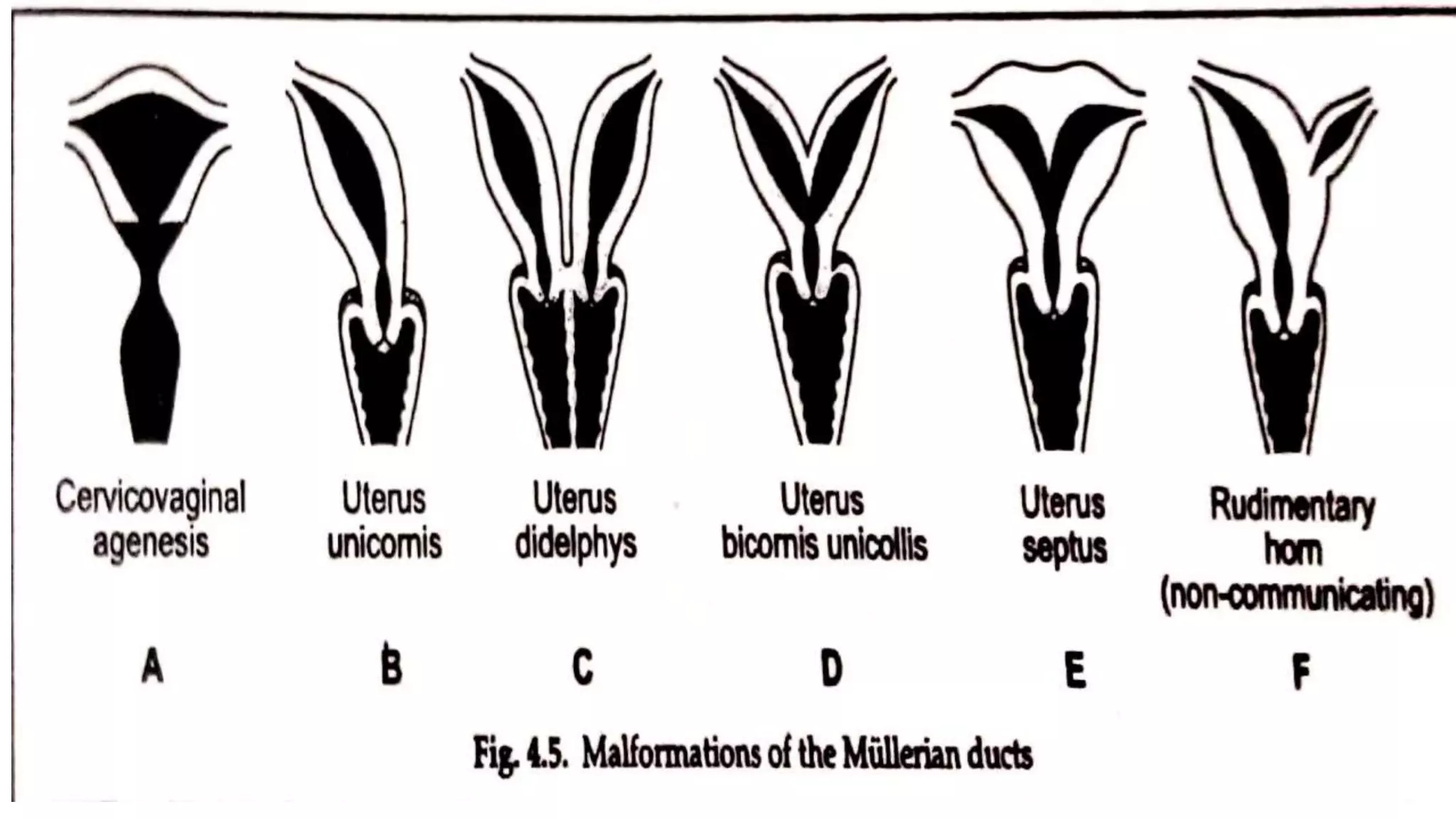
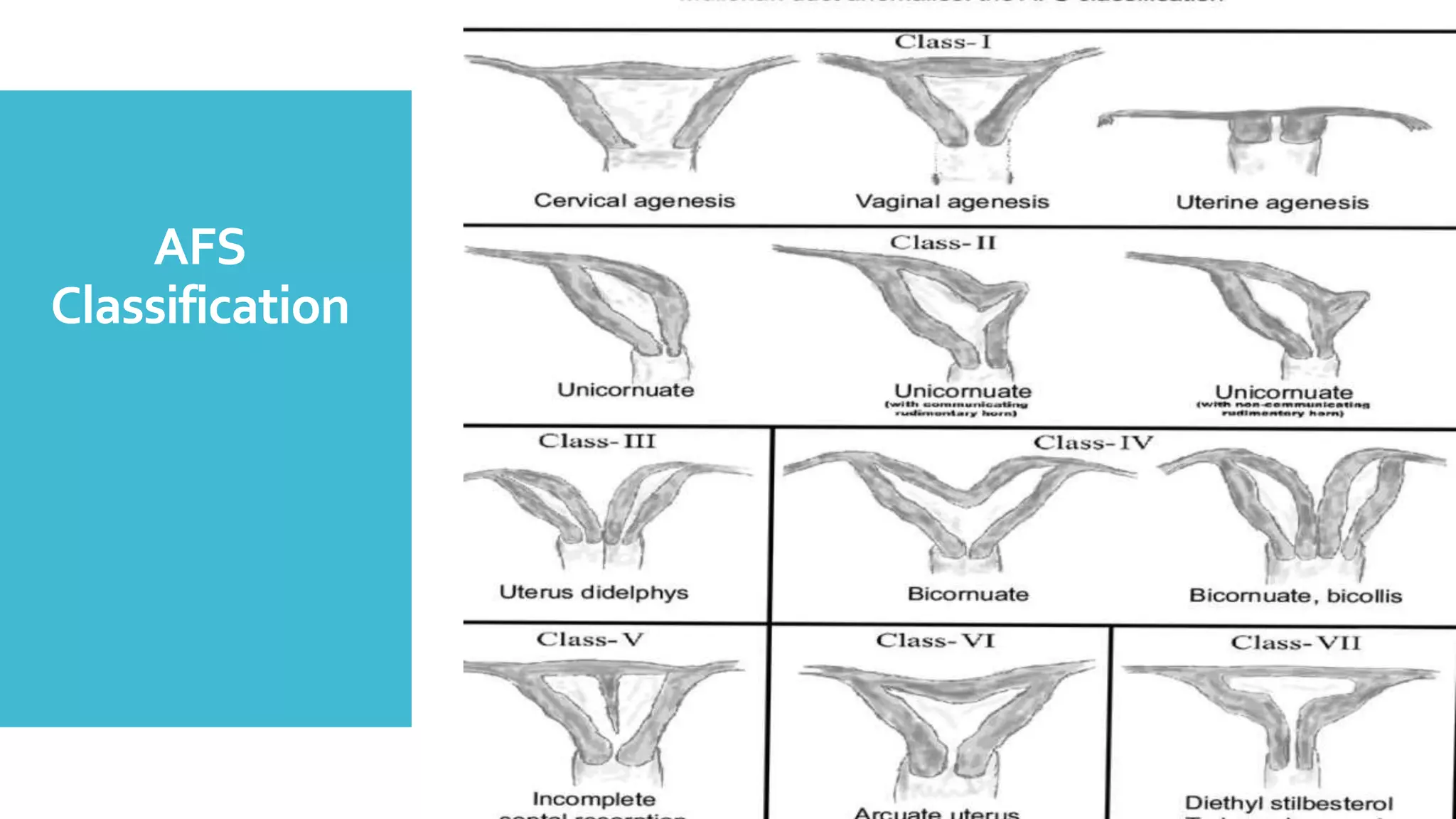
Uterine
Malformations
Susmita Haldar
Sister tutor
School of nursing Asia foundation
Introduction
For pregnancy and labour to
be achieved with minimal
difficulty a woman must
have normal reproductive
anatomy.
When abnormality of pelvic
organ exist it can lead to An
extra burden on mother as
well as the fetus.
Definition
An uterine malformation is
a result of abnormal
development of mullerian
duct during embryogenesis
Uterine malformations are
often associated with
vaginal maldevelopment
Incidence
Embryological
development
ofuterus
Related
anatomyand
physiologyof
uterus
The uterus is a hollow pyriform muscular
organ situated in the pelvis between the
bladder informed and the rectum in behind
The normal position is one of anteversion
and anteflexion the uterus usually inclines
to the right (dextrorotation) so that the
cervix is directed to the left
(levorotation)and comes in close to relation
with the left ureter.
The uterus measures about 8 cm long 5 cm
wide at the fundus and its walls are 1.25 CM
thick
It waits around 50 to 80 grams.
Related
anatomyand
physiologyof
uterus
Uterus has got following part
1.Bodyy or Corpus is father divided into,
 fundus the part which lies above the
openings of the uterine tube
The body is proper triangular and lies
between the opening of the tubes and the
isthmus.
2. Isthmus is constricted part between the
body and the cervix
3. Service is the lowermost part of uterus
Partsof
uterus
Supportsof
uterus
 Middle tier constitutes the strongest support of uterus
1. Paracervical ring is the coller of fibroelastic
connective tissue encircling the supravaginal cervix. It
is connected with the pubo cervical ligaments And
physical vaginal septum anteriorly cardinal ligaments
laterally and Ritu vaginal septum posteriorly. It’s
stabilizer service at the level of interspinous diameter
along the other ligaments.
2. Pelvic cellular tissues and the in endopelvic fascia
consists of connective tissue and smooth muscles .
the blood vessels nerves supplying the uterus bladder
and vagina pass through it from the lateral pelvic wall.
As they passed the pelvic cellular tissues condense
surrounding them and give them direct support to the
viscera.
Supportsof
uterus
Inferior tier give indirect support to the the Uterus.The
support is principally given by the pelvic floor muscles.
Supportsof
vagina
Supports of anterior vaginal wall
1. Positional support in the erect foster the vagina
makes an angle of 45 degree to the horizontal.
Normal vaginal axis is horizontal in the upper
two third and vertical in the lower third
2. the vagina is insured by strong condensation of
pelvic cellular tissue called endopelvic fascia.
Supports of posterior vaginal wall
1. Endopelvic fascia sheath covering the vagina
and the rectuM.
2. Attachment of the uterus after ligament to the
lateral wall of the vault
I Müllerian agenesis/ Hypoplasia- segmental
II Unicornuate Uterus
III Didelphys Uterus
IV Bicornuate Uterus
V Septate Uterus
VI Arcutate Uterus
VII Diethyl stilboestrol (DES) Related abnormality
AmericanFertility
Society(AFS)
Classificationof
Müllerian
Anomalies(1988)
AFS
Classification
Mullerian abnormalities
Incidence of
Müllerian
abnormalities
Varies between 3 to 4 %
The incidence is found to be high in
women suffering from recurrent
miscarriage or preterm deliveries.
Failure of
development
of one/both
Müllerian
ducts
The absence of both drugs leads to
absence of uterus including oviducts.
There is absence of vagina as well. Primary
amenorrhea is chief complaint.Absence of
one dog leads to unicornuate uterus with a
single oviduct.
Failure of
recanalization
of mullerian
ducts
Agencies of upper vagina or of the
cervix may lead to hematometra as
the uterus is functioning
Failure of
fusion of
mullerian
ducts
In majority in the presence of deformity
escape attention. In some detection is
made accidentally during investigation of
infertility or repeated pregnancy wastage.
In others the diagnosis is made during
dilation and evacuation operation manual
removal of placenta or during cesarean
section.
Fusion anomalies
Arcutate
(18%)
The cornual parts of the uterus means
separated.The uterine fundus looks concave
with heart shaped cavity outline
Uterine
didelphys 8%
There is complete lack of fusion of mullerian ducts
with the w2s double cervix and double vagina
Septate
Uterus 35%
The two Mullerian ducts fuse together but there is
persistence of September 2 into other partially or
completely
Uterus
Bicornis
26%
Uterus Bicornis Bicolis – there are
two uterine cavities with double
cervix with or without vaginal
septum
Uterus Bicornis unicolis – there are
two uterine cavity is with one
service the horns maybe equal or
one-horned maybe rudimentary
and have no communication with
developed horn
Uterus
Unicornis 10%
Failure of development of one Müllerian duct
DES related
abnormality
Due to DES exposure during intrauterine life
varieties of malformations are included
Vagina- adenosis, Adenocarcinoma
Cervix – Cockscomb Cervix, Cervical Collar
Uterus – Hypoplasia, t shaped cavity, uterine
synaechiae
Fallopian tube- cornual budding, abnormal
fimbriae
Clinical
features
Gynaecological
Infertility and dyspareunia often related in
association with vaginal septum
Dysmenorrhea in by convert uterus or due
to cryptomenorrhea pent-up menstrual
blood in rudimentary horn
Menstrual disorders like menorrhagia
cryptomenorrhea are seen menorrhagia is
due to increased surface area in bi cornate
uterus
Clinical
features
Obstetrical
 MidTrimester abortion which may be recurrent
 Rudimentary hornpregnancy mein orchid due to transfer
internal migration of sperm for ovum from the opposite side.
Cornual pregnancy in rapture around 16th week
 Cervical incompetence
 Increased incidence of Mal presentation like transverse lie in
arcuate or subseptate breech in bicornuate with or complete
septate uterus
 Preterm labour intrauterine growth retardation intrauterine
and death
 Prolonged labour due to incoordinate uterine action
 Obstructed labour due to obstruction by the non gravid horn of
bicornuate uterus or rudimentary horn
 Retained placenta and postpartum hemorrhage where
placenta is implanted over the uterine septum
Investigation
 Internal examination reveals vagina and 2 cervices
 Passage of sound can diagnose to separate cavities.
Infact significant number of cases clinical diagnosis
is made during uterine curettage manual removal of
placenta cesarean section.
 For exact diagnosis internal as well as external
architecture of uterus mass be visualised leave for
following investigations are carried out
 Hysteography/ hysteroscopy / LAPAROSCOPY
 Ultrasound with vaginal probe
 MRI
Management
Mere presence of
uterine anomalies are
not indication of surgical
correction usually it is
asymptomatic
Reproductive
Outcome
Better Obstratic outcomes in septate uterus
86% bicornuate uterus 50% unicornuate
Uterus 40% pregnancy outcome. No
treatment is generally effective. Uterine
didelphys has best possiblity of successful
pregnancy. Unification operation is
generally not needed.Other causes of
infirtility or recurrent fetal loss must be
excluded
Rudimentary horn should be excised to
reduce the risk of ectopic pregnancy
Surgical
management
ofpelVicorgan
prolapse
Unification operation is therefore
indicated in otherwise unexplained cases
with uterine malformation. Abdominal
metroplasty should be done either by
excising septum ( Strassman Jones and
Jones ) or by incising the septum.
Success rate of abdominal metroplasty is
in terms of Live birth is high 5-75%
Surgical
management
ofpelVicorgan
prolapse
Hysteroscopic metroplasty is more commonly
done
Resection of septum can be done either by
resectoscope or by lasers
Advantages
 High success rate 80- 90%
 Short hospital stay
 Reduced postoperative morbidity ( infections or
adhesion)
 Subsequent chances of vaginal delivery is high
compared to abdominal metroplasty where
cessation section is mandatory
ABNORMALITIES
of FallopianTubes
The tubes maybe unduly elongated,may
have accessory ostia or diverticula. Rarely
the tube may be absent on one side.These
conditions may lower fertility or favour
ectopic pregnancy
Anomalies of the
Ovaries
There maybe streak gonadsvor gonadal
dysgenesis which are usually associated with
errors off sex chromosomal pattern. No
treatment is of any help. Accessory ovary
(division of original ovary into two) maybe rarely
( 1 in 93000) present. Rarely supernumerary
ovaries maybe found ( 1 in 29000) in broad
ligament or elsewhere.This can explain a rare
event where menstruation continues even after
removal of two ovaries.
Wolffian Remnant
Abnormalities
The outer end ofWolffian duct may be cystic size of
pea, often penductulated ( Hydatid of Morgagani) and
attached near the outer end of the tube.The tubules of
the Gartner’s duct maybe cystic the outer one’s are
Kobelts tubules the middle set epoophoron and the
proximal set the parophoron. Small cyst may arise
from any of the tubules . A cystic swelling from the
Gartner’s duct may appear in the anterolateral wall of
vagina which maybe confused with cystocele
ParovarianCyst
It arises from the vestigial remanence of wolffian
tissue situated in the mesosalpinx between the
tube and the ovary.This can attend a big size.
The cyst is unilocular the wall is thin and
contains clear translucent fluid.The ovary
inferior with ovary is chest over the cyst.The
world consists of connective tissue line by single
layer of lower columnar epithelium.
Other
Abnormalities
1. Labia Minora
A. True- due to developmental defect
B. Inflammatory
2. Labia Majora
A. Hyperplasic or hypoplasic labia
B. Abnormal fusion in adrenogenital syndrome
3. Clitoris- Clitorial hypertrophy – often associated with various
intersex problems
4. Perineum- perineum differentes from the area of contact
between the urorectal septum ( mesoderm) and dorsal wall of
cloaca( endodrem) at 7th week.This site of contact between
the two is the perineal body. Malformations of the perineum
are rare. Imperforate anus anal stenosis or fistula are result of
abnormal development of urorectal septum.This is due to
posterior deviation of septum as it approaches cloacal
memberane. Anal fistula may open into posterior aspect of
vestibule of the vagina ( anovestibular Fistula)
Key
points
📝
 While minor abnormality at skips attention it is
moderate or severe from which will produce
gynaecology and obstetrics problems. For exact
diagnosis of malformation both the internal and
external architecture of uterus must be viewed.
Failure of fusion of mullerian duct leads to arcuate
bicornuate septate uterus
 While gynaecological symptoms are far and few but at
times they may produce infertility objective problems
like recurrent miscarriage for no pregnancy preterm
labour or even obstructed labour
 Nearly 15 to 20% of women with recurrent miscarriage
are associated with malformation of uterus
Thank you 🙂

Uterine malformations

  • 1.
  • 2.
    Introduction For pregnancy andlabour to be achieved with minimal difficulty a woman must have normal reproductive anatomy. When abnormality of pelvic organ exist it can lead to An extra burden on mother as well as the fetus.
  • 3.
    Definition An uterine malformationis a result of abnormal development of mullerian duct during embryogenesis Uterine malformations are often associated with vaginal maldevelopment
  • 4.
  • 5.
  • 7.
    Related anatomyand physiologyof uterus The uterus isa hollow pyriform muscular organ situated in the pelvis between the bladder informed and the rectum in behind The normal position is one of anteversion and anteflexion the uterus usually inclines to the right (dextrorotation) so that the cervix is directed to the left (levorotation)and comes in close to relation with the left ureter. The uterus measures about 8 cm long 5 cm wide at the fundus and its walls are 1.25 CM thick It waits around 50 to 80 grams.
  • 8.
    Related anatomyand physiologyof uterus Uterus has gotfollowing part 1.Bodyy or Corpus is father divided into,  fundus the part which lies above the openings of the uterine tube The body is proper triangular and lies between the opening of the tubes and the isthmus. 2. Isthmus is constricted part between the body and the cervix 3. Service is the lowermost part of uterus
  • 9.
  • 10.
    Supportsof uterus  Middle tierconstitutes the strongest support of uterus 1. Paracervical ring is the coller of fibroelastic connective tissue encircling the supravaginal cervix. It is connected with the pubo cervical ligaments And physical vaginal septum anteriorly cardinal ligaments laterally and Ritu vaginal septum posteriorly. It’s stabilizer service at the level of interspinous diameter along the other ligaments. 2. Pelvic cellular tissues and the in endopelvic fascia consists of connective tissue and smooth muscles . the blood vessels nerves supplying the uterus bladder and vagina pass through it from the lateral pelvic wall. As they passed the pelvic cellular tissues condense surrounding them and give them direct support to the viscera.
  • 11.
    Supportsof uterus Inferior tier giveindirect support to the the Uterus.The support is principally given by the pelvic floor muscles.
  • 12.
    Supportsof vagina Supports of anteriorvaginal wall 1. Positional support in the erect foster the vagina makes an angle of 45 degree to the horizontal. Normal vaginal axis is horizontal in the upper two third and vertical in the lower third 2. the vagina is insured by strong condensation of pelvic cellular tissue called endopelvic fascia. Supports of posterior vaginal wall 1. Endopelvic fascia sheath covering the vagina and the rectuM. 2. Attachment of the uterus after ligament to the lateral wall of the vault
  • 13.
    I Müllerian agenesis/Hypoplasia- segmental II Unicornuate Uterus III Didelphys Uterus IV Bicornuate Uterus V Septate Uterus VI Arcutate Uterus VII Diethyl stilboestrol (DES) Related abnormality AmericanFertility Society(AFS) Classificationof Müllerian Anomalies(1988)
  • 15.
  • 16.
  • 17.
    Incidence of Müllerian abnormalities Varies between3 to 4 % The incidence is found to be high in women suffering from recurrent miscarriage or preterm deliveries.
  • 18.
    Failure of development of one/both Müllerian ducts Theabsence of both drugs leads to absence of uterus including oviducts. There is absence of vagina as well. Primary amenorrhea is chief complaint.Absence of one dog leads to unicornuate uterus with a single oviduct.
  • 19.
    Failure of recanalization of mullerian ducts Agenciesof upper vagina or of the cervix may lead to hematometra as the uterus is functioning
  • 20.
    Failure of fusion of mullerian ducts Inmajority in the presence of deformity escape attention. In some detection is made accidentally during investigation of infertility or repeated pregnancy wastage. In others the diagnosis is made during dilation and evacuation operation manual removal of placenta or during cesarean section.
  • 21.
  • 23.
    Arcutate (18%) The cornual partsof the uterus means separated.The uterine fundus looks concave with heart shaped cavity outline
  • 24.
    Uterine didelphys 8% There iscomplete lack of fusion of mullerian ducts with the w2s double cervix and double vagina
  • 26.
    Septate Uterus 35% The twoMullerian ducts fuse together but there is persistence of September 2 into other partially or completely
  • 27.
    Uterus Bicornis 26% Uterus Bicornis Bicolis– there are two uterine cavities with double cervix with or without vaginal septum Uterus Bicornis unicolis – there are two uterine cavity is with one service the horns maybe equal or one-horned maybe rudimentary and have no communication with developed horn
  • 29.
    Uterus Unicornis 10% Failure ofdevelopment of one Müllerian duct
  • 31.
    DES related abnormality Due toDES exposure during intrauterine life varieties of malformations are included Vagina- adenosis, Adenocarcinoma Cervix – Cockscomb Cervix, Cervical Collar Uterus – Hypoplasia, t shaped cavity, uterine synaechiae Fallopian tube- cornual budding, abnormal fimbriae
  • 32.
    Clinical features Gynaecological Infertility and dyspareuniaoften related in association with vaginal septum Dysmenorrhea in by convert uterus or due to cryptomenorrhea pent-up menstrual blood in rudimentary horn Menstrual disorders like menorrhagia cryptomenorrhea are seen menorrhagia is due to increased surface area in bi cornate uterus
  • 33.
    Clinical features Obstetrical  MidTrimester abortionwhich may be recurrent  Rudimentary hornpregnancy mein orchid due to transfer internal migration of sperm for ovum from the opposite side. Cornual pregnancy in rapture around 16th week  Cervical incompetence  Increased incidence of Mal presentation like transverse lie in arcuate or subseptate breech in bicornuate with or complete septate uterus  Preterm labour intrauterine growth retardation intrauterine and death  Prolonged labour due to incoordinate uterine action  Obstructed labour due to obstruction by the non gravid horn of bicornuate uterus or rudimentary horn  Retained placenta and postpartum hemorrhage where placenta is implanted over the uterine septum
  • 34.
    Investigation  Internal examinationreveals vagina and 2 cervices  Passage of sound can diagnose to separate cavities. Infact significant number of cases clinical diagnosis is made during uterine curettage manual removal of placenta cesarean section.  For exact diagnosis internal as well as external architecture of uterus mass be visualised leave for following investigations are carried out  Hysteography/ hysteroscopy / LAPAROSCOPY  Ultrasound with vaginal probe  MRI
  • 35.
    Management Mere presence of uterineanomalies are not indication of surgical correction usually it is asymptomatic
  • 36.
    Reproductive Outcome Better Obstratic outcomesin septate uterus 86% bicornuate uterus 50% unicornuate Uterus 40% pregnancy outcome. No treatment is generally effective. Uterine didelphys has best possiblity of successful pregnancy. Unification operation is generally not needed.Other causes of infirtility or recurrent fetal loss must be excluded Rudimentary horn should be excised to reduce the risk of ectopic pregnancy
  • 38.
    Surgical management ofpelVicorgan prolapse Unification operation istherefore indicated in otherwise unexplained cases with uterine malformation. Abdominal metroplasty should be done either by excising septum ( Strassman Jones and Jones ) or by incising the septum. Success rate of abdominal metroplasty is in terms of Live birth is high 5-75%
  • 39.
    Surgical management ofpelVicorgan prolapse Hysteroscopic metroplasty ismore commonly done Resection of septum can be done either by resectoscope or by lasers Advantages  High success rate 80- 90%  Short hospital stay  Reduced postoperative morbidity ( infections or adhesion)  Subsequent chances of vaginal delivery is high compared to abdominal metroplasty where cessation section is mandatory
  • 40.
    ABNORMALITIES of FallopianTubes The tubesmaybe unduly elongated,may have accessory ostia or diverticula. Rarely the tube may be absent on one side.These conditions may lower fertility or favour ectopic pregnancy
  • 41.
    Anomalies of the Ovaries Theremaybe streak gonadsvor gonadal dysgenesis which are usually associated with errors off sex chromosomal pattern. No treatment is of any help. Accessory ovary (division of original ovary into two) maybe rarely ( 1 in 93000) present. Rarely supernumerary ovaries maybe found ( 1 in 29000) in broad ligament or elsewhere.This can explain a rare event where menstruation continues even after removal of two ovaries.
  • 42.
    Wolffian Remnant Abnormalities The outerend ofWolffian duct may be cystic size of pea, often penductulated ( Hydatid of Morgagani) and attached near the outer end of the tube.The tubules of the Gartner’s duct maybe cystic the outer one’s are Kobelts tubules the middle set epoophoron and the proximal set the parophoron. Small cyst may arise from any of the tubules . A cystic swelling from the Gartner’s duct may appear in the anterolateral wall of vagina which maybe confused with cystocele
  • 43.
    ParovarianCyst It arises fromthe vestigial remanence of wolffian tissue situated in the mesosalpinx between the tube and the ovary.This can attend a big size. The cyst is unilocular the wall is thin and contains clear translucent fluid.The ovary inferior with ovary is chest over the cyst.The world consists of connective tissue line by single layer of lower columnar epithelium.
  • 44.
    Other Abnormalities 1. Labia Minora A.True- due to developmental defect B. Inflammatory 2. Labia Majora A. Hyperplasic or hypoplasic labia B. Abnormal fusion in adrenogenital syndrome 3. Clitoris- Clitorial hypertrophy – often associated with various intersex problems 4. Perineum- perineum differentes from the area of contact between the urorectal septum ( mesoderm) and dorsal wall of cloaca( endodrem) at 7th week.This site of contact between the two is the perineal body. Malformations of the perineum are rare. Imperforate anus anal stenosis or fistula are result of abnormal development of urorectal septum.This is due to posterior deviation of septum as it approaches cloacal memberane. Anal fistula may open into posterior aspect of vestibule of the vagina ( anovestibular Fistula)
  • 45.
    Key points 📝  While minorabnormality at skips attention it is moderate or severe from which will produce gynaecology and obstetrics problems. For exact diagnosis of malformation both the internal and external architecture of uterus must be viewed. Failure of fusion of mullerian duct leads to arcuate bicornuate septate uterus  While gynaecological symptoms are far and few but at times they may produce infertility objective problems like recurrent miscarriage for no pregnancy preterm labour or even obstructed labour  Nearly 15 to 20% of women with recurrent miscarriage are associated with malformation of uterus
  • 46.