
Recommended
More Related Content
Similar to Congenital anomalies of female genital tract.pptx
Similar to Congenital anomalies of female genital tract.pptx (20)
Recently uploaded
Recently uploaded (20)
Congenital anomalies of female genital tract.pptx
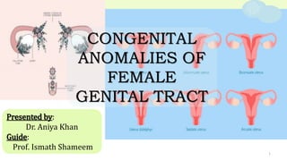
- 1. 1 Presented by: Dr. Aniya Khan Guide: Prof. Ismath Shameem CONGENITAL ANOMALIES OF FEMALE GENITAL TRACT
- 2. 2 CONTENTS 1. embyology overview 2. MULLERIAN DUCT Anomalies I. AGENESIS II. Errors in vertical fusion III. errors in lateral fusion 3. Anomalies of UROGENITAL SINUS
- 3. 4. anomalies of EXTERNAL GENITALIA 3 CONTENTS 5. ANOMALIES OF OVARies 8. Unani Concept 7. Case scenario
- 4. 4 EMBRYOLOGY Cellular differentiation Migration Fusion Canalization
- 5. 5 Testicular determining factor Testicular determining factor AMH Testes Undifferentiate d gonad Undifferentiate d gonad Ovary
- 6. 6 DHT DHT
- 8. 8 6 WK 7 WK 8-9 WK 9 WK
- 11. 11 Failure of initial descent (defect in formation) Agenesis Failure of vertical fusion Failure of lateral fusion or resorption transverse vaginal septum IMPERFORATE HYMEN Duplication defects
- 12. 12 Agenesis Mayer–Rokitansky–Küster–Hauser syndrome Unicornuate uterus Segmental hypoplasia/agenesis of mullerian duct
- 13. 13 Mayer–Rokitansky–Küster–Hauser syndrome (MRKH- Mullerian agenesis) Prevalence - 1 in 5000 live female births 2nd m/c/c of pathological 1 ֯amenorrhea Karyotype - 46XX females Presentation • 1 ֯amenorrhea • Normal sec. sex characters Examination • Normal breast development and pubic hair • Normal height • No vaginal opening or blind vaginal pouch with no cx at apex • Normal external genitalia • Uterus not palpated on palpation
- 14. 14 Diagnosis • History • Clinical examination • Investigations - USG pelvis: no uterus or vaginal canal or fallopian tubes with normal ovaries MRI (gold standard): Determines the presence of rudimentary uterine buds or complete uterovaginal agenesis Hormonal assay: Normal Karyotyping: 46XX Renal ultrasound, spine radiograph, auditory testing Class: Type I - isolated uterovaginal aplasia Type II - associated with extragenital manifestations: renal, skeletal, ear, or cardiac malformations.
- 15. 15 DiFFERENTIAL DIAGNOSIS • Congenital adrogen insensitivity syndrome(Morris syndrome) • Imperforate hymen • Transverse vaginal septum Management • Counselling and eductation • Correction of vaginal agenesis in MRKH syndrome with creation of a functional neovagina - non surgical or surgical methods • Treatment of infertility
- 16. 16 Non surgical methods 1st line treatment Vaginal dilators Dilatation by intercoarse Daily 10-30 mins once to thrice a day for 6-12 months Daily More advantageous Low cost Less complication
- 17. 17 Surgical methods Autografts Split skin graft Bowel graft Peritoneal graft Labia majora flaps Cultured grafts Autologous vulval tissue Tissue engineered biomaterial A space is created digitally between bladder and rectum. Graft is used over mold and kept in situ
- 18. Treatment for infertility Uterus transplantation 18 IVF f/b gestational surrogacy Legal adoption
- 19. 19 Unicornuate uterus Prevalence - 5–20% of congenital uterine anomalies (4.3% of population) Classification Unicornuate with rudimentary horn (TYPE-A) Unicornuate without rudimentary horn (TYPE-B) + Functioning endometrium (TYPE-A1) + No cavity/functioning endometrium (TYPE-A2) Communicating (TYPE-A1a) Non Communicating (TYPE-A1b)
- 20. 20 Presentation • Asymptomatic • Infertility • Dysmenorrhea • Endometriosis(non comunicating) Examination • Uterus is markedly deviated • Other findings are normal Diagnosis • HSG - Deviated banana shaped cavity with single fallopian tube • USG - Rudimentary horn is best confirmed • Cornual pregnancy • Obstructed labor
- 21. 21
- 22. 22 Rudimentary uterine horn. (a) Abdominal ultrasound showing longitudinal uterine image. (b) Abdominal ultrasound showing a cross-sectional image of the uterus and rudimentary uterine horn with a hypoechoic ribbon connecting the horn with the main body of the uterus. (c) Transvaginal ultrasound showing rudimentary horn of the uterus and its endometrial echo
- 23. 23 TYPE A1b
- 24. 24
- 25. 25
- 26. 26 FUSION DEFECTS LATERAL FUSION DEFECTS VERTICAL FUSION DEFECTS COMPLETE PARTIAL CANALIZATION/ RESORPTION DEFECTS TRANSVERSE VAGINAL SEPTUM IMPERFORATE HYMEN ACCESSORY FT DIDELPHYS UT DOUBLE VAGINA BICORNUATE UT ARCUATE UT SEPTATE UT SEPTATE VAGINA
- 27. 27 ACCESSORY FALLOPIAN TUBE/ ACCESSORY OSTIA Incidence of 1.9% Result from the bifurcation of the proximal ends of the müllerian ducts Presentation:Dysmenorrhea or primary infertility Complication: Ectopic pregnancy, pyosalpinx, infertility A/W Endometriosis Management: monofilament purse-string suture at the base of the accessory tube/ostium followed by resection or electrocoagulation of stump. Laproscopic diagnosis
- 28. 28 Uterus Didelphys Prevalence - 8% Associated with VS in most cases Occasionally hemivagina is obstructed by LVS/oblique septum- hematocolpos, hydrocolpos and endometriosis Examination • Lump in lower abdomen or u/l vaginal mass i/c/o obstructed one uterine horn • On P/V- vaginal septum & 2 cervices • Passage of sound - 2 uterine cavities Obstructive- u/l vaginal / pelvic pain
- 29. 29 Diagnosis • USG-Separate divergent uterine horns are identified with a large fundal cleft. Endometrial cavities are uniformly separate. • HSG - to identify possiblity of communication between uteri. If associated with an obstructed longitudinal vaginal septum mimicking a unicornuate uterus.
- 30. 30 Abdominal ultrasound showing double uterine abnormalities. It shows both the left and right uteri, with uterine body completely separated and two separate endometrial echoes
- 31. 31 DiFFERENTIAL DIAGNOSIS • Uterus Bicornis Bicollis (separation of horns only) • Septate uterus (midline uterine septum) Uterus didelphys Uterus bicornis bicollis The uterine horns are separate and divergent with a large fundal cleft
- 32. 32 Uterus Bicornis Prevalence - 26% Classification Uterus bicornis bicollis Uterus bicornis unicollis Diagnosis • HSG - an angle of more than 105° between uterine horns • USG - confirm anatomy by showing a deep (>1 cm) fundal cleft in the outer uterine contour and an intercornual distance of >4 cm. Examination • On P/V- 2 cervices • Uterine sound - 2 cavities
- 33. 33 Bicornuate uterus: an abdominal scan showing an abnormal uterine cross-sectional image, displaying a widened fundus with a saddle-shaped depression at the middle
- 34. 34
- 35. 35
- 36. 36 24 YO with C/O Anxious to conceive since 4 yrs M/H : Reg cycles ; 4-5/ 28-30 days 12/04/23
- 37. 37 Septate uterus Prevalence - 35% • HSG - An angle of less than 75° between the uterine horns is suggestive of a septate uterus. • MRI - uterus normal in size and each endometrial cavity appears smaller than the configuration of a normal cavity. The septum may be composed of fibrous tissue (low T2 signal intensity), myometrial tissue (intermediate signal) or both • USG - The echogenic endometrial stripe is separated at the fundus by the intermediate echogenicity septum (which is isoechoic to myometrium).The external uterine contour convex, flat, or mildly concave (ideally <1 cm) configuration. Intercornual dist <4cm • Color Doppler: may show vascularity in the septum (70% of cases) which, if present, may be associated with a higher rate of obstetric complications Diagnosis m/c Mullerian anomaly
- 38. 38 Septate uterus: (a) an abdominal ultrasound showing a cross-sectional image of two endometrial echoes (marked by arrows) separated by a septum at the middle of the uterus. (b) A central septum (marked by an arrow) in the middle of the uterus
- 39. 39 Incomplete septate uterus: transvaginal 3D ultrasound showing endometrial echo as a “Y”-shaped image. The uterine cavity is separated in its upper half and shows a midline echo from its middle downward Complete septate uterus: transvaginal 3D ultrasound showing endometrial echo as a “V”-shaped image; the uterine cavity with a hypoechoic middle septum.
- 40. Arcuate uterus 40 USG - In an arcuate uterus, fundus of the uterus is indented with depth of indentation less than 1 cm giving a heart shaped appearance. Prevalence - 18% Diagnosis Mild varient- little or no impact on reproductive outcomes
- 41. 41 Arcuate uterus: transvaginal 3D ultrasound image showing muscular thickening at the central part of the uterine fundus, slightly protruding into the uterine cavity
- 42. 42 Mullerian uterine defects Presentation • Midtrimester miscarriages (esp unicornuate and septate) • Cervical incompetence • Malpresentations • IUGR, IUD, Preterm labor • Obstructed labor (uniconuate with horn and bicornuate) • Prolonged labor • PPH & Retained placenta • Infertility • Dyspareunia • Menstrual disorder- menorrhagia(more surface area), cryptomenorrhea Associated anomalies • Urinary tract abnormalities (40%)- renal agenesis • Skeletal abnormalities (12%)
- 43. 43 • Better neonatal outcome, pregnancy success rates are seen with ARCUATE UTERUS. • More cases of placental abruptions are reported with arcuate and septate, but not with didelphys • Septate is a/w reccurent abortions. • Didelphic uterus has the better reproductive prognosis compared to unicornuate. Uterine didelphys probably has an improved blood supply through collateral connections between the two horns. Alternatively, improved fetal survival may be secondary to earlier diagnosis of the uterine didelphys (because of the vaginal septum) Prognosis Diagnosis • USG 2D & 3D • HSG • Laproscopy • Hysteroscopy • MRI • Combined hystero-laproscopy to differentiate septate from bicornuate and arcuate • Diagnosis of associated defects by radiography and usg Mostly diagnosed accidently during D&C, C-Section and manual removal of placenta
- 44. 44 • USG- used as the first line for evaluating the uterus. The sensitivity 88% to 93%, and the specificity 94% to 99%. • Sonohysterography- By infusing saline solution into the uterine cavity as a contrast medium, excellent appraisal of the uterine cavity can be obtained, most useful in delineating uterine anomalies with sensitivity of 97% and specificity of 11%.
- 45. 45 • HSG- Diagnostic accuracy of HSG in differentiating septate from bicornuate uterus is as low as 55%. • Laproscopy &Hysteroscopy: evaluation of the adnexae and pelvic cavity for possible pelvic adhesions, tubal or ovarian distortions, and endometriosis that could be treated during the same session.Combined hystero-laproscopy to differentiate septate from bicornuate • Diagnosis of associated defects by radiography and usg
- 46. 46 • MRI- not frequently used for evaluation of uterine anomalies because of its cost. Its accuracy and lack of ionizing radiation make this method invaluable in particular in difficult and complex situations and when HSG or sonography fail to provide the proper and precise diagnosis.
- 47. 47 Management • Asymptomatic cases- no treatment • Rudimentary horn should be excised to reduce the risk of ectopic pregnancy • Unification operation (bicornuate/septate uterus) - Abdominal or hysterosopic metroplasty. • Hysteroscopic metroplasty is more commonly done as it avoids a uterine scar and need for elective caesarean section. • Resection of the septum can be done either by a resectoscope or by laser • Postoperative management: Oral oestrogen for 3 months Insertion of a Foley catheter with its bulb distended with 4–8 mL of sterile water for 5–7 days to keep the uterine cavity open and prevent intrauterine adhesions. Antibiotics (doxycycline 100 mg b.i.d. for 5–7 days) and NSAID for pain Asherman syndrome with uterine adhesions and adherent placenta are the late complications
- 48. 48 Hysteroscopic Transcervical Resection of Uterine Septum Anesthesia Cervical dilatation using cervical dilators 5% dextrose instilled Uterine distention @ 150mmhg Electro-Resectoscope (location, size, range of septum) Incision by needle electrode under USG
- 49. 49 Strassman technique repair of a Bicornuate uterus Epinephrine solution (30 mL) infiltrated subserosally along medial aspect of horns Incision through myometrium from supero-medial aspect of each horn, about 2 cm medial and caudal to origin of FT till the base of horn Suturing of opposing myometrial edges (Interrupted- vicryl 0); excluding endometrium
- 50. 50 Wedge resection for septate
- 51. 51
- 52. 52 Longitudinal vaginal septum Generally seen with partial or complete duplication of the cervix and uterus Presentation: • Asymptomatic • Dyspareunia • Vaginal bleeding despite tampon placement • In case of obstructive lesion: Unilateral vaginal / pelvic pain • Obstructed labor Rarely, septum may deviate from the centre and fuse with one lateral vaginal wall
- 53. 53 O/E: • P/v Septum can be felt • Obstructed vagina and cervix with unilateral vaginal mass or pelvic mass in case of obstructed variety Diagnosis: • History • Examination • USG, MRI, HSG - esp to look for associated bicornuate of didelphic uterus
- 54. 54 Management - LVS • Vaginal septum may get torn with sexual activity thus creating one vagina • Treatment in symptomatic cases involves complete removal of the septum • The tissue should be completely excised, since remaining fragments of the septum may cause dyspareunia. • The septal tissue is resected and the normal vaginal mucosa on each vaginal wall is sutured (continuous suture) together along the length of the defect made by the resection.
- 55. 55
- 56. 56
- 57. 57
- 58. 58
- 59. 59 Transverse vaginal septum Failure of the vaginal plate to regress completely Some have small perforations that allow prolonged menstrual blood flow In case of no opening - Accumulation of blood/mucus and distension of upper reproductive tract Incidence of 1 in 70,000 females More common in upper vagina Typically septum in thin, but seldom thickness- 5-6cm O/E: • Palpable mass at suprapubic region • Foreshortened vagina • Inability to identify the cervix. • P/R: Bulged vagina
- 60. 60 Imperforate hymen Imperforate hymen results from failure of the hymen to canalize during the perinatal period Diagnosed after appearance of symptoms usually during adolescence In infants distension from mucus accumulation(hydromucocolpos) O/E: • Bluish bulge at the introitus • Palpable mass at suprapubic region • P/R: bulged vagina Incidence of 1 in 2,000 females
- 61. 61 ANOMALIES OF urogenital sinus Vaginal Atresia Vaginal Agenesis
- 62. 62 Vaginal Agenesis / Atresia Agenesis: Failure of the urogenital sinus to contribute its expected caudal portion of the vagina Atresia: Involves lower portion of the vagina- one fifth to one third of the total length, is replaced by 2 to 3 cm of fibrous tissue O/E: • Normal external genitalia and upper reproductive tract organs • Normal breast and pubic hair • Vaginal dimple or small pouch (1.5 inches) is seen beyond the hymeneal ring • Rectoabdominal examination confirms the presence of midline structures Prevalence : 1 in 5000
- 63. 63 Presentation- TVS / Imperforate hymen/ V. Atresia Primary amenorrhea Cyclic abdominal pain Supra-pubic bulge urinary symptoms- frequency, dysuria, retention Dysfunction of defecation No opening Opening + (TVS/Microperforate hymen) Abnormal menstrual flow Painful intercoarse Difficulty in placing or removing tampon Obstructed labor
- 64. 64 Complications- TVS / Imperforate hymen / V. Atresia Hematometra/salpinx Lymphatic obstruction Endometriosis Acute urinary retention Hydro ureteronephrosis Bacterial nephritis Kidney failure
- 65. 65 Diagnosis- TVS / Imperforate hymen / V. Atresia History Examination USG MRI • TVS - Done prior to surgery to determine the thickness and depth of the transverse septum • Identify if a cervix is present differentiating a high vaginal septum from cervical agenesis • In case of atresia - Length of the atresia, the amount of upper vaginal dilatation, and the presence or absence of a cervix can be identified.
- 66. 66 Management - Transverse vaginal septum Candidate for TVS&IH repair Mature adolescent/young adult Estrogen- improves healing Postoperative vaginal dilatation to prevent stricture Low Vaginal septum High up Vaginal septum Retractors placed to reveal upper extent of vagina Diagnostic needle aspiration of suspected hematocolpos Vaginal vault transverse incision
- 67. 67 Transverse vaginal septum Vaginal vault transverse incision Dissection Identification of cervix Wide excision of septum till base Wound closure by suturing cephalad v. mucosa to caudad edge Circumferential ring- interrupted sutures (2-0 absorbable suture) Stent placement Thin septum Thick septum Skin graft
- 68. 68 Management - Imperforate hymen Cruciate hymen incision (from 10-4 o’Clock & 2-8 o’Clock) Escape of dark menstrual blood/mucoid fluid Trimming of hymeneal leaflets from hymeneal ring Irrigation of vagina by saline Oversewing of cut edges of leaflet base (3-0 or 4-0 absorbable suture) making ring of sutures Intraoperative evaluation of upper vagina/uterus - discourgaged (d/t thinning of walls by hematocolpos or hematometra there is risk for perforation
- 70. 70 Ovotestis Fertilization by a sperm carrying one ‘X’ chromosome, which contains some male determining material from ‘Y’ Presentation: • Unilateral ovotestis with a contralateral ovary or testis or bilateral ovotestes • Ambiguity of external genitalia • Internal structures depend on the degree of differentiation of the associated gonad on that side • 75% cases develop gynecomastia • 50% menstruate
- 71. 71
- 72. 72
- 73. 73
- 74. 74 streak gonads/GONADAL DYSGENESIS A/W errors of sex chromosomal pattern - Turner synd Due to cytogenetic abnormality, the germ cells either fail to develop or fail to reach the gonads Gonads are represented by white streaks without any germ cell Turner synd 1 in every 2500 live female births Work up - FSH, LH, Sr. E2, Pelvic USG, Gonadal biopsy, Karyotyping Prenatally - cell free DNA/ USG/CVS Presentation: primary Amenorrhea with failure to develop sec. sexual characters, turner stigma Counselling and management of associated disorders (cardiac, renal) HRT- growth hormone/ estrogen
- 75. 75 Accessory & supernumerary ovaries Presence of excess ovarian tissue near a normally placed ovary which is connected to it - Accessory ovary A/w abnormalities of genitourinary tract Supernumerary ovary is an ectopic ovary Aberrant migration of part of the gonadal ridge after the incorporation of germ cells followed by inductive formation of the surrounding tissue into ovarian stroma and hence ectopic ovary Enlargement or torsion of a supernumerary ovary, however, can lead to symptoms such as abdominal pain Diag: CT, MRI, ultrasonography, and color Doppler. If the margin is defined or a thick-walled cystic lesion can be seen on CT or MRI, with no visible vascular connection on doppler, a supernumerary can be suspected.
- 77. 77 AMBIGUOUS GENITALIA External genitals that do not appear clearly as male or female or have features of both the genders CATEGORIES Category - I Category - II Category - III Female pseudohermaphroditism Male pseudohermaphroditism True hermaphroditism
- 78. 78 Female Pseudohermaphroditism Adrenogenital Syndrome / Congenital Adrenal Hyperplasia Exposure to increased androgen in maternal circulation • AR • Prev. : 1 in 10,000 • Inborn error of adrenal steroid metabolism (21- hydroxylase deficiency) • Lack of cortisol -> excess ACTH -> Excess androgens • Use of Danazol / norethisterone in pregnancy • Adrenal tumor, androgen secreting tumor or Cushing’s syndrome of the mother Presentation: Enlarged clitoris, presence of penile urethra or hypospadius & fusion of the labia minora
- 79. 79
- 81. 81 Clitoral index (width × length in mm) more than 10 mm2 Congenital adrenal hyperplasia (m/c/c) or excess androgen in maternal circulation ( virilizing tumors or administration of hormonal medications - androgens, estrogens, or progestins) Paradoxical effect of estrogens on female genitalia is difficult to explain. It may conceivably be ascribed to a temporary adrenal hyperplasia caused by their action. Exposure to androgens between 8 and 12 weeks of pregnancy produces labioscrotal fusion and clitoral hypertrophy After 12 weeks it only induces clitoromegaly.
- 82. Age Crosswise width of glans Length of hood 0–3 years >5 mm >12.6 mm 4–8 years >6 mm >18.8 mm 9–12 years >7 mm >24.2 mm 13–16 years >8 mm >27.4 mm lengthwise width >5–6 mm 82
- 83. 83 Associated Symptoms • Discomfort • Errections • Chafing Investigations R/O DSD • USG - Reproductive tract • Karyotyping • Sr. Hormonal profile - Testosterone, 17 - hydroxyprogesterone • Gonadal biopsy
- 84. 84 Congenital clitoromegaly in premature females Rare phenomenon Transient finding of uncertain etiology Spontaneous resolution during childhood Lack of vulval fat and labial edema in preterm girls makes the clitoris appear relatively larger DSD should be excluded Conservative MX with reassurance Avoidance of soap in the genital area & use of emollients
- 85. 85 CAH: Replacement therapy with cortisol or aldosterone reduces excess adrenocorticotropic hormone - dec. adrogen production from adrenal gland Associated metabolic abnormality such as salt wasting (hyponatremia, hyperkalemia) and hypotension requires lifelong monitoring and treatment accordingly Reassurance & general advice Clitoral reduction surgery Surgery in children with DSD leads to a loss of sensation and a potential detrimental impact on future sexual function Surgery in childhood is essentially cosmetic Ideally - psychological support and general help with symptoms rather than surgery
- 86. 86 Genital tubercle is formed by fusion of the anterior segments of the cloaca folds, which when fail to fuse form a bifid clitoris Associated with rare genitourinary anomalies - Female epispadias, imperforate hymen, anorectal malformation, sacral anomalies & split symphysis pubis Cases may present with incontinence leading to perineal skin erosion and uriniferous odor • Not associated with incontinence -> only vulvoplasty • With anomalies of the urethra or bladder or associated incontinence -> surgical correction Preva : 1 in 480,000 females
- 87. 87 Accessory Phallic Urethra Abnormal descent of the Müllerian ducts results in posterior displacement of the vaginal introitus allowing the accessory phallic urethra to form Extremely rare condition Extreme degree: phallus develops into penile structure with the urethra pursuing a normal course and opening at the tip of the glans and with fusion of the labia to resemble a scrotum Associated with : Urinary tract obstruction Management: • The therapy of choice - reconstruction of the urethra and reforming the bladder neck • Should not be carried out before the age of 3 years (postoperative bladder continence training is important) • Lasting urinary continence can only be expected in less than 50 % of the cases
- 89. Management - Surgical correction in case of aesthetic concerns, or other symptoms Asymmetric or protuberant labia minora can be corrected by excising or trimming the excess areas and over- sewing for closure 89 Labial hypertrophy Uni / bi-lateral; Usually isolated Cause- Idiopathic mostly - May be a normal varient • Genetics, Androgenic Hormones (both labia majora and minora hypertrophy is common with cases of adrenogenital hyperplasia) Presention- Chronic local irritation, problems with hygiene, discomfort, recurrent local infections and dyspareunia Transient hypertrophy due to transient hyperestrogenemia
- 90. 90 Labial hypoplasia Associated with popliteal pterygium syndrome Short labia with hypoplastic or absent posterior part, covering only the anterior two-thirds of the introitus (not forming posterior commissure) May be evident in childhood or may only be evident through development in puberty Doesnt affect sexual function or doesnt cause any symptom No treatment is required
- 91. 91 Labial fusion Partial or complete adherence of the labia minora Labial fusion is so nearly complete as to make the girl having no vaginal orifice or misdiagnosed as a case of intersex In severe cases- urinary outflow obstruction with resultant bladder distention or hydronephrosis Associated with virilization from congenital adrenal hyperplasia.
- 92. 92 Asymptomatic cases - no treatment required. Most separate naturally over time Application of topical estrogen cream onto the areas of adhesion Severe cases - Surgical separation Recurrence is prevented by maintaining hygiene, application of estrogen or steroid cream Fused labia with imperforate anus
- 93. 93 Case scenario
- 94. 94
- 95. 95
- 96. 96 UNANI CONCEPT
- 99. 99 Ratqa Al-Hawi
- 100. 10 0 Al-Hawi
- 103. 103 REFERENCES • Tagatz GE, Kopher RA, Nagel TC, Okagaki T. The clitoral index: a bioassay of androgenic stimulation. Obstet Gynecol. 1979;54(5):562–64. • Gupta M, Saini V, Poddar A, Kumari S, Maitra A. Acquired Clitoromegaly: A Gynaecological Problem or an Obstetric Complication? J Clin Diagn Res. 2016 Dec;10(12):QD10-QD11. doi: 10.7860/JCDR/2016/23212.9072. Epub 2016 Dec 1. PMID: 28208951; PMCID: PMC5296524. • Iezzi ML, Lasorella S, Varriale G, Zagaroli L, Ambrosi M, Verrotti A. Clitoromegaly in Childhood and Adolescence: Behind One Clinical Sign, a Clinical Sea. Sex Dev. 2018;12(4):163-174. doi: 10.1159/000489385. Epub 2018 May 26. PMID: 29804109. • Herlin MK, Petersen MB, Brännström M. Mayer-Rokitansky-Küster-Hauser (MRKH) syndrome: a comprehensive update. Orphanet J Rare Dis. 2020 Aug 20;15(1):214. doi: 10.1186/s13023-020-01491-9. PMID: 32819397; PMCID: PMC7439721. • Jayaprakasan K, Ojha K. Diagnosis of Congenital Uterine Abnormalities: Practical Considerations. J Clin Med. 2022 Feb 25;11(5):1251. doi: 10.3390/jcm11051251. PMID: 35268343; PMCID: PMC8911320. • Caserta D, Mallozzi M, Meldolesi C, Bianchi P, Moscarini M. Pregnancy in a unicornuate uterus: a case report. J Med Case Rep. 2014 Apr 29;8:130. doi: 10.1186/1752-1947-8-130. PMID: 24779751; PMCID: PMC4031931. • Stearns K, Al Khabbaz A. Bicornuate Bicollis Uterus with Obstruction of the Lower Uterine Segment and Cervical Prolapse Complicating Pregnancy. Case Rep Obstet Gynecol. 2018 Aug 13;2018:8910976. doi: 10.1155/2018/8910976. PMID: 30186650; PMCID: PMC6109999. • Hosseinirad H, Yadegari P, Falahieh FM, Shahrestanaki JK, Karimi B, Afsharzadeh N, Sadeghi Y. The impact of congenital uterine abnormalities on pregnancy and fertility: a literature review. JBRAAssist Reprod. 2021 Oct 4;25(4):608-616. doi: 10.5935/1518-0557.20210021. PMID: 34224238; PMCID: PMC8489822. • Wang S, Shi X, Hua X, Gu X, Yang D. Hysteroscopic transcervical resection of uterine septum. JSLS. 2013 Oct- Dec;17(4):517-20. doi: 10.4293/108680813X13753907291954. PMID: 24398191; PMCID: PMC3866053.
- 104. 104 • Valle RF, Ekpo GE. Hysteroscopic metroplasty for the septate uterus: review and meta-analysis. J Minim Invasive Gynecol. 2013 Jan-Feb;20(1):22-42. doi: 10.1016/j.jmig.2012.09.010. Erratum in: J Minim Invasive Gynecol. 2013 Nov- Dec;20(6):917-8. PMID: 23312243. • de França Neto AH, Nóbrega BV, Clementino Filho J, do Ó TC, de Amorim MM. Intrapartum diagnosis and treatment of longitudinal vaginal septum. Case Rep Obstet Gynecol. 2014;2014:108973. doi: 10.1155/2014/108973. Epub 2014 May 7. PMID: 24891963; PMCID: PMC4033546. • Williams CE, Nakhal RS, Achermann JC, Creighton SM. Persistent unexplained congenital clitoromegaly in females born extremely prematurely. J Pediatr Urol. 2013 Dec;9(6 Pt A):962-5. doi: 10.1016/j.jpurol.2013.03.001. Epub 2013 Apr 22. PMID: 23619354; PMCID: PMC3857598. • Rawat J, Singh S. Sensation-preserving clitoral reduction surgery: A preliminary report of our experience. Afr J Paediatr Surg. 2022 Jan-Mar;19(1):23-26. doi: 10.4103/ajps.AJPS_32_21. PMID: 34916347; PMCID: PMC8759416. • Agarwal M, Sinha S, Sharma S, Singh S, Roy S. A Case Report on Ambiguous Genitalia: A Diagnostic, Therapeutic, and Cosmetic Challenge. Cureus. 2023 Jun 29;15(6):e41142. doi: 10.7759/cureus.41142. PMID: 37485217; PMCID: PMC10361632. • Pereira N, Kligman I. Clinical implications of accessory fallopian tube ostium in endometriosis and primary infertility. Womens Health (Lond). 2016 Jul;12(4):404-6. doi: 10.1177/1745505716658897. PMID: 27638894; PMCID: PMC5373279. • Shaw W, Padubidri V, Daftary S, Howkins J, Bourne G. Shaw’s textbook of gynaecology. 16th edition. New Delhi: Reed Elsevier India Private Limited; 2015. • Dutta DC. DC Dutta’s textbook of gynecology: including contraception. Enlarged&revised reprint of sixth edition. Konar H, editor. New Delhi: Jaypee Brothers Medical Publishers (P) Ltd; 2014. • Singh V. Textbook of clinical embryology. First edition. New Delhi, India: Elsevier; 2014.
- 105. Discussion 105
- 106. Thank You 106