SlideShare a Scribd company logo
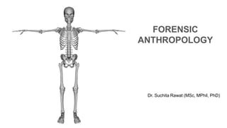
FORENSIC
ANTHROPOLOGY
Dr. Suchita Rawat (MSc, MPhil, PhD)
Analysing the differences between animal and human
bones.
● Differential Skeletal Anatomy of Humans and Animals: Cranium
● Differential Skeletal Anatomy of Humans and Animals: Dentition
● Differential Skeletal Anatomy of Humans and Animals: Post cranium
HUMAN ANIMAL
Large bulbous vault, small face Small vault, large face
Vault relatively smooth Pronounced muscle markings, sagittal crest Inferior
Inferior Foramen Magnum Posterior Foramen Magnum
Chin present Chin absent
Orbits at front, above nasal aperture Orbits at sides, posterior to nasal aperture
"U"- shaped mandible (no midline separation) "V"- shaped mandible (separates at midline)
Differential Skeletal Anatomy of Humans and Animals: Cranium
HUMAN ANIMAL
Omnivorous Carnivorous; Herbivorous; Omnivorou
Dental formula 2:1:2:3 Basic dental formula 3:1:4:3
incisors (maxillary) are larger than other
mammals
Horse maxillary incisors are larger
than human
incisors
Canines small Carnivores have large conical
canines; Herbivores have small or
missing canines
Premolars and molars have low,
rounded cusps divided by distinct
grooves
Carnivores have sharp, pointed
molars and premolar teeth;
Herbivores have broad, flat
premolars and molars with parallel
furrows and ridges
Differential Skeletal Anatomy of Humans and Animals: Dentition
Differential Skeletal Anatomy of Humans and Animals: Post cranium
Human Animal
Upper limbs less robust Robust upper limbs
Radius and ulna are separate bones Radius and ulna often fused
Large, flat and broad vertebral bodies with short
spinous
processes
Small vertebral bodies with convex/ concave
surfaces and
long spinous processes
Sacrum with 5 fused vertebrae, short and broad Sacrum with 3 or 4 fused vertebrae, long and
narrow
Pelvis is broad and short, bowl - shaped Pelvis is long and narrow, blade - shaped
Femur is longest bone in body, lineaaspera is
singular
feature
Femur is similar length to other limb bones,
lineaaspera
double or plateau
Separate tibia and fibula Tibia and fibula are often fused
Foot is long and narrow, weight borne on heel
and toes
Foot is broad, weight borne mainly on toes
TRAIT MALE FEMALE
General size Larger, more massive Smaller, slender
Long bones Ridges, depression and process are
more prominent. Bones of arms and
legs are 8% longer
Less prominent
Shaft of long bones Rougher Smoother, thinner with relatively wider
medullary cavity
Articular surface Larger Smaller
Metacarpal bones Longer and broader Shorter and narrower
Weight 4.5 kg 2.75 kg
Comparison of Male and Female Skeleton
TRAIT MALE FEMALE
General Appearance Larger, longer Smaller, lighter, walls thinner
Architecture Rugged, muscle ridges more marked,
especially in occipital and temporal areas
Smooth
Glabella More prominent Small or absent
Forehead Steeper less rounded Vertical, round, full
Orbits Square, set lower on the face, relatively
smaller, rounded margins
Rounded higher, relatively larger, sharper
margins
Supraorbital ridges Prominent Less prominent or absent
Zygomatic arch More prominent Less prominent
Nasal aperture Higher and narrower. Margins sharp Lower and broader
Frontal eminences Small Large
Parietal eminences Small Large
Occipital area Muscle lines and protuberance prominent Not prominent
Comparison of Male and Female Skull
TRAIT MALE FEMALE
Occipital area Muscle lines and protuberance
prominent
Not prominent
Mastoid process Medium to large, round, blunt. Small to medium, smooth, pointed
Palate Larger, broader, tends more to be u-
shape.
Smaller, tends more to be parabolic
shape
Foramen magnum Relatively large and long Relatively small and round
Mandible Larger and thicker Smaller and thinner
Ascending ramus Greater breath Smaller breath
Mandible Condyles Larger Smaller
Angle of body and
ramus
Less obtuse (under 125º) More obtuse, and not prominent
Teeth Larger Smaller
Comparison of Male and Female Skull
https://www.youtube.com/watch?v=o3GvTnCuOIY
Comparison of Male and Female Pelvis
Trait Male Female
Bony framework Massive, rougher, marked
muscle sites.
Less massive, slender,
smoother
General Deep funnel Flat bowl
Acetabulum Large and directed laterally Small and directed antero-
laterally
Obturator Foramen Large, often oval with base
upward
Small, triangular with apex
forward
Body of pubis Narrow, triangular Broad, square, pits on
posterior surface if borne
children
Ramus of pubis It is like continuation of body of
pubis.
Has a constricted or
narrowed appearance
and is short and thick
Sacrum Longer, narrower, with more
evenly distributed curvatures,
prominently well marked.
Body of first sacral vertebra
larger. Shorter, wider,
upper half almost straight,
curve forward in lower half,
prominently less marked.
Coccyx Less movable More movable
Comparison of Male and Female Pelvis
Trait Male Female
Pre-auricular sulcus
(attachment of anterior
sacroiliac ligament)
Not frequent, narrow, shallow More frequent, broad and deep
Greater sciatic notch Smaller, narrower, deeper Larger, wider, shallower
Comparison of Male and Female Pelvis
Trait Male Female
Symphysis High Low and distance between
two pubic tubercles greater.
The dorsal border is irregular
and shows depressions or
pits (scars of parturition)
Subpubic
angle
V-shaped, sharp angle 70º
to 75º
U-shaped, rounded, broader
angle, 90 to 100º
Pelvic brim Heart shaped Circular or elliptical, more
spacious, diameter longer
Pelvic inlet Conical and funnel shaped Broad and rounded
https://www.youtube.com/watch?v=K4jShowlAOU
Sex Determination from Long Bone
Measurements & Morphology
Male vs. female (greater in males)
Maximum length
Maximum Vertical Diameter of the Head
Maximum Transverse Diameter of the Head
Bone weight (in gm)
Male vs. female (greater in males)
Maximum length
Maximum Vertical Diameter of the Head
Maximum Transverse Diameter of the
Less curve of bone for males than female
Male vs. female (greater in males)
Maximum length
Bone weight
Size
Shape of head of bone less globular
Male vs. female (greater in males)
Maximum length
Maximum Vertical Diameter of the Head
Bicondylar width
Maximum Trochanteric length
Angle formed by neck & Shaft axis If low
denotes masculine character
Male vs. female (greater in males)
Maximum length
Bicondylar width
Weight
Male vs. female (greater in males)
Diameter of middle of bone
Bone weight
Male vs. female (greater in males)
Glenoid breadth
Total spine length
Weight
Male Female
The thoracic cage is longer
and narrower.
It is shorter and wider.
Ribs are thicker and
comparatively massive in
texture.
Ribs are thinner and
delicate in texture.
Ribs have lesser curvatures. Ribs have greater
curvatures.
Manubrium Manubrium is
somewhat smaller.
It is somewhat bigger.
 Sutures of the skull, also known as cranial
sutures, are fibrous joints with a fracture-like
appearance found between the bones of the skull.
 Sutures are formed during embryonic
development.
 They are sites for bone expansion, ensuring
craniofacial growth during the embryonic,
postnatal, and later growth periods.
 The cranial sutures ossify at different rates, but
most sutures have ossified by the age of 20.
 Sutures of an adult skull are categorized
as synarthroses ( a type of joint that, under
normal circumstances, is immobile.)
Figuíe 1: Vault sutuíe closuíe stages. Illustíation of degíees of closuíe foí
sagittal sutuíe at obelion.
0 open; there is no evidence of any
ectocranial closure
1 minimal closure; the score is
assigned to any minimal to moderate
closure, from single bony bridge to
about 50% synostosis
2 significant closure; there is a
marked degree of closure but some
portions still not completely fused
3 complete obliteration; the site is
completely fused.
 In neonates, the sutures are incompletely fused,
leaving membranous gaps
called fontanelles.
 Fontanelles are also often called soft spots.
Fontanelle Location closure
frontal fontanelle
(anterior
fontanelle)*
at the junction of the
coronal and sagittal
sutures.
closes between 12
and 18 months of
age
occipital fontanelle
( posterior
fontanelle)*
at the junction
between the sagittal
and lambdoid
suture
closes at 6-8 month
of birth
sphenoid
fontanelle (2)
located between
the sphenoid, temp
oral, frontal,
and parietal bones.
closes at 2 months
after birth
mastoid
fontanelle (2)
situated between the
temporal, occipital,
and parietal bones.
closes at 2 months
after birth
 The metopic suture is present in
newborns.
 The metopic suture divides the
frontal
bone along the midline.
 Metopic suture closes at 2-4 years
but may extend up to six years.
Sutures of neurocranium
Sutures Location ossification
sagittal suture (2) formed by the
two parietal
bones articulating
with each other.
Fusion begins
around 25 years &
completed by 30 -
35 Years
Coronal suture (2) formed at the junction
between the parietal
bones and frontal
bone.
Fusion begins around
28 years & completed
by 35 - 40 Years
lambdoid
suture (2)
formed at the
articulation
between the
occipital bone
and parietal
bones.
Fusion begins
around 30 years &
completed by 50 -
55 Years
squamous suture (2) formed by the
parietal bone and
temporal bone.
Fusion begins around
50 - 55 years &
completed by 70 Years
Sutures Location ossification
Occipito-mastoid
suture (2)
Formed by the
articulation of
the occipital
bone and the
temporal
bone's
mastoid part
Fusion begins
around 60 - 65
years &
completed by 80
Years
Parieto-mastoid
suture (2)
formed at the
Junction
between the
parietal and
temporal
bones.
Fusion begins
around 60 - 65years
& completed by 80 -
82 Years
Spheno
Parietal suture
(2)
suture between
parietalbone and
the sphenoid
bone.
Fusion begins
around 60 - 65
years &
completed by 80
- 85 Years
Landmark location Fusion
bregma It is the intersection of the
coronal and sagittal sutures.
site of the frontal fontanelle in
neonates and young children,
which usually fuses around
the age of 2.
lambda formed at the convergence
between the sagittal and
lambdoid sutures.
site for the occipital
fontanelle was located, which
usually closes around 2
months of age.
obelion is formed at the intersection of
the sagittal suture and an
imaginary line that connects
the two parietal foramina.
-
suture-associated landmarks
Landmark location
Asterion (2) site of
the previously located
mastoid fontanelle
at the junction of the
parietomastoid,
occipitomastoid and
lambdoid sutures.
closes by 80 years
Pterion (2) H-shaped point of junction
between four bones:
the sphenoid, temporal, fr
ontal and parietal bone.
starts closing at 40 years and
completely closes by 65 years
Asterion
suture-associated landmarks
Skeletal Age and Ossification
 The human bones develop from a number of ossification centers.
 At 11- 12th week of intrauterine life, there are 806 ossification centers that at
birth are reduced to about 450.
 Adult human is made up of 206 bones.
determination
of age
time of appearance of center of ossification
process of union of the epiphysis with the
diaphysis at the metaphysis
Limitations: Hereditary factors Growth and
development Geographical variation Climate
Dietary habits Association with diseases
Ossification of bones
Centers of bones Appearance Fusion
Clavicle - Medial
end
15-19 years 20-22 years
Centers of bones Appearance Fusion
Sternum 5 month IUL 60-70 years
Manubrium Body
 Ist segment)
 IInd segment
 IIIrd segment
 IVth segment
5 month IUL
7 month IUL
7 month IUL
10 month IUL
14-25 years from
below upwards;
3rd and 4th -15
years
2 nd & 3rd -20
years
1 st & 2nd -25
years
Xiphoid process 3 years >40 years with
the body
Centers of bones Appearance Fusion
Humerus (upper end)
 Head
 Greater tubercle
 Lesser tubercle
1 year
3 years
5 years
18 years
4-5 years with head
5-7 years with
greater tubercle
Humerus (Lower end)
 Medial Epicondyle
 Capitulum
 Trochlea
 Lateral Epicondyle
5-6 years
1 year
9-10 years
10-12 years
Capitulum, trochlea
& lateral epicondyle
form conjoint tendon
at 14 years, unites
with shaft at 15
years Medial
epicondyle unites at
16 years
Centers of bones Appearanc
e
Fusion
Scapula
Coracoid base
Acromion
process
10-11 year
14-15 year
14-15 years
17-18 years
Centers of
bones
Appearance Fusion
Radius
Upper end
Lower end
5-6 years
1-2 years
15-16 years
18-19 years
Ulna
Upper end
Lower end
8-9 years
5-6 years
16-17 years
18-19 years
Centers of bones Appearance Fusion
Head of Ist
metacarpal
Head other
metacarpals
2 years
1½ to 2½
years
15-17 years
15-19 years
Centers of bones Appearance Fusion
Hip bone
• Iliac crest
• Ischial tuberosity
• Sacrum
14-15 years
15-16 years
8 months
IUL
18-20
years 20-
22 years
25 years
This Photo by Unknown Author is licensed under CC BY
Centers of bones Appearance Fusion
Femur (Upper end)
• Head
• Greater trochanter
• Lesser trochanter
Femur (Lower end)
1 year
4 years
14 years
9 month IUL
17-18 years
17 years
15-17 years
17-18 years
Centers of
bones
Appearance Fusion
Tibia
• Upper end
• Lower end
9 month IUL
1 year
16-17 years
16 years
Type of bone Age of ossification
Trapezium
Trapezoid
Capitate
Hamate
Pisiform
Triquetrum
Lunate
Capitate
4-5 months
4-5 months
2 months
3 months
9-12 years
3 months
4 months
2 months
2 months
Type of bone Age of ossification
Calcaneum
Cuboid
Lateral cuneiform
Medial cuneiform
Intermediate
cuneiform
Navicular
Talus
5 months
10 months
1 year
2 years
3 years
3 years
7 months
 Non dominant hand
 Widely spread of fingers
 Focus at carpal
 Maturation (proximal to
distal) & fusion (distal
side)
 Carpal
 Phalanges
 Radius ulna (terminal
fusion)
 Girls have advance age 1-2
Source: https://www.youtube.com/watch?v=uxJ11zyhQ1Q
Distal Phalanx (appearance)
Source: https://www.youtube.com/watch?v=uxJ11zyhQ1Q
Exceptions
Source: https://www.youtube.com/watch?v=uxJ11zyhQ1Q
ULNA
Source: https://www.youtube.com/watch?v=uxJ11zyhQ1Q
RADIUS
Maturation score for estimation of skeletal age:
● To minimize the errors of epiphyseal union, Mckern and Stewart in 1957 suggested a
scheme of scoring involving seven combinations of various segments.
● The total score is applied to the prediction equation for more accurate age estimation.
Degree of union Scoring
No union
¼ th union
½ union
¾ th union
Complete union
1
2
3
4
5
Racial Characteristics of the Skull
Trait Mongoloid Caucasoid Negroid
Skull Length Long Short Long
Skull Breadth Broad Broad Narrow
Skull Height Middle High Low
Sagittal Contour Arched Arched Flat
Face Breadth Very wide Wide Narrow
Face Height High High Low
Orbital Opening Rounded Rounded Rectangular
Racial Characteristics of the Skull
Trait Mongoloid Caucasoid Negroid
Nasal Opening Narrow Moderately Wide Wide
Nasal Bones Wide Flat Narrow
Arched
Narrow
Lower Nasal Margin Sharp Sharp Troughed
Facial Profile Straight Straight Downward
slant
Palate Shape Broad U-shaped V-shaped U-shaped
Shovel-shaped
incisors
90% Less than 5 % Less than 5%
measuring all bones constituting the components of
stature, summing those measurements and correcting
for the missing soft tissue
employing a regression formula with the
measurement of a complete bone.
employing incomplete limb bones, non-limb bones
and alternative statistical methods
Alternate statistical approaches (e.g., maximum
likelihood estimation) exist to estimate stature.
Method Do’s
Anatomical
Method
(Complete Skeleton
Method)
 skeletal elements constituting
stature minimally damaged.
 ancestry and sex of the
individual cannot be estimated
 anomalous number of
vertebrae
 individual’s limb bones appear
to be atypical in length.
 Bones typically measured in these methods are the
 height of the skull
 the heights of each of the vertebrae (a missing vertebra estimate the height by
averaging the heights of the vertebra immediately above and below)
 the lengths of the femur and tibia
 and the height of the ankle
Christensen, A. M., Passalacqua, N. V., & Bartelink, E. J. (2019). Stature estimation and other skeletal
metrics. Forensic Anthropology, 351–368. doi:10.1016/b978-0-12-815734-3.00011-7
Christensen, A. M., Passalacqua, N. V., & Bartelink, E. J. (2019). Stature estimation and other skeletal
metrics. Forensic Anthropology, 351–368. doi:10.1016/b978-0-12-815734-3.00011-7
Method Do’s
Complete Limb
Bones
(Mathematical
Method or
Regression
Approach)
 limb bone length or bone lengths
 selecting the most appropriate
regression formula by sex and
ancestry, inserting the
measurement into the formula, and
calculating the estimated stature
 limb bone measurements are
usually maximum lengths
 he formula with the smallest
prediction interval should be the
most accurate and precise, and
should be employed in the
stature estimation
Christensen, A. M., Passalacqua, N. V., & Bartelink, E. J. (2019). Stature estimation and other skeletal
metrics. Forensic Anthropology, 351–368. doi:10.1016/b978-0-12-815734-3.00011-7
Method Do’s
Fragmentary Limb
Bones
 Some of these methods require
estimating bone length and then
estimating stature based on the
estimated bone length, thus
compounding the error present in the
estimation.
 fragmentary remains estimate stature
directly from the fragment, without
requiring the second step of the
previous method.
Method Do’s
Non-Limb Bones  Non-limb bones (e.g., skulls,
innominates, and bones of the
hands or feet) may also be
used to estimate stature
https://fac.utk.edu/fordisc-3-1-personal-computer-forensic-discriminant-functions/
This Photo by Unknown Author is licensed under CC BY-SA

More Related Content

Similar to MSCIII_Forensic anthropology_Personal Identification.pptx

تشريح الهيكل المحوري.pdf
تشريح الهيكل المحوري.pdfتشريح الهيكل المحوري.pdf
تشريح الهيكل المحوري.pdf
fathiwww2
 
axial skeleton human anatomy. Mbbs....pdf
axial skeleton human anatomy. Mbbs....pdfaxial skeleton human anatomy. Mbbs....pdf
axial skeleton human anatomy. Mbbs....pdf
siddhimeena3
 
THE SKELETAL SYSTEM (ANATOMY).pptx
THE SKELETAL SYSTEM (ANATOMY).pptxTHE SKELETAL SYSTEM (ANATOMY).pptx
THE SKELETAL SYSTEM (ANATOMY).pptx
SatyadeepBanerjee
 
GENERAL OSTEOLOGY.pptx
GENERAL OSTEOLOGY.pptxGENERAL OSTEOLOGY.pptx
GENERAL OSTEOLOGY.pptx
DrRashidKaziMahbubur
 
Determination of sex from skeletal remains
Determination of sex from skeletal remains Determination of sex from skeletal remains
Determination of sex from skeletal remains
PallaviKumari112
 
THE SKELETAL SYSTEM.pdf
THE SKELETAL SYSTEM.pdfTHE SKELETAL SYSTEM.pdf
THE SKELETAL SYSTEM.pdf
IvetteGonzlez13
 
THE SKELETAL SYSTEM.pdf
THE SKELETAL SYSTEM.pdfTHE SKELETAL SYSTEM.pdf
THE SKELETAL SYSTEM.pdf
UmaymaMatoobe
 
Head and neck anat fin
Head and neck anat finHead and neck anat fin
Head and neck anat finEmiru Tilahun
 
Anatomy Lab exercise 8
Anatomy Lab exercise 8 Anatomy Lab exercise 8
Anatomy Lab exercise 8
Jhillock
 
Fetal head diameters
Fetal head diametersFetal head diameters
Fetal head diameters
mohammad tailakh
 
Appendicular skeleton
Appendicular skeletonAppendicular skeleton
Appendicular skeleton
PARUL UNIVERSITY
 
POSTURAL-ANALYSIS-REPORT-FINAL.pptx
POSTURAL-ANALYSIS-REPORT-FINAL.pptxPOSTURAL-ANALYSIS-REPORT-FINAL.pptx
POSTURAL-ANALYSIS-REPORT-FINAL.pptx
ReschaelGiango
 
The Human Skeletal System & Joints for B.Pharm and Pharm.D
The Human Skeletal System & Joints for B.Pharm and Pharm.DThe Human Skeletal System & Joints for B.Pharm and Pharm.D
The Human Skeletal System & Joints for B.Pharm and Pharm.D
Kameshwaran Sugavanam
 
Appendicular Skeleton combined skeletal.ppt
Appendicular Skeleton combined skeletal.pptAppendicular Skeleton combined skeletal.ppt
Appendicular Skeleton combined skeletal.ppt
ssuser48ded4
 
Anatomy for Art
Anatomy for ArtAnatomy for Art
Anatomy for Art
ManamiIshimura
 
SKELETAL SYSTEM.pptx
SKELETAL SYSTEM.pptxSKELETAL SYSTEM.pptx
SKELETAL SYSTEM.pptx
JainabaKandeh
 
skeletal system
skeletal systemskeletal system
skeletal system
Devu67
 

Similar to MSCIII_Forensic anthropology_Personal Identification.pptx (20)

تشريح الهيكل المحوري.pdf
تشريح الهيكل المحوري.pdfتشريح الهيكل المحوري.pdf
تشريح الهيكل المحوري.pdf
 
axial skeleton human anatomy. Mbbs....pdf
axial skeleton human anatomy. Mbbs....pdfaxial skeleton human anatomy. Mbbs....pdf
axial skeleton human anatomy. Mbbs....pdf
 
THE SKELETAL SYSTEM (ANATOMY).pptx
THE SKELETAL SYSTEM (ANATOMY).pptxTHE SKELETAL SYSTEM (ANATOMY).pptx
THE SKELETAL SYSTEM (ANATOMY).pptx
 
GENERAL OSTEOLOGY.pptx
GENERAL OSTEOLOGY.pptxGENERAL OSTEOLOGY.pptx
GENERAL OSTEOLOGY.pptx
 
Determination of sex from skeletal remains
Determination of sex from skeletal remains Determination of sex from skeletal remains
Determination of sex from skeletal remains
 
THE SKELETAL SYSTEM.pdf
THE SKELETAL SYSTEM.pdfTHE SKELETAL SYSTEM.pdf
THE SKELETAL SYSTEM.pdf
 
THE SKELETAL SYSTEM.pdf
THE SKELETAL SYSTEM.pdfTHE SKELETAL SYSTEM.pdf
THE SKELETAL SYSTEM.pdf
 
Femur
FemurFemur
Femur
 
Head and neck anat fin
Head and neck anat finHead and neck anat fin
Head and neck anat fin
 
Femur
FemurFemur
Femur
 
Anatomy Lab exercise 8
Anatomy Lab exercise 8 Anatomy Lab exercise 8
Anatomy Lab exercise 8
 
Lp 5 bones 2008 no path
Lp 5 bones 2008 no pathLp 5 bones 2008 no path
Lp 5 bones 2008 no path
 
Fetal head diameters
Fetal head diametersFetal head diameters
Fetal head diameters
 
Appendicular skeleton
Appendicular skeletonAppendicular skeleton
Appendicular skeleton
 
POSTURAL-ANALYSIS-REPORT-FINAL.pptx
POSTURAL-ANALYSIS-REPORT-FINAL.pptxPOSTURAL-ANALYSIS-REPORT-FINAL.pptx
POSTURAL-ANALYSIS-REPORT-FINAL.pptx
 
The Human Skeletal System & Joints for B.Pharm and Pharm.D
The Human Skeletal System & Joints for B.Pharm and Pharm.DThe Human Skeletal System & Joints for B.Pharm and Pharm.D
The Human Skeletal System & Joints for B.Pharm and Pharm.D
 
Appendicular Skeleton combined skeletal.ppt
Appendicular Skeleton combined skeletal.pptAppendicular Skeleton combined skeletal.ppt
Appendicular Skeleton combined skeletal.ppt
 
Anatomy for Art
Anatomy for ArtAnatomy for Art
Anatomy for Art
 
SKELETAL SYSTEM.pptx
SKELETAL SYSTEM.pptxSKELETAL SYSTEM.pptx
SKELETAL SYSTEM.pptx
 
skeletal system
skeletal systemskeletal system
skeletal system
 

More from Suchita Rawat

MSC IV_Forensic medicine -sexual offence.pdf
MSC IV_Forensic medicine -sexual offence.pdfMSC IV_Forensic medicine -sexual offence.pdf
MSC IV_Forensic medicine -sexual offence.pdf
Suchita Rawat
 
MSC IV_Forensic medicine -child offence.pdf
MSC IV_Forensic medicine -child offence.pdfMSC IV_Forensic medicine -child offence.pdf
MSC IV_Forensic medicine -child offence.pdf
Suchita Rawat
 
MSC IV_Forensic medicine - Mechanical injuries.pdf
MSC IV_Forensic medicine - Mechanical injuries.pdfMSC IV_Forensic medicine - Mechanical injuries.pdf
MSC IV_Forensic medicine - Mechanical injuries.pdf
Suchita Rawat
 
MSC IV_Forensic medicine - Thermal and firearm injuries.pdf
MSC IV_Forensic medicine - Thermal and firearm injuries.pdfMSC IV_Forensic medicine - Thermal and firearm injuries.pdf
MSC IV_Forensic medicine - Thermal and firearm injuries.pdf
Suchita Rawat
 
MSC IV_Forensic medicine - autopsy.pdf
MSC IV_Forensic  medicine -  autopsy.pdfMSC IV_Forensic  medicine -  autopsy.pdf
MSC IV_Forensic medicine - autopsy.pdf
Suchita Rawat
 
MSCII_ FCT UNIT 5 TOXICOLOGY.pdf
MSCII_              FCT UNIT 5 TOXICOLOGY.pdfMSCII_              FCT UNIT 5 TOXICOLOGY.pdf
MSCII_ FCT UNIT 5 TOXICOLOGY.pdf
Suchita Rawat
 
Unit 1 _ Analysis of Adulteration.pdf
Unit 1   _ Analysis  of Adulteration.pdfUnit 1   _ Analysis  of Adulteration.pdf
Unit 1 _ Analysis of Adulteration.pdf
Suchita Rawat
 
FORENSIC CHEMISTRY ARSON INVESTIGATION.pdf
FORENSIC CHEMISTRY ARSON INVESTIGATION.pdfFORENSIC CHEMISTRY ARSON INVESTIGATION.pdf
FORENSIC CHEMISTRY ARSON INVESTIGATION.pdf
Suchita Rawat
 
MSC IV SEMESTER_DNA Profiling - DNA quantification.pdf
MSC IV SEMESTER_DNA Profiling - DNA quantification.pdfMSC IV SEMESTER_DNA Profiling - DNA quantification.pdf
MSC IV SEMESTER_DNA Profiling - DNA quantification.pdf
Suchita Rawat
 
MSC IV SEMESTER_DNA Profiling -DNA introduction and extraction.pdf
MSC IV SEMESTER_DNA Profiling -DNA introduction and extraction.pdfMSC IV SEMESTER_DNA Profiling -DNA introduction and extraction.pdf
MSC IV SEMESTER_DNA Profiling -DNA introduction and extraction.pdf
Suchita Rawat
 
MSC IV SEMESTER_ DNA Profiling -PCR.pdf
MSC IV SEMESTER_  DNA Profiling -PCR.pdfMSC IV SEMESTER_  DNA Profiling -PCR.pdf
MSC IV SEMESTER_ DNA Profiling -PCR.pdf
Suchita Rawat
 
MSC IV SEMESTER_DNA Profiling - CE.pdf
MSC   IV SEMESTER_DNA Profiling - CE.pdfMSC   IV SEMESTER_DNA Profiling - CE.pdf
MSC IV SEMESTER_DNA Profiling - CE.pdf
Suchita Rawat
 
MSC IV SEMESTER_DNA Profiling quality assurance.pdf
MSC IV SEMESTER_DNA Profiling quality assurance.pdfMSC IV SEMESTER_DNA Profiling quality assurance.pdf
MSC IV SEMESTER_DNA Profiling quality assurance.pdf
Suchita Rawat
 
MSC IV SEMESTER_DNA Profiling - STR biology and artifacts.pdf
MSC IV SEMESTER_DNA Profiling - STR biology and artifacts.pdfMSC IV SEMESTER_DNA Profiling - STR biology and artifacts.pdf
MSC IV SEMESTER_DNA Profiling - STR biology and artifacts.pdf
Suchita Rawat
 
MSC IV SEMESTER_DNA Profiling DNA national institute and DNA database.pdf
MSC IV SEMESTER_DNA Profiling DNA national institute and DNA database.pdfMSC IV SEMESTER_DNA Profiling DNA national institute and DNA database.pdf
MSC IV SEMESTER_DNA Profiling DNA national institute and DNA database.pdf
Suchita Rawat
 
MSC IV SEMESTER_DNA Profiling - DNA Sequencing.pdf
MSC IV SEMESTER_DNA Profiling - DNA Sequencing.pdfMSC IV SEMESTER_DNA Profiling - DNA Sequencing.pdf
MSC IV SEMESTER_DNA Profiling - DNA Sequencing.pdf
Suchita Rawat
 
MSC IV_Forensic medicine - Thanatology.pdf
MSC IV_Forensic medicine - Thanatology.pdfMSC IV_Forensic medicine - Thanatology.pdf
MSC IV_Forensic medicine - Thanatology.pdf
Suchita Rawat
 
Forensic examination of Blood semen saliva.pptx
Forensic examination of Blood semen saliva.pptxForensic examination of Blood semen saliva.pptx
Forensic examination of Blood semen saliva.pptx
Suchita Rawat
 
Forensic identification of uncommon body fluids.pptx
Forensic identification of uncommon body fluids.pptxForensic identification of uncommon body fluids.pptx
Forensic identification of uncommon body fluids.pptx
Suchita Rawat
 
Species origin.pptx
Species origin.pptxSpecies origin.pptx
Species origin.pptx
Suchita Rawat
 

More from Suchita Rawat (20)

MSC IV_Forensic medicine -sexual offence.pdf
MSC IV_Forensic medicine -sexual offence.pdfMSC IV_Forensic medicine -sexual offence.pdf
MSC IV_Forensic medicine -sexual offence.pdf
 
MSC IV_Forensic medicine -child offence.pdf
MSC IV_Forensic medicine -child offence.pdfMSC IV_Forensic medicine -child offence.pdf
MSC IV_Forensic medicine -child offence.pdf
 
MSC IV_Forensic medicine - Mechanical injuries.pdf
MSC IV_Forensic medicine - Mechanical injuries.pdfMSC IV_Forensic medicine - Mechanical injuries.pdf
MSC IV_Forensic medicine - Mechanical injuries.pdf
 
MSC IV_Forensic medicine - Thermal and firearm injuries.pdf
MSC IV_Forensic medicine - Thermal and firearm injuries.pdfMSC IV_Forensic medicine - Thermal and firearm injuries.pdf
MSC IV_Forensic medicine - Thermal and firearm injuries.pdf
 
MSC IV_Forensic medicine - autopsy.pdf
MSC IV_Forensic  medicine -  autopsy.pdfMSC IV_Forensic  medicine -  autopsy.pdf
MSC IV_Forensic medicine - autopsy.pdf
 
MSCII_ FCT UNIT 5 TOXICOLOGY.pdf
MSCII_              FCT UNIT 5 TOXICOLOGY.pdfMSCII_              FCT UNIT 5 TOXICOLOGY.pdf
MSCII_ FCT UNIT 5 TOXICOLOGY.pdf
 
Unit 1 _ Analysis of Adulteration.pdf
Unit 1   _ Analysis  of Adulteration.pdfUnit 1   _ Analysis  of Adulteration.pdf
Unit 1 _ Analysis of Adulteration.pdf
 
FORENSIC CHEMISTRY ARSON INVESTIGATION.pdf
FORENSIC CHEMISTRY ARSON INVESTIGATION.pdfFORENSIC CHEMISTRY ARSON INVESTIGATION.pdf
FORENSIC CHEMISTRY ARSON INVESTIGATION.pdf
 
MSC IV SEMESTER_DNA Profiling - DNA quantification.pdf
MSC IV SEMESTER_DNA Profiling - DNA quantification.pdfMSC IV SEMESTER_DNA Profiling - DNA quantification.pdf
MSC IV SEMESTER_DNA Profiling - DNA quantification.pdf
 
MSC IV SEMESTER_DNA Profiling -DNA introduction and extraction.pdf
MSC IV SEMESTER_DNA Profiling -DNA introduction and extraction.pdfMSC IV SEMESTER_DNA Profiling -DNA introduction and extraction.pdf
MSC IV SEMESTER_DNA Profiling -DNA introduction and extraction.pdf
 
MSC IV SEMESTER_ DNA Profiling -PCR.pdf
MSC IV SEMESTER_  DNA Profiling -PCR.pdfMSC IV SEMESTER_  DNA Profiling -PCR.pdf
MSC IV SEMESTER_ DNA Profiling -PCR.pdf
 
MSC IV SEMESTER_DNA Profiling - CE.pdf
MSC   IV SEMESTER_DNA Profiling - CE.pdfMSC   IV SEMESTER_DNA Profiling - CE.pdf
MSC IV SEMESTER_DNA Profiling - CE.pdf
 
MSC IV SEMESTER_DNA Profiling quality assurance.pdf
MSC IV SEMESTER_DNA Profiling quality assurance.pdfMSC IV SEMESTER_DNA Profiling quality assurance.pdf
MSC IV SEMESTER_DNA Profiling quality assurance.pdf
 
MSC IV SEMESTER_DNA Profiling - STR biology and artifacts.pdf
MSC IV SEMESTER_DNA Profiling - STR biology and artifacts.pdfMSC IV SEMESTER_DNA Profiling - STR biology and artifacts.pdf
MSC IV SEMESTER_DNA Profiling - STR biology and artifacts.pdf
 
MSC IV SEMESTER_DNA Profiling DNA national institute and DNA database.pdf
MSC IV SEMESTER_DNA Profiling DNA national institute and DNA database.pdfMSC IV SEMESTER_DNA Profiling DNA national institute and DNA database.pdf
MSC IV SEMESTER_DNA Profiling DNA national institute and DNA database.pdf
 
MSC IV SEMESTER_DNA Profiling - DNA Sequencing.pdf
MSC IV SEMESTER_DNA Profiling - DNA Sequencing.pdfMSC IV SEMESTER_DNA Profiling - DNA Sequencing.pdf
MSC IV SEMESTER_DNA Profiling - DNA Sequencing.pdf
 
MSC IV_Forensic medicine - Thanatology.pdf
MSC IV_Forensic medicine - Thanatology.pdfMSC IV_Forensic medicine - Thanatology.pdf
MSC IV_Forensic medicine - Thanatology.pdf
 
Forensic examination of Blood semen saliva.pptx
Forensic examination of Blood semen saliva.pptxForensic examination of Blood semen saliva.pptx
Forensic examination of Blood semen saliva.pptx
 
Forensic identification of uncommon body fluids.pptx
Forensic identification of uncommon body fluids.pptxForensic identification of uncommon body fluids.pptx
Forensic identification of uncommon body fluids.pptx
 
Species origin.pptx
Species origin.pptxSpecies origin.pptx
Species origin.pptx
 

Recently uploaded

Lab report on liquid viscosity of glycerin
Lab report on liquid viscosity of glycerinLab report on liquid viscosity of glycerin
Lab report on liquid viscosity of glycerin
ossaicprecious19
 
platelets- lifespan -Clot retraction-disorders.pptx
platelets- lifespan -Clot retraction-disorders.pptxplatelets- lifespan -Clot retraction-disorders.pptx
platelets- lifespan -Clot retraction-disorders.pptx
muralinath2
 
GBSN- Microbiology (Lab 3) Gram Staining
GBSN- Microbiology (Lab 3) Gram StainingGBSN- Microbiology (Lab 3) Gram Staining
GBSN- Microbiology (Lab 3) Gram Staining
Areesha Ahmad
 
Structures and textures of metamorphic rocks
Structures and textures of metamorphic rocksStructures and textures of metamorphic rocks
Structures and textures of metamorphic rocks
kumarmathi863
 
(May 29th, 2024) Advancements in Intravital Microscopy- Insights for Preclini...
(May 29th, 2024) Advancements in Intravital Microscopy- Insights for Preclini...(May 29th, 2024) Advancements in Intravital Microscopy- Insights for Preclini...
(May 29th, 2024) Advancements in Intravital Microscopy- Insights for Preclini...
Scintica Instrumentation
 
Circulatory system_ Laplace law. Ohms law.reynaults law,baro-chemo-receptors-...
Circulatory system_ Laplace law. Ohms law.reynaults law,baro-chemo-receptors-...Circulatory system_ Laplace law. Ohms law.reynaults law,baro-chemo-receptors-...
Circulatory system_ Laplace law. Ohms law.reynaults law,baro-chemo-receptors-...
muralinath2
 
Penicillin...........................pptx
Penicillin...........................pptxPenicillin...........................pptx
Penicillin...........................pptx
Cherry
 
Predicting property prices with machine learning algorithms.pdf
Predicting property prices with machine learning algorithms.pdfPredicting property prices with machine learning algorithms.pdf
Predicting property prices with machine learning algorithms.pdf
binhminhvu04
 
Hemostasis_importance& clinical significance.pptx
Hemostasis_importance& clinical significance.pptxHemostasis_importance& clinical significance.pptx
Hemostasis_importance& clinical significance.pptx
muralinath2
 
Nutraceutical market, scope and growth: Herbal drug technology
Nutraceutical market, scope and growth: Herbal drug technologyNutraceutical market, scope and growth: Herbal drug technology
Nutraceutical market, scope and growth: Herbal drug technology
Lokesh Patil
 
insect morphology and physiology of insect
insect morphology and physiology of insectinsect morphology and physiology of insect
insect morphology and physiology of insect
anitaento25
 
Mammalian Pineal Body Structure and Also Functions
Mammalian Pineal Body Structure and Also FunctionsMammalian Pineal Body Structure and Also Functions
Mammalian Pineal Body Structure and Also Functions
YOGESH DOGRA
 
Anemia_ different types_causes_ conditions
Anemia_ different types_causes_ conditionsAnemia_ different types_causes_ conditions
Anemia_ different types_causes_ conditions
muralinath2
 
GBSN - Biochemistry (Unit 5) Chemistry of Lipids
GBSN - Biochemistry (Unit 5) Chemistry of LipidsGBSN - Biochemistry (Unit 5) Chemistry of Lipids
GBSN - Biochemistry (Unit 5) Chemistry of Lipids
Areesha Ahmad
 
Earliest Galaxies in the JADES Origins Field: Luminosity Function and Cosmic ...
Earliest Galaxies in the JADES Origins Field: Luminosity Function and Cosmic ...Earliest Galaxies in the JADES Origins Field: Luminosity Function and Cosmic ...
Earliest Galaxies in the JADES Origins Field: Luminosity Function and Cosmic ...
Sérgio Sacani
 
Richard's entangled aventures in wonderland
Richard's entangled aventures in wonderlandRichard's entangled aventures in wonderland
Richard's entangled aventures in wonderland
Richard Gill
 
ESR_factors_affect-clinic significance-Pathysiology.pptx
ESR_factors_affect-clinic significance-Pathysiology.pptxESR_factors_affect-clinic significance-Pathysiology.pptx
ESR_factors_affect-clinic significance-Pathysiology.pptx
muralinath2
 
Structural Classification Of Protein (SCOP)
Structural Classification Of Protein  (SCOP)Structural Classification Of Protein  (SCOP)
Structural Classification Of Protein (SCOP)
aishnasrivastava
 
insect taxonomy importance systematics and classification
insect taxonomy importance systematics and classificationinsect taxonomy importance systematics and classification
insect taxonomy importance systematics and classification
anitaento25
 
Astronomy Update- Curiosity’s exploration of Mars _ Local Briefs _ leadertele...
Astronomy Update- Curiosity’s exploration of Mars _ Local Briefs _ leadertele...Astronomy Update- Curiosity’s exploration of Mars _ Local Briefs _ leadertele...
Astronomy Update- Curiosity’s exploration of Mars _ Local Briefs _ leadertele...
NathanBaughman3
 

Recently uploaded (20)

Lab report on liquid viscosity of glycerin
Lab report on liquid viscosity of glycerinLab report on liquid viscosity of glycerin
Lab report on liquid viscosity of glycerin
 
platelets- lifespan -Clot retraction-disorders.pptx
platelets- lifespan -Clot retraction-disorders.pptxplatelets- lifespan -Clot retraction-disorders.pptx
platelets- lifespan -Clot retraction-disorders.pptx
 
GBSN- Microbiology (Lab 3) Gram Staining
GBSN- Microbiology (Lab 3) Gram StainingGBSN- Microbiology (Lab 3) Gram Staining
GBSN- Microbiology (Lab 3) Gram Staining
 
Structures and textures of metamorphic rocks
Structures and textures of metamorphic rocksStructures and textures of metamorphic rocks
Structures and textures of metamorphic rocks
 
(May 29th, 2024) Advancements in Intravital Microscopy- Insights for Preclini...
(May 29th, 2024) Advancements in Intravital Microscopy- Insights for Preclini...(May 29th, 2024) Advancements in Intravital Microscopy- Insights for Preclini...
(May 29th, 2024) Advancements in Intravital Microscopy- Insights for Preclini...
 
Circulatory system_ Laplace law. Ohms law.reynaults law,baro-chemo-receptors-...
Circulatory system_ Laplace law. Ohms law.reynaults law,baro-chemo-receptors-...Circulatory system_ Laplace law. Ohms law.reynaults law,baro-chemo-receptors-...
Circulatory system_ Laplace law. Ohms law.reynaults law,baro-chemo-receptors-...
 
Penicillin...........................pptx
Penicillin...........................pptxPenicillin...........................pptx
Penicillin...........................pptx
 
Predicting property prices with machine learning algorithms.pdf
Predicting property prices with machine learning algorithms.pdfPredicting property prices with machine learning algorithms.pdf
Predicting property prices with machine learning algorithms.pdf
 
Hemostasis_importance& clinical significance.pptx
Hemostasis_importance& clinical significance.pptxHemostasis_importance& clinical significance.pptx
Hemostasis_importance& clinical significance.pptx
 
Nutraceutical market, scope and growth: Herbal drug technology
Nutraceutical market, scope and growth: Herbal drug technologyNutraceutical market, scope and growth: Herbal drug technology
Nutraceutical market, scope and growth: Herbal drug technology
 
insect morphology and physiology of insect
insect morphology and physiology of insectinsect morphology and physiology of insect
insect morphology and physiology of insect
 
Mammalian Pineal Body Structure and Also Functions
Mammalian Pineal Body Structure and Also FunctionsMammalian Pineal Body Structure and Also Functions
Mammalian Pineal Body Structure and Also Functions
 
Anemia_ different types_causes_ conditions
Anemia_ different types_causes_ conditionsAnemia_ different types_causes_ conditions
Anemia_ different types_causes_ conditions
 
GBSN - Biochemistry (Unit 5) Chemistry of Lipids
GBSN - Biochemistry (Unit 5) Chemistry of LipidsGBSN - Biochemistry (Unit 5) Chemistry of Lipids
GBSN - Biochemistry (Unit 5) Chemistry of Lipids
 
Earliest Galaxies in the JADES Origins Field: Luminosity Function and Cosmic ...
Earliest Galaxies in the JADES Origins Field: Luminosity Function and Cosmic ...Earliest Galaxies in the JADES Origins Field: Luminosity Function and Cosmic ...
Earliest Galaxies in the JADES Origins Field: Luminosity Function and Cosmic ...
 
Richard's entangled aventures in wonderland
Richard's entangled aventures in wonderlandRichard's entangled aventures in wonderland
Richard's entangled aventures in wonderland
 
ESR_factors_affect-clinic significance-Pathysiology.pptx
ESR_factors_affect-clinic significance-Pathysiology.pptxESR_factors_affect-clinic significance-Pathysiology.pptx
ESR_factors_affect-clinic significance-Pathysiology.pptx
 
Structural Classification Of Protein (SCOP)
Structural Classification Of Protein  (SCOP)Structural Classification Of Protein  (SCOP)
Structural Classification Of Protein (SCOP)
 
insect taxonomy importance systematics and classification
insect taxonomy importance systematics and classificationinsect taxonomy importance systematics and classification
insect taxonomy importance systematics and classification
 
Astronomy Update- Curiosity’s exploration of Mars _ Local Briefs _ leadertele...
Astronomy Update- Curiosity’s exploration of Mars _ Local Briefs _ leadertele...Astronomy Update- Curiosity’s exploration of Mars _ Local Briefs _ leadertele...
Astronomy Update- Curiosity’s exploration of Mars _ Local Briefs _ leadertele...
 

MSCIII_Forensic anthropology_Personal Identification.pptx

  • 2. Analysing the differences between animal and human bones. ● Differential Skeletal Anatomy of Humans and Animals: Cranium ● Differential Skeletal Anatomy of Humans and Animals: Dentition ● Differential Skeletal Anatomy of Humans and Animals: Post cranium
  • 3. HUMAN ANIMAL Large bulbous vault, small face Small vault, large face Vault relatively smooth Pronounced muscle markings, sagittal crest Inferior Inferior Foramen Magnum Posterior Foramen Magnum Chin present Chin absent Orbits at front, above nasal aperture Orbits at sides, posterior to nasal aperture "U"- shaped mandible (no midline separation) "V"- shaped mandible (separates at midline) Differential Skeletal Anatomy of Humans and Animals: Cranium
  • 4. HUMAN ANIMAL Omnivorous Carnivorous; Herbivorous; Omnivorou Dental formula 2:1:2:3 Basic dental formula 3:1:4:3 incisors (maxillary) are larger than other mammals Horse maxillary incisors are larger than human incisors Canines small Carnivores have large conical canines; Herbivores have small or missing canines Premolars and molars have low, rounded cusps divided by distinct grooves Carnivores have sharp, pointed molars and premolar teeth; Herbivores have broad, flat premolars and molars with parallel furrows and ridges Differential Skeletal Anatomy of Humans and Animals: Dentition
  • 5. Differential Skeletal Anatomy of Humans and Animals: Post cranium Human Animal Upper limbs less robust Robust upper limbs Radius and ulna are separate bones Radius and ulna often fused Large, flat and broad vertebral bodies with short spinous processes Small vertebral bodies with convex/ concave surfaces and long spinous processes Sacrum with 5 fused vertebrae, short and broad Sacrum with 3 or 4 fused vertebrae, long and narrow Pelvis is broad and short, bowl - shaped Pelvis is long and narrow, blade - shaped Femur is longest bone in body, lineaaspera is singular feature Femur is similar length to other limb bones, lineaaspera double or plateau Separate tibia and fibula Tibia and fibula are often fused Foot is long and narrow, weight borne on heel and toes Foot is broad, weight borne mainly on toes
  • 6.
  • 7. TRAIT MALE FEMALE General size Larger, more massive Smaller, slender Long bones Ridges, depression and process are more prominent. Bones of arms and legs are 8% longer Less prominent Shaft of long bones Rougher Smoother, thinner with relatively wider medullary cavity Articular surface Larger Smaller Metacarpal bones Longer and broader Shorter and narrower Weight 4.5 kg 2.75 kg Comparison of Male and Female Skeleton
  • 8. TRAIT MALE FEMALE General Appearance Larger, longer Smaller, lighter, walls thinner Architecture Rugged, muscle ridges more marked, especially in occipital and temporal areas Smooth Glabella More prominent Small or absent Forehead Steeper less rounded Vertical, round, full Orbits Square, set lower on the face, relatively smaller, rounded margins Rounded higher, relatively larger, sharper margins Supraorbital ridges Prominent Less prominent or absent Zygomatic arch More prominent Less prominent Nasal aperture Higher and narrower. Margins sharp Lower and broader Frontal eminences Small Large Parietal eminences Small Large Occipital area Muscle lines and protuberance prominent Not prominent Comparison of Male and Female Skull
  • 9. TRAIT MALE FEMALE Occipital area Muscle lines and protuberance prominent Not prominent Mastoid process Medium to large, round, blunt. Small to medium, smooth, pointed Palate Larger, broader, tends more to be u- shape. Smaller, tends more to be parabolic shape Foramen magnum Relatively large and long Relatively small and round Mandible Larger and thicker Smaller and thinner Ascending ramus Greater breath Smaller breath Mandible Condyles Larger Smaller Angle of body and ramus Less obtuse (under 125º) More obtuse, and not prominent Teeth Larger Smaller Comparison of Male and Female Skull
  • 10.
  • 12.
  • 13.
  • 14. Comparison of Male and Female Pelvis Trait Male Female Bony framework Massive, rougher, marked muscle sites. Less massive, slender, smoother General Deep funnel Flat bowl Acetabulum Large and directed laterally Small and directed antero- laterally Obturator Foramen Large, often oval with base upward Small, triangular with apex forward Body of pubis Narrow, triangular Broad, square, pits on posterior surface if borne children Ramus of pubis It is like continuation of body of pubis. Has a constricted or narrowed appearance and is short and thick Sacrum Longer, narrower, with more evenly distributed curvatures, prominently well marked. Body of first sacral vertebra larger. Shorter, wider, upper half almost straight, curve forward in lower half, prominently less marked. Coccyx Less movable More movable
  • 15. Comparison of Male and Female Pelvis Trait Male Female Pre-auricular sulcus (attachment of anterior sacroiliac ligament) Not frequent, narrow, shallow More frequent, broad and deep Greater sciatic notch Smaller, narrower, deeper Larger, wider, shallower
  • 16. Comparison of Male and Female Pelvis Trait Male Female Symphysis High Low and distance between two pubic tubercles greater. The dorsal border is irregular and shows depressions or pits (scars of parturition) Subpubic angle V-shaped, sharp angle 70º to 75º U-shaped, rounded, broader angle, 90 to 100º Pelvic brim Heart shaped Circular or elliptical, more spacious, diameter longer Pelvic inlet Conical and funnel shaped Broad and rounded
  • 18.
  • 19.
  • 20. Sex Determination from Long Bone Measurements & Morphology
  • 21. Male vs. female (greater in males) Maximum length Maximum Vertical Diameter of the Head Maximum Transverse Diameter of the Head Bone weight (in gm)
  • 22. Male vs. female (greater in males) Maximum length Maximum Vertical Diameter of the Head Maximum Transverse Diameter of the Less curve of bone for males than female
  • 23. Male vs. female (greater in males) Maximum length Bone weight Size Shape of head of bone less globular
  • 24. Male vs. female (greater in males) Maximum length Maximum Vertical Diameter of the Head Bicondylar width Maximum Trochanteric length Angle formed by neck & Shaft axis If low denotes masculine character
  • 25. Male vs. female (greater in males) Maximum length Bicondylar width Weight
  • 26. Male vs. female (greater in males) Diameter of middle of bone Bone weight
  • 27. Male vs. female (greater in males) Glenoid breadth Total spine length Weight
  • 28. Male Female The thoracic cage is longer and narrower. It is shorter and wider. Ribs are thicker and comparatively massive in texture. Ribs are thinner and delicate in texture. Ribs have lesser curvatures. Ribs have greater curvatures. Manubrium Manubrium is somewhat smaller. It is somewhat bigger.
  • 29.  Sutures of the skull, also known as cranial sutures, are fibrous joints with a fracture-like appearance found between the bones of the skull.  Sutures are formed during embryonic development.  They are sites for bone expansion, ensuring craniofacial growth during the embryonic, postnatal, and later growth periods.  The cranial sutures ossify at different rates, but most sutures have ossified by the age of 20.  Sutures of an adult skull are categorized as synarthroses ( a type of joint that, under normal circumstances, is immobile.)
  • 30. Figuíe 1: Vault sutuíe closuíe stages. Illustíation of degíees of closuíe foí sagittal sutuíe at obelion. 0 open; there is no evidence of any ectocranial closure 1 minimal closure; the score is assigned to any minimal to moderate closure, from single bony bridge to about 50% synostosis 2 significant closure; there is a marked degree of closure but some portions still not completely fused 3 complete obliteration; the site is completely fused.
  • 31.  In neonates, the sutures are incompletely fused, leaving membranous gaps called fontanelles.  Fontanelles are also often called soft spots. Fontanelle Location closure frontal fontanelle (anterior fontanelle)* at the junction of the coronal and sagittal sutures. closes between 12 and 18 months of age occipital fontanelle ( posterior fontanelle)* at the junction between the sagittal and lambdoid suture closes at 6-8 month of birth sphenoid fontanelle (2) located between the sphenoid, temp oral, frontal, and parietal bones. closes at 2 months after birth mastoid fontanelle (2) situated between the temporal, occipital, and parietal bones. closes at 2 months after birth
  • 32.  The metopic suture is present in newborns.  The metopic suture divides the frontal bone along the midline.  Metopic suture closes at 2-4 years but may extend up to six years.
  • 33. Sutures of neurocranium Sutures Location ossification sagittal suture (2) formed by the two parietal bones articulating with each other. Fusion begins around 25 years & completed by 30 - 35 Years Coronal suture (2) formed at the junction between the parietal bones and frontal bone. Fusion begins around 28 years & completed by 35 - 40 Years lambdoid suture (2) formed at the articulation between the occipital bone and parietal bones. Fusion begins around 30 years & completed by 50 - 55 Years squamous suture (2) formed by the parietal bone and temporal bone. Fusion begins around 50 - 55 years & completed by 70 Years
  • 34. Sutures Location ossification Occipito-mastoid suture (2) Formed by the articulation of the occipital bone and the temporal bone's mastoid part Fusion begins around 60 - 65 years & completed by 80 Years Parieto-mastoid suture (2) formed at the Junction between the parietal and temporal bones. Fusion begins around 60 - 65years & completed by 80 - 82 Years Spheno Parietal suture (2) suture between parietalbone and the sphenoid bone. Fusion begins around 60 - 65 years & completed by 80 - 85 Years
  • 35. Landmark location Fusion bregma It is the intersection of the coronal and sagittal sutures. site of the frontal fontanelle in neonates and young children, which usually fuses around the age of 2. lambda formed at the convergence between the sagittal and lambdoid sutures. site for the occipital fontanelle was located, which usually closes around 2 months of age. obelion is formed at the intersection of the sagittal suture and an imaginary line that connects the two parietal foramina. - suture-associated landmarks
  • 36. Landmark location Asterion (2) site of the previously located mastoid fontanelle at the junction of the parietomastoid, occipitomastoid and lambdoid sutures. closes by 80 years Pterion (2) H-shaped point of junction between four bones: the sphenoid, temporal, fr ontal and parietal bone. starts closing at 40 years and completely closes by 65 years Asterion suture-associated landmarks
  • 37. Skeletal Age and Ossification  The human bones develop from a number of ossification centers.  At 11- 12th week of intrauterine life, there are 806 ossification centers that at birth are reduced to about 450.  Adult human is made up of 206 bones. determination of age time of appearance of center of ossification process of union of the epiphysis with the diaphysis at the metaphysis Limitations: Hereditary factors Growth and development Geographical variation Climate Dietary habits Association with diseases
  • 38. Ossification of bones Centers of bones Appearance Fusion Clavicle - Medial end 15-19 years 20-22 years
  • 39. Centers of bones Appearance Fusion Sternum 5 month IUL 60-70 years Manubrium Body  Ist segment)  IInd segment  IIIrd segment  IVth segment 5 month IUL 7 month IUL 7 month IUL 10 month IUL 14-25 years from below upwards; 3rd and 4th -15 years 2 nd & 3rd -20 years 1 st & 2nd -25 years Xiphoid process 3 years >40 years with the body
  • 40. Centers of bones Appearance Fusion Humerus (upper end)  Head  Greater tubercle  Lesser tubercle 1 year 3 years 5 years 18 years 4-5 years with head 5-7 years with greater tubercle Humerus (Lower end)  Medial Epicondyle  Capitulum  Trochlea  Lateral Epicondyle 5-6 years 1 year 9-10 years 10-12 years Capitulum, trochlea & lateral epicondyle form conjoint tendon at 14 years, unites with shaft at 15 years Medial epicondyle unites at 16 years
  • 41. Centers of bones Appearanc e Fusion Scapula Coracoid base Acromion process 10-11 year 14-15 year 14-15 years 17-18 years
  • 42. Centers of bones Appearance Fusion Radius Upper end Lower end 5-6 years 1-2 years 15-16 years 18-19 years Ulna Upper end Lower end 8-9 years 5-6 years 16-17 years 18-19 years
  • 43. Centers of bones Appearance Fusion Head of Ist metacarpal Head other metacarpals 2 years 1½ to 2½ years 15-17 years 15-19 years
  • 44. Centers of bones Appearance Fusion Hip bone • Iliac crest • Ischial tuberosity • Sacrum 14-15 years 15-16 years 8 months IUL 18-20 years 20- 22 years 25 years This Photo by Unknown Author is licensed under CC BY
  • 45. Centers of bones Appearance Fusion Femur (Upper end) • Head • Greater trochanter • Lesser trochanter Femur (Lower end) 1 year 4 years 14 years 9 month IUL 17-18 years 17 years 15-17 years 17-18 years
  • 46. Centers of bones Appearance Fusion Tibia • Upper end • Lower end 9 month IUL 1 year 16-17 years 16 years
  • 47. Type of bone Age of ossification Trapezium Trapezoid Capitate Hamate Pisiform Triquetrum Lunate Capitate 4-5 months 4-5 months 2 months 3 months 9-12 years 3 months 4 months 2 months 2 months
  • 48. Type of bone Age of ossification Calcaneum Cuboid Lateral cuneiform Medial cuneiform Intermediate cuneiform Navicular Talus 5 months 10 months 1 year 2 years 3 years 3 years 7 months
  • 49.  Non dominant hand  Widely spread of fingers  Focus at carpal  Maturation (proximal to distal) & fusion (distal side)  Carpal  Phalanges  Radius ulna (terminal fusion)  Girls have advance age 1-2 Source: https://www.youtube.com/watch?v=uxJ11zyhQ1Q
  • 51. Distal Phalanx (appearance) Source: https://www.youtube.com/watch?v=uxJ11zyhQ1Q
  • 58. Maturation score for estimation of skeletal age: ● To minimize the errors of epiphyseal union, Mckern and Stewart in 1957 suggested a scheme of scoring involving seven combinations of various segments. ● The total score is applied to the prediction equation for more accurate age estimation. Degree of union Scoring No union ¼ th union ½ union ¾ th union Complete union 1 2 3 4 5
  • 59.
  • 60. Racial Characteristics of the Skull Trait Mongoloid Caucasoid Negroid Skull Length Long Short Long Skull Breadth Broad Broad Narrow Skull Height Middle High Low Sagittal Contour Arched Arched Flat Face Breadth Very wide Wide Narrow Face Height High High Low Orbital Opening Rounded Rounded Rectangular
  • 61. Racial Characteristics of the Skull Trait Mongoloid Caucasoid Negroid Nasal Opening Narrow Moderately Wide Wide Nasal Bones Wide Flat Narrow Arched Narrow Lower Nasal Margin Sharp Sharp Troughed Facial Profile Straight Straight Downward slant Palate Shape Broad U-shaped V-shaped U-shaped Shovel-shaped incisors 90% Less than 5 % Less than 5%
  • 62.
  • 63. measuring all bones constituting the components of stature, summing those measurements and correcting for the missing soft tissue employing a regression formula with the measurement of a complete bone. employing incomplete limb bones, non-limb bones and alternative statistical methods Alternate statistical approaches (e.g., maximum likelihood estimation) exist to estimate stature.
  • 64. Method Do’s Anatomical Method (Complete Skeleton Method)  skeletal elements constituting stature minimally damaged.  ancestry and sex of the individual cannot be estimated  anomalous number of vertebrae  individual’s limb bones appear to be atypical in length.
  • 65.  Bones typically measured in these methods are the  height of the skull  the heights of each of the vertebrae (a missing vertebra estimate the height by averaging the heights of the vertebra immediately above and below)  the lengths of the femur and tibia  and the height of the ankle
  • 66. Christensen, A. M., Passalacqua, N. V., & Bartelink, E. J. (2019). Stature estimation and other skeletal metrics. Forensic Anthropology, 351–368. doi:10.1016/b978-0-12-815734-3.00011-7
  • 67. Christensen, A. M., Passalacqua, N. V., & Bartelink, E. J. (2019). Stature estimation and other skeletal metrics. Forensic Anthropology, 351–368. doi:10.1016/b978-0-12-815734-3.00011-7
  • 68. Method Do’s Complete Limb Bones (Mathematical Method or Regression Approach)  limb bone length or bone lengths  selecting the most appropriate regression formula by sex and ancestry, inserting the measurement into the formula, and calculating the estimated stature  limb bone measurements are usually maximum lengths  he formula with the smallest prediction interval should be the most accurate and precise, and should be employed in the stature estimation
  • 69. Christensen, A. M., Passalacqua, N. V., & Bartelink, E. J. (2019). Stature estimation and other skeletal metrics. Forensic Anthropology, 351–368. doi:10.1016/b978-0-12-815734-3.00011-7
  • 70. Method Do’s Fragmentary Limb Bones  Some of these methods require estimating bone length and then estimating stature based on the estimated bone length, thus compounding the error present in the estimation.  fragmentary remains estimate stature directly from the fragment, without requiring the second step of the previous method.
  • 71. Method Do’s Non-Limb Bones  Non-limb bones (e.g., skulls, innominates, and bones of the hands or feet) may also be used to estimate stature
  • 72.
  • 73.
  • 74.
  • 75.
  • 77. This Photo by Unknown Author is licensed under CC BY-SA