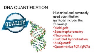
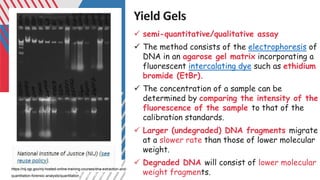
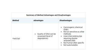
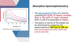
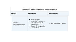
DNA Profiling
DNA QUANTIFICATION
Historical and commonly
used quantitation
methods include the
following:
•Yield gels
•Spectrophotometry
•Fluorometry
•Slot blot hybridization
•AluQuant®
•Quantitative PCR (qPCR)
Yield Gels
✓ semi-quantitative/qualitative assay
✓ The method consists of the electrophoresis of
DNA in an agarose gel matrix incorporating a
fluorescent intercalating dye such as ethidium
bromide (EtBr).
✓ The concentration of a sample can be
determined by comparing the intensity of the
fluorescence of the sample to that of the
calibration standards.
✓ Larger (undegraded) DNA fragments migrate
at a slower rate than those of lower molecular
weight.
✓ Degraded DNA will consist of lower molecular
weight fragments.
https://nij.ojp.gov/nij-hosted-online-training-courses/dna-extraction-and-
quantitation-forensic-analysts/quantitation/yield-gels
Absorption Spectrophotometry
• The optical density (O.D.) of a solution
containing 50 µg/mL of double-stranded
DNA or 40 µg/µl of single-stranded
DNA is 1.00 at a wavelength of 260nm.
• The quality or purity of the sample can
be determined by comparing the
measurements at 260 and at 280 nm
(the wavelengths for which DNA and
protein absorb)
Fluorometry
• the molecules lose some of the absorbed
energy by irradiating light of a longer
wavelength. This property is known as
fluorescence.
• There are several dyes : fluorescence
enhancement when bound to double-
stranded DNA.
• PicoGreen® is one of the more common
fluorescent dyes that can be used in this
way.
• The fluorescence is easily measured using
a fluorometer.
Fluorometry
Slot Blot Hybridization
• The majority of laboratories used the
commercially available QuantiBlot® kit,
which employs the following procedures:
Extracted DNA is
denatured to single-
stranded DNA is bound to
a positively charged nylon
membrane
a probe complementary to
the D17Z1 locus (present
in high quantities in higher
primates) is applied and
allowed to hybridize to
the DNA.
hybridized complex is
detection i.e
colorimetry/Chemiluminesc
ence,
Detection methods include:
•Colorimetry: tetramethylbenzidine
(TMB), which yields a blue color when
oxidized by hydrogen peroxide.
•Chemiluminescence: ACES (Gibco BRL)
and (Amersham Biosciences). The
chemiluminescent reactions cause the
release of photons that are captured on
film or a digital imaging device.
•Chemiluminescence is more sensitive
than colorimetry and can detect down to
10 to 20 pg of DNA
AluQuant
• Alu sequences are abundant in the human genome numbering approximately
• Alu repeats (500,000 to 1,000,000 copies per genome. )were probed in the AluQuant
Human DNA Quantitation System.
denaturing the sample
and incubating it with the
AluQuant Enzyme and
AluQ Probe solutions
production of adenosine
triphosphate (ATP)
(correlated with the
amount of DNA present)
ATP was determined by
using a luminometer
Reference
https://nij.ojp.gov/nij-hosted-online-training-courses
This Photo by Unknown Author is licensed under CC BY-SA

MSC IV SEMESTER_DNA Profiling - DNA quantification.pdf

  • 1.
  • 2.
    DNA QUANTIFICATION Historical andcommonly used quantitation methods include the following: •Yield gels •Spectrophotometry •Fluorometry •Slot blot hybridization •AluQuant® •Quantitative PCR (qPCR)
  • 3.
    Yield Gels ✓ semi-quantitative/qualitativeassay ✓ The method consists of the electrophoresis of DNA in an agarose gel matrix incorporating a fluorescent intercalating dye such as ethidium bromide (EtBr). ✓ The concentration of a sample can be determined by comparing the intensity of the fluorescence of the sample to that of the calibration standards. ✓ Larger (undegraded) DNA fragments migrate at a slower rate than those of lower molecular weight. ✓ Degraded DNA will consist of lower molecular weight fragments. https://nij.ojp.gov/nij-hosted-online-training-courses/dna-extraction-and- quantitation-forensic-analysts/quantitation/yield-gels
  • 5.
    Absorption Spectrophotometry • Theoptical density (O.D.) of a solution containing 50 µg/mL of double-stranded DNA or 40 µg/µl of single-stranded DNA is 1.00 at a wavelength of 260nm. • The quality or purity of the sample can be determined by comparing the measurements at 260 and at 280 nm (the wavelengths for which DNA and protein absorb)
  • 8.
    Fluorometry • the moleculeslose some of the absorbed energy by irradiating light of a longer wavelength. This property is known as fluorescence. • There are several dyes : fluorescence enhancement when bound to double- stranded DNA. • PicoGreen® is one of the more common fluorescent dyes that can be used in this way. • The fluorescence is easily measured using a fluorometer.
  • 9.
  • 11.
    Slot Blot Hybridization •The majority of laboratories used the commercially available QuantiBlot® kit, which employs the following procedures: Extracted DNA is denatured to single- stranded DNA is bound to a positively charged nylon membrane a probe complementary to the D17Z1 locus (present in high quantities in higher primates) is applied and allowed to hybridize to the DNA. hybridized complex is detection i.e colorimetry/Chemiluminesc ence,
  • 12.
    Detection methods include: •Colorimetry:tetramethylbenzidine (TMB), which yields a blue color when oxidized by hydrogen peroxide. •Chemiluminescence: ACES (Gibco BRL) and (Amersham Biosciences). The chemiluminescent reactions cause the release of photons that are captured on film or a digital imaging device. •Chemiluminescence is more sensitive than colorimetry and can detect down to 10 to 20 pg of DNA
  • 14.
    AluQuant • Alu sequencesare abundant in the human genome numbering approximately • Alu repeats (500,000 to 1,000,000 copies per genome. )were probed in the AluQuant Human DNA Quantitation System. denaturing the sample and incubating it with the AluQuant Enzyme and AluQ Probe solutions production of adenosine triphosphate (ATP) (correlated with the amount of DNA present) ATP was determined by using a luminometer
  • 16.
  • 17.
    This Photo byUnknown Author is licensed under CC BY-SA