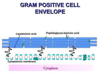
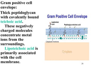
The Bacterial Cell


MORPHOLOGY OF BACTERIA


                         1
Key Words
Prokaryote                            Outer membrane
Eubacteria (Bacteria)                 Periplasmic space
Archaebacteria (Archaea)              Oxidative phosphorylation
Eukaryote                             Spheroplast/protoplast
Plasmid                               Flagella
Chromosome                            Chemotaxis
Ribosome                              Axial filament
Peptidoglycan (murein, mucopeptide)   Storage Granules
Gram stain
Gram negative                         Pili (fimbriae)
                                      Endospore (spore)
Gram positive
Cell envelope                         Capsule (slime layer, glycocalyx)
Cell membrane
Cell wall
                                                                  2
EUKARYOTES

  PROKARYOTES


EUBACTERIA   ARCHAEA

                       3
Prokaryotes (Bacteria)
• Eubacter "True" bacteria
  –   human pathogens
  –   clinical or environmental
  –   one kingdom
• Archaea
  – Environmental organisms
  – second kingdom
                                  4
Introduction
• Bacteria despite their simplicity, contain a
  well developed cell structure which is
  responsible for many of their unique
  biological properties. Many structural
  features are unique to bacteria and are not
  found among eukaryotes.
Cell Morphology
• The most elemental structural property of bacteria is cell
  morphology (shape).
• Bacteria are classified by shape into three basic groups:
      • cocci (spherical)
      • bacilli (rod-like)
      • spirochetes (spiral)
• Some bacteria are variable in shape and said to
  pleomorphic.
• The shape of a bacteria is determined by its rigid cell wall.
• The microscopic appearance of a bacterium is one of the
  most important criteria used in identification.
Bacteria come in a wide variety of shapes
• The arrangement of bacteria is important.
  Example:
     • Cocci in pairs (diplococci)
     • Cocci in chains (streptococci)
     • Cocci in clusters (staphylococci)
Bacteria Size
• Bacteria range in size from about 0.2 to 5
  um.
• The smallest bacteria (Mycoplasma) are the
  same size as a large virus.
• The longest bacteria rods are the same size
  as some yeasts and human red blood cells.
CLASSIFICATION OF
           BACTERIA
•   ON BASIS OF SHAPE
•   ON BASIS OF ARRANGEMENT
•   ON BASIS OF STAINING
•   ON BASIS OF OXYGEN
    REQUIREMENT


                              10
MORPHOLOGY OF BACTERIA
             (SHAPE)
•   COCCI -- round
•   BACILLI-- rods
•   SPIROCHETES-- spiral
•   PLEOMORPHIC


                               11
ARRANGEMENT OF
          BACTERIA

•   CHAINS
•   CLUSTERS
•   PAIRS
•   TETRADS


                        12
STAINING OF BACTERIA
• GRAM’S STAINING
  Gram positive
  Gram negative
  Gram variable


                         13
ON BASIS OF OXYGEN
        REQUIREMENT
• Aerobes
• Anaerobes
• Facultative anaerobes
 etc


                          14
STRUCTURE OF BACTERIA


• ESSENTIAL STRUCTURES
• NON-ESSENTIAL STRUCTURES


                             15
Essential structures

• Cell envelope:
   Cell wall (except Mycoplasma)
   Cell membrane
• Ribosome
• Nucleoid
• Mesosome
• Periplasm ( in gram’s negative bacteria)
                                         16
Non Essential structures

•   Capsule
•   Pilus
•   Flagellum
•   Spore
•   Plasmid , Transposons
•   Granule
•   Glycocalyx
                                      17
Bacterial Structure
Cell Structure
CELL ENVELOPE
   KEY WORDS
Cell envelope
• Cell wall
• Cell membrane


                       20
Cell envelope
– The cell envelope: Consists
  of cell membrane and cell
  wall plus an outer
  membrane if present.
– Most bacterial cell
  envelopes fall into two
  major categories: Gram
  positive and Gram
  negative based on Gram
  staining characteristics
  that reflect major
  structural differences            21
  between the two groups
GRAM POSITIVE CELL
            ENVELOPE


     Lipoteichoic acid    Peptidoglycan-teichoic acid


Cytoplasmic membrane

                         Cytoplasm
                                                        22
GRAM NEGATIVE
              CELL ENVELOPE
Outer Membrane
(Major permeability barrier)                                 Lipopolysaccharide
                                                  Porin


                          Braun lipoprotein


Periplasmic space

                               Periplasmic binding protein
Inner (cytoplasmic) membrane                                   Permease


                               Cytoplasm                                  23
Cell Walls


 Depending on Structure of their cell walls, the bacteria absorb either the
 purple dye or the pink dye.
Prokaryotic cell

Cell membrane                         Cell wall


                                      Gram +


                                       Gram -


 Cell (inner) membrane         Outer membrane
                         Cell wall       25
Cell wall
• Cell wall consists of
 the peptidoglycan layer
 attached structures
  (eg: capsule, pili etc)


                             26
PEPTIDOGLYCANS ( murein/ mucopeptide)
– Multilayered in gram positive bacteria.
– Surrounds the bacterial cell membrane.
– Provides rigidity. It is huge (billions in molecular weight)

– Consists of a glycan (polysaccharide) backbone with peptide
  side chains.
– PG is found in all bacteria except Chlamydia and
  Mycoplasma.


                                                           27
Gram positive cell
envelope:
Thick peptidoglycan
with covalently bound
teichoic acid.
   These negatively
charged molecules
concentrate metal
ions from the
surroundings.
   Lipoteichoic acid is
primarily associated
with the cell
membrane.                 28
Gram negative cell envelope

• Outer membrane present
  ( lipopolysachride, lipoprotein &phospholipid) ---
  endotoxin
  Lipid A --- toxic
  Polysaccharide core
  Outer polysaccharide -- antigenic
• Thin peptidoglycan layer
• No teichoic acid
• Periplasmic space : b/w two membranes                29
Gram negative cell envelope


• Porin protein


                              30
GRAM POSITIVE
                            Lipoteichoic acid          Peptidoglycan-teichoic acid


                    Cytoplasmic membrane

                                                   Cytoplasm

                                     GRAM NEGATIVE                Porin
                                                                                Lipopolysaccharide
Periplasmic space


                    Outer Membrane                        Braun lipoprotein


                    Inner (cytoplasmic) membrane
                                                   Cytoplasm                                   31
CELL MEMBRANE
                   .


Cell Wall
                 Cytoplasm
                                      Cell membrane


•Oxidative phosphorylation (no mitochondria).
•Synthesis of cell wall precursor.
•Active transport
•Synthesis of enzymes.                      32
CYTOPLASM
• Mesosome
 Cytoplasmic membrane invagination.
 Cell division-----DNA binding site.
 Ribosomes
 70S in size (50S & 30S)
 Protein synthesis.
  Nucleoid
• No nuclear membrane.
• DNA---Single, circular, about 2000 genes.
• No introns in DNA, no mitotic spindle, no   33
  nucleolus, no histones
Non Essential structures

•   Capsule
•   Pilus
•   Flagellum
•   Spore
•   Plasmid , Transposons
•   Granule
•   Glycocalyx
                                      34
Capsules and slime layers
                     (Glycocalyx)
• well defined: capsule
• outside cell envelope
• usually polysaccharide (in B. anthracis– D
  glutamate)
•    Gives virulance (antiphagocytic).
•    Antigenic (vaccine formation).
•    Adherence.
•    Quellung reaction (Identification).
2.   not well defined-- slime layer or glycocalyx
o    Adherence
                                                    35
o    S.mutans (Plaque formation)
FLAGELLA
•   For motility – long & whip like
•    Subunits—Flagellin
•   Respond to food/poison –chemotaxis
•   Never present in cocci.
•   Use ATPs – proton motive force
•   Number and location of flagella.
    (Peritrichous, monotrichous,
    lophotrichous, axial).         36
• Flagella
  –embedded in cell membrane
  –project as strand
  –Flagellin (protein) subunits
  –move cell by propeller like action
  –*axial filament
  –*flagellar antigen
                                    37
Flagella


A-Monotrichous; B-Lophotrichous;
C-Amphitrichous; D-Peritrichous;
Axial filaments

– spirochetes
– similar function to flagella
– run lengthwise along cell
– snake-like movement

                                 39
Pili (fimbriae)
•Short , hair-like projections of the cell
•Protein--pilin
•Mostly on gram-ve bacteria
•adhesion to host epithelium
•Two types– ordinary pili
              sex pili
•sexual conjugation (sex pili)
                                      40
Endospores (spores)
• Metabolically dormant cell
• Produced when adverse conditions eg starved –
  sporulation – bacterial DNA, cytoplasm, cell
  membrane, peptidoglycans, water & keratin- like coat
• Resistant to adverse conditions
  - high temperatures (not killed by boiling),
  - radiation, dehydration
  - organic solvents
• Killed by autoclaving (121 C for 30 min)
• contain dipicolinic acid
• Bacillus (central) and Clostridium (terminal)
• Survival for many years                          41
CYTOPLASM
  Granules
  – Storage of nutrients
  – Stained with dyes
  Transposons
 Jumping genes – within DNA or between DNAs of
  bacteria, plasmids & bacteriophages
 Pieces of DNA
 No independent replication.
 Can be more than one in 1 DNA.
 For toxins, enzymes, antibiotic resistance etc.
 4 domains– inverted repeats, transposase, repressor,
  resistance.                                       42
Plasmids
 Multiple copies in number
 Extra-chromosomal DNA, double
  stranded, circular
 Can be incorporated in DNA
 Coding pathogenesis and antibiotic
  resistance, heavy metal & U-V light
  resistance, toxins, pili etc.
 Replication independent of bacterial
  chromosome.
Transmissable & Non transmissable.      43
Making Wall-less forms
• Result from action of:
  – enzymes lytic for cell wall (eg lyzozyme)
  – antibiotics inhibiting peptidoglycan biosynthesis

• Wall-less bacteria that don’t replicate:
  – spheroplasts (with outer membrane) gram - ve
  – protoplasts (no outer membrane) gram +ve.

• Wall-less bacteria that replicate
  – L forms ( relative resistance to antibiotics)

                                                    44
Naturally Wall-less Genus
    • Mycloplasma


                            45
The Cell Envelope


Gram Positive   Gram Negative


                                46
GRAM STAIN
• Gram positive
• Gram negative
• Gram variable


                       47
Procedure
           of Gram Staining
• Developed by Christian Gram
• In 1884
• 4 steps:
 Stain with Crystal voilet (primary stain)
 Then pour Gram,s iodine (mordant)
 Decolourize
 Pour Safranin (counter stain)
                                              48
Gram negative                Gram positive
                Heat/Dry
         Crystal violet stain

                Iodine Fix

           Alcohol de-stain
                   de

            Safranin stain
                                             49
Gram stain morphology
• Shape
   – cocci (round)
   – bacilli (rods)
   – spiral or curved (e.g. spirochetes)

• Single or multiple cells
   – clusters (e.g. staphylococci)
   – chains (e.g. streptococci)

• Gram positive or negative
                                           50
51
52

Morphoofbacteria 110725164319-phpapp01

  • 1.
  • 2.
    Key Words Prokaryote Outer membrane Eubacteria (Bacteria) Periplasmic space Archaebacteria (Archaea) Oxidative phosphorylation Eukaryote Spheroplast/protoplast Plasmid Flagella Chromosome Chemotaxis Ribosome Axial filament Peptidoglycan (murein, mucopeptide) Storage Granules Gram stain Gram negative Pili (fimbriae) Endospore (spore) Gram positive Cell envelope Capsule (slime layer, glycocalyx) Cell membrane Cell wall 2
  • 3.
  • 4.
    Prokaryotes (Bacteria) • Eubacter"True" bacteria – human pathogens – clinical or environmental – one kingdom • Archaea – Environmental organisms – second kingdom 4
  • 5.
    Introduction • Bacteria despitetheir simplicity, contain a well developed cell structure which is responsible for many of their unique biological properties. Many structural features are unique to bacteria and are not found among eukaryotes.
  • 6.
    Cell Morphology • Themost elemental structural property of bacteria is cell morphology (shape). • Bacteria are classified by shape into three basic groups: • cocci (spherical) • bacilli (rod-like) • spirochetes (spiral) • Some bacteria are variable in shape and said to pleomorphic. • The shape of a bacteria is determined by its rigid cell wall. • The microscopic appearance of a bacterium is one of the most important criteria used in identification.
  • 7.
    Bacteria come ina wide variety of shapes
  • 8.
    • The arrangementof bacteria is important. Example: • Cocci in pairs (diplococci) • Cocci in chains (streptococci) • Cocci in clusters (staphylococci)
  • 9.
    Bacteria Size • Bacteriarange in size from about 0.2 to 5 um. • The smallest bacteria (Mycoplasma) are the same size as a large virus. • The longest bacteria rods are the same size as some yeasts and human red blood cells.
  • 10.
    CLASSIFICATION OF BACTERIA • ON BASIS OF SHAPE • ON BASIS OF ARRANGEMENT • ON BASIS OF STAINING • ON BASIS OF OXYGEN REQUIREMENT 10
  • 11.
    MORPHOLOGY OF BACTERIA (SHAPE) • COCCI -- round • BACILLI-- rods • SPIROCHETES-- spiral • PLEOMORPHIC 11
  • 12.
    ARRANGEMENT OF BACTERIA • CHAINS • CLUSTERS • PAIRS • TETRADS 12
  • 13.
    STAINING OF BACTERIA •GRAM’S STAINING Gram positive Gram negative Gram variable 13
  • 14.
    ON BASIS OFOXYGEN REQUIREMENT • Aerobes • Anaerobes • Facultative anaerobes etc 14
  • 15.
    STRUCTURE OF BACTERIA •ESSENTIAL STRUCTURES • NON-ESSENTIAL STRUCTURES 15
  • 16.
    Essential structures • Cellenvelope: Cell wall (except Mycoplasma) Cell membrane • Ribosome • Nucleoid • Mesosome • Periplasm ( in gram’s negative bacteria) 16
  • 17.
    Non Essential structures • Capsule • Pilus • Flagellum • Spore • Plasmid , Transposons • Granule • Glycocalyx 17
  • 18.
  • 19.
  • 20.
    CELL ENVELOPE KEY WORDS Cell envelope • Cell wall • Cell membrane 20
  • 21.
    Cell envelope – Thecell envelope: Consists of cell membrane and cell wall plus an outer membrane if present. – Most bacterial cell envelopes fall into two major categories: Gram positive and Gram negative based on Gram staining characteristics that reflect major structural differences 21 between the two groups
  • 22.
    GRAM POSITIVE CELL ENVELOPE Lipoteichoic acid Peptidoglycan-teichoic acid Cytoplasmic membrane Cytoplasm 22
  • 23.
    GRAM NEGATIVE CELL ENVELOPE Outer Membrane (Major permeability barrier) Lipopolysaccharide Porin Braun lipoprotein Periplasmic space Periplasmic binding protein Inner (cytoplasmic) membrane Permease Cytoplasm 23
  • 24.
    Cell Walls Dependingon Structure of their cell walls, the bacteria absorb either the purple dye or the pink dye.
  • 25.
    Prokaryotic cell Cell membrane Cell wall Gram + Gram - Cell (inner) membrane Outer membrane Cell wall 25
  • 26.
    Cell wall • Cellwall consists of  the peptidoglycan layer  attached structures (eg: capsule, pili etc) 26
  • 27.
    PEPTIDOGLYCANS ( murein/mucopeptide) – Multilayered in gram positive bacteria. – Surrounds the bacterial cell membrane. – Provides rigidity. It is huge (billions in molecular weight) – Consists of a glycan (polysaccharide) backbone with peptide side chains. – PG is found in all bacteria except Chlamydia and Mycoplasma. 27
  • 28.
    Gram positive cell envelope: Thickpeptidoglycan with covalently bound teichoic acid. These negatively charged molecules concentrate metal ions from the surroundings. Lipoteichoic acid is primarily associated with the cell membrane. 28
  • 29.
    Gram negative cellenvelope • Outer membrane present ( lipopolysachride, lipoprotein &phospholipid) --- endotoxin Lipid A --- toxic Polysaccharide core Outer polysaccharide -- antigenic • Thin peptidoglycan layer • No teichoic acid • Periplasmic space : b/w two membranes 29
  • 30.
    Gram negative cellenvelope • Porin protein 30
  • 31.
    GRAM POSITIVE Lipoteichoic acid Peptidoglycan-teichoic acid Cytoplasmic membrane Cytoplasm GRAM NEGATIVE Porin Lipopolysaccharide Periplasmic space Outer Membrane Braun lipoprotein Inner (cytoplasmic) membrane Cytoplasm 31
  • 32.
    CELL MEMBRANE . Cell Wall Cytoplasm Cell membrane •Oxidative phosphorylation (no mitochondria). •Synthesis of cell wall precursor. •Active transport •Synthesis of enzymes. 32
  • 33.
    CYTOPLASM • Mesosome  Cytoplasmicmembrane invagination.  Cell division-----DNA binding site. Ribosomes  70S in size (50S & 30S)  Protein synthesis. Nucleoid • No nuclear membrane. • DNA---Single, circular, about 2000 genes. • No introns in DNA, no mitotic spindle, no 33 nucleolus, no histones
  • 34.
    Non Essential structures • Capsule • Pilus • Flagellum • Spore • Plasmid , Transposons • Granule • Glycocalyx 34
  • 35.
    Capsules and slimelayers (Glycocalyx) • well defined: capsule • outside cell envelope • usually polysaccharide (in B. anthracis– D glutamate) • Gives virulance (antiphagocytic). • Antigenic (vaccine formation). • Adherence. • Quellung reaction (Identification). 2. not well defined-- slime layer or glycocalyx o Adherence 35 o S.mutans (Plaque formation)
  • 36.
    FLAGELLA • For motility – long & whip like • Subunits—Flagellin • Respond to food/poison –chemotaxis • Never present in cocci. • Use ATPs – proton motive force • Number and location of flagella. (Peritrichous, monotrichous, lophotrichous, axial). 36
  • 37.
    • Flagella –embedded in cell membrane –project as strand –Flagellin (protein) subunits –move cell by propeller like action –*axial filament –*flagellar antigen 37
  • 38.
  • 39.
    Axial filaments – spirochetes –similar function to flagella – run lengthwise along cell – snake-like movement 39
  • 40.
    Pili (fimbriae) •Short ,hair-like projections of the cell •Protein--pilin •Mostly on gram-ve bacteria •adhesion to host epithelium •Two types– ordinary pili sex pili •sexual conjugation (sex pili) 40
  • 41.
    Endospores (spores) • Metabolicallydormant cell • Produced when adverse conditions eg starved – sporulation – bacterial DNA, cytoplasm, cell membrane, peptidoglycans, water & keratin- like coat • Resistant to adverse conditions - high temperatures (not killed by boiling), - radiation, dehydration - organic solvents • Killed by autoclaving (121 C for 30 min) • contain dipicolinic acid • Bacillus (central) and Clostridium (terminal) • Survival for many years 41
  • 42.
    CYTOPLASM Granules – Storage of nutrients – Stained with dyes Transposons  Jumping genes – within DNA or between DNAs of bacteria, plasmids & bacteriophages  Pieces of DNA  No independent replication.  Can be more than one in 1 DNA.  For toxins, enzymes, antibiotic resistance etc.  4 domains– inverted repeats, transposase, repressor, resistance. 42
  • 43.
    Plasmids  Multiple copiesin number  Extra-chromosomal DNA, double stranded, circular  Can be incorporated in DNA  Coding pathogenesis and antibiotic resistance, heavy metal & U-V light resistance, toxins, pili etc.  Replication independent of bacterial chromosome. Transmissable & Non transmissable. 43
  • 44.
    Making Wall-less forms •Result from action of: – enzymes lytic for cell wall (eg lyzozyme) – antibiotics inhibiting peptidoglycan biosynthesis • Wall-less bacteria that don’t replicate: – spheroplasts (with outer membrane) gram - ve – protoplasts (no outer membrane) gram +ve. • Wall-less bacteria that replicate – L forms ( relative resistance to antibiotics) 44
  • 45.
    Naturally Wall-less Genus • Mycloplasma 45
  • 46.
    The Cell Envelope GramPositive Gram Negative 46
  • 47.
    GRAM STAIN • Grampositive • Gram negative • Gram variable 47
  • 48.
    Procedure of Gram Staining • Developed by Christian Gram • In 1884 • 4 steps:  Stain with Crystal voilet (primary stain)  Then pour Gram,s iodine (mordant)  Decolourize  Pour Safranin (counter stain) 48
  • 49.
    Gram negative Gram positive Heat/Dry Crystal violet stain Iodine Fix Alcohol de-stain de Safranin stain 49
  • 50.
    Gram stain morphology •Shape – cocci (round) – bacilli (rods) – spiral or curved (e.g. spirochetes) • Single or multiple cells – clusters (e.g. staphylococci) – chains (e.g. streptococci) • Gram positive or negative 50
  • 51.
  • 52.

Editor's Notes

  • #10 the bacterial cell wall provides structural integrity to the cell. In prokaryotes, the primary function of the cell wall is to protect the cell from internal turgor pressure caused by the much higher concentrations of proteins and other molecules inside the cell compared to its external environment. The bacterial cell wall differs from that of all other organisms by the presence of peptidoglycan (poly- N -acetyl glucosamine and  N -acetylmuramic acid), which is located immediately outside of the cytoplasmic membrane. Peptidoglycan is responsible for the rigidity of the bacterial cell wall and for the determination of cell shape. It is relatively porous and is not considered to be a permeability barrier for small substrates. While all bacterial cell walls (with a few exceptions e.g. intracellular parasites such as  Mycoplasma ) contain peptidoglycan, not all cell walls have the same overall structures. Since the cell wall is required for bacterial survival, but is absent in eukaryotes, several antibiotics (penicillins and cephalosporins) stop bacterial infections by interfering with cell wall synthesis, while having no effects on human cells.