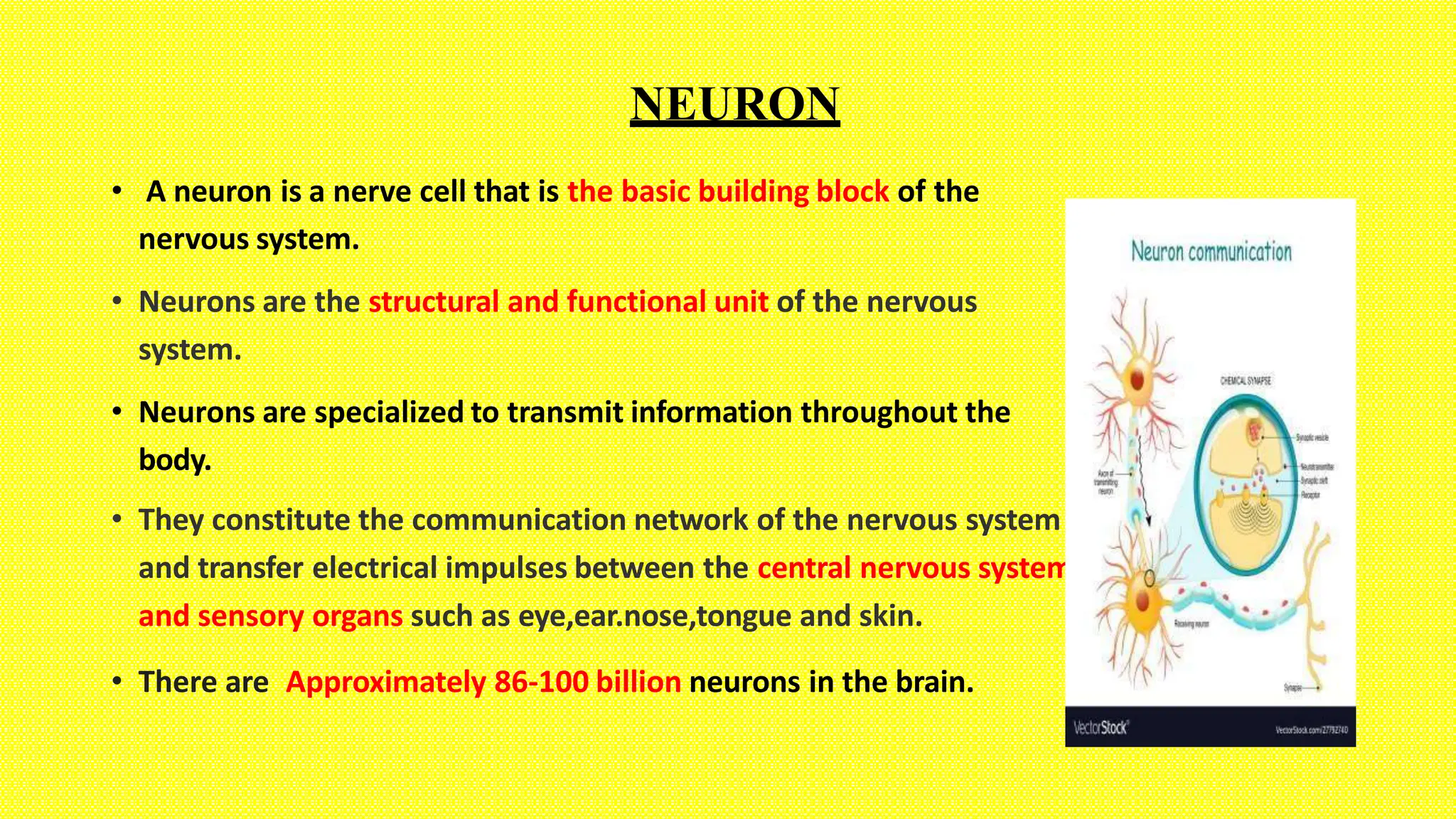
THE NERVOUS SYSTEM CONTAINS TWO MAIN TYPES OF CELLS:
NEURONS GLIAL CELLS
NEURON
• A neuron is a nerve cell that is the basic building block of the
nervous system.
• Neurons are the structural and functional unit of the nervous
system.
• Neurons are specialized to transmit information throughout the
body.
• They constitute the communication network of the nervous system
and transfer electrical impulses between the central nervous system
and sensory organs such as eye,ear.nose,tongue and skin.
• There are Approximately 86-100 billion neurons in the brain.
PARTS OF NEURON
1. DENDRITES
 Dendrites are the tree-like branched structures that arise from the nerve cell body.
• Apart from the main dendrite branches, dendrites may contain additional protrusions
known as dendrite spines.
 These small membranous protrusions receive input from the axon of another cell and
thus play an important role in the transmission of nerve impulses .
 As an expansion of the cell body, dendrites and dendrite spines also contain
cytoplasm and different types of organelles.
 In particular, dendrite spines contain a variety of microtubules and some
neurofilaments that contribute to the changes observed in their shape.
2. AXON HILLOCK
• The axon hillock is a specialized region from which the
axon extends.
 It is the area at which the axon is attached to the cell
body.
 Initial segment - this is the region between theAxon
and Hillock and the front part of the myelin sheath.
 This region is said to be the area for the initiation of
action potential.[Electrical impulse]
 TheAxon Hillock is cone-shaped.
3.THE AXON
 The axon is a single elongated tubal structure that extends from the Axon Hillock.
 Each neuron has a single axon that extends and branches at its end.
 The axon contains microtubules (along the length of the axon) and specialized,
insulating substances known as myelin on its surface that boost the transmission of
nerve impulses.
 Myeline sheath is covering the axon of the nerves in brain and spinal cord.
 Myelin sheath is made up of cells (Schwann cells) wrapping themselves around the
axon.
 These cells produce the fatty and protein substance called myelin.
 The inner most Plasma membrane around the axon is Axolemma.
 Neurilemma is the plasma membrane of schwann cells .
 The spaces/gaps between the Schwann cells are known as the nodes of
Ranvier and they serve to propagate electrical signals along the axon.
 Myelin incisures (also known as Schmidt-Lanterman clefts, Schmidt-Lanterman
incisures] are small pockets of cytoplasm left behind during the Schwann cell
myelination process.
 Thebranched end of the axon is known as the axon terminal[arborization] and
branches at the middle of the axon is axon collaterals .
4. NERVE ENDING/ AXON TERMINAL
 This is the distal part of the axon that comes in
contact with other cells. Also called as terminal
boutons.
 This part of the axon is largely involved in the
release of the neurotransmitter,
[EX: acetylcholine for learning, glutamate for
memory, dopamine for pleasure, adrenaline for
mitochondria
fight or flight etc].
 It contains a large number of that
produce the energy required to facilitate the
process.
5. THE CELL BODY OR SOMA
6. Ribosomes,
7. Lysosomes,
8. Endosomes,
9.And Peroxisomes.
 The cell body, also called the soma, is the spherical part of the neuron
that contains the nucleus ,cytoplasm and organelles.
 The cell body connects to the dendrites, and send information to the
axon depending on the strength of the signal.
 The neuronal cytoplasm have the following
organelles
1. The Nucleus,
2. Nucleolus,
3. Endoplasmic Reticulum,
4. Golgi Apparatus,
5. Mitochondria,
Functions of the Cell Body.
 The job of the cell body is to control all of the functions of the cell.
It contains several important organelles that help it do this.
 Organelles are tiny organs in the cell that each do a specific job.
 The most important organelle in the cell body is the nucleus.
The nucleus contains the cell's DNA and regulates all processes in
the cell.
 The cell needs DNA to act as a blueprint to direct cellular activity.
 The nucleus also contains the nucleolus, which makes ribosomes
needed for protein production.
 Endoplasmic reticulum plays role in synthesis, folding,
modification and transport of protein.
 Mitochondria are powerhouses and provide energy.
 Lysosome breakdown the bacteria and digest them and do
protective function.
 Ribosomes produce ribo neuclic acid and ribo proteins.
 Endosomes do protein trafficking/ storing.
 Peroxisomes will breakdown toxic materials by using
various enzymes.
TYPES OF CELL BODIES.
1. A unipolar neuron is a type of neuron in which only one process called a
neurite extends from the cell body.
2. A bipolar neuron is a type of neuron which has two extensions (one axon
and one dendrite).
3. A multipolar neuron is a type of neuron that possesses a single axon and
many dendrites (and dendritic branches), allowing for the integration of a
great deal of information from other neurons.
4. A pseudounipolar neuron is a type of neuron which has one extension from
its cell body. This type of neuron contains an axon that has split into two
branches; one branch travels to the PNS and the other to the CNS.
TYPES OF NEURON ACCORDING TO IT’S FUNCTION
They are three types of neurons based on the function as follows:
• Sensory Neuron
• Inter-Neuron
• Motor Neuron
The dorsal (posterior) or sensory root bears a dorsal root ganglion (DRG) containing the cell bodies of
the sensory neurons.
The ventral (anterior) or motor root consists of axons from the lower motor neurons in the ventral horn of
the spinal cord
Sensory Neuron Motor Neuron
• Neurons that carry sensory impulse from
sensory organs to the central nervous
system are known as sensory neurons
• A neuron that carries motor impulses
from the central nervous system to
specific effectors is known as motor
neurons.
• They are located in the dorsal root ganglion
of the spinal nerve
• They are located in the ventral root
ganglion of the spinal cord.
• It is unipolar • It is multipolar
• Comprises of a short axon • Comprises of a long axon
• An adult has an average of 10 million
sensory nerves in the body
• Half million of motor[5million] neurons
are found in the body
• Found in eyes, skin, ears, tongue and nose • Found in muscles and glands
INTERNEURON:
 Interneurons (also known as association neurons) are neurons that are
found exclusively in the CNS
 Generally it can be refered to any neuron that acts as an intermediary
in passing signals between two neurons.
 Interneurons are the central nodes of neural circuits, enabling
communication between sensory or motor neurons and the (CNS).
MYELINATED AND UNMYELINATED NEURON
MYELINATED VS UNMYELINATED NEURON
BASIS OF
COMPARISON
MYELINATED NERVE FIBER UNMYELINATED NERVE FIBER
Description
• Myelinated Nerve Fibers are
nerve fibers that are insulated by
a myelin sheath.
• Unmyelinated Nerve fibers are
nerve fibers that do not have a
myelin sheath.
Color
• The myelinated nerve fibers are
white in color.
• The unmyelinated nerve fibers are
gray in color.
Nodes of
Ranvier
• The myelinated nerve fibers have
nodes of Ranvier.
• Unmyelinated nerve fibers do not
have nodes of Ranvier.
Speed of
Transmission
• Due to presence of nodes of
Ranvier on myelinated nerve
fibers, the speed of transmission
of nerve impulses is high in
myelinated nerve fibers.
• Unmyelinated nerve fibers do not
have myelin insulations, and
therefore, the speed of the
transmission of the nerve impulses
is low.
Location
• Most neurons in the central and
few in peripheral nervous
system are myelinated because
they require fast conduction
speed such as neuron involved
in spinal reflexes.[stretch and
withdrawal]
• found in both the peripheral and
central nervous system in the group c
nerve fibers, responsible for
transmission of secondary pain or
itch.
• More in pns
[After a chemical burn first severe
burning pain followed by mild burning
sensation.]
Impulse
Conduction
• Due to presence of myelin
sheath, myelinated nerves do
not lose the impulse during
conduction
• Unmyelinated nerve fibers can lose
the nerve impulse during conduction.
Axons
• The nerve fibers with long axons
are myelinated.
• The short axon nerve fibers are
unmyelinated.
GLIAL CELLS
Glial cells (named from the Greek word for "glue") are non-
neuronal cells that
• provide support and nutrition,
• maintain homeostasis,
• form myelin,
• and participate in signal transmission in the nervous system.
THERE ARE SIX TYPES OF NEUROGLIA.
The four types of neuroglia found in the central
nervous system are
1,Astrocytes
2. Microglial Cells
3.Ependymal Cells
4.And Oligodendrocytes.
The two types of neuroglia found in the
peripheral nervous system are
1.Satellite Cells 2. And Schwann Cells.
1.ASTROCYTES
• A STAR-SHAPED GLIAL CELL OF THE CENTRAL NERVOUS
SYSTEM
• These cells fill in the spaces between neurons and
provide for structural integrity.
• They also have processes which join to the capillary
blood vessels. These are known as perivascular end
feet.
• Therefore, with their close apposition to blood vessels,
they are also thought to be responsible for metabolite
exchange between the neurons and the vasculature.
• THEYARE FOUND IN THE CNS.
• Microglia are a type of
neuroglia (glial cell) located
throughout the brain and spinal
cord.
• Microglia account for 10–15%
of all cells found within the brain.
• As the resident macrophage
cells, they act as the first and
main form of active immune
defense in the central nervous
system (CNS).
2.MICROGLIA
3.Ependymal cells
• The ependyma is the thin neuro-epithelial lining of
the ventricular system of the brain and the central
canal of the spinal cord.
• There are three types of ependymal cells—
1.The ependymocytes allow for the free movement of
molecules between the cerebrospinal fluid (CSF) and the
neurons.
2.Tanycytes are generally found in the third ventricle and can
be involved in responding to changing hormonal levels of the
blood derived hormones in the CSF. [prolactin,Hcg]
3.Choroidal epithelial cells are the cells which control the
chemical composition of the CSF. THEY ARE FOUND IN THE CNS.
4.Oligodendrocytes
with
similar to an
LESS
A glial cell
astrocyte but
protuberances, concerned with
the production of myelin in the
central nervous system.
THEYARE FOUND IN THE CNS.
5. SCHWANN CELLS
a type of
• Schwann cells are
glial cells of the PNS.
• Help to form the myelin sheath
around the nerve fibers.
schwann cell envelops and
• when a
rotates
myelin
around the axon forming
sheath, now axon is
myelinated.
• They also have an additional role
in phagocytosis of any debris; therefore
help to clean the surrounding
environment.
6.SATELLITE CELLS
Satellite cells are small, flattened cells
,these cells surround those neurons of
the PNS.
Satellite cells also act as protective,
cushioning cells.
They maintain a stable chemical
balance of the surrounding environment
to the neurons.
They are therefore found in the PNS.
RADIAL GLIA
• Radial glial cells are progenitor cells
that can generate neurons, astrocytes
and oligodendrocytes.
• Radial glia are specialized cells in the
developing nervous system of all
vertebrates, and are characterized by
long radial processes.
ENTERIC GLIAL CELLS
• Enteric glial cells are found in the nerves in the
digestive system.
• Beneath the intestinal epithelial cells lies a
number of of astrocyte-like cells that are known
as enteric glia.
• The enteric nervous system (ENS), also known
as second brain, innervates our gastrointestinal
tract controlling its functions, such as motility,
fluid secretion, nutrient absorption, and even
involvement in the control of immunity and
inflammatory processes.
.
NERVE CELLS FINAL( NEURON AND GLIAL CELLS.pptx FOR NURSING STUDENTS

NERVE CELLS FINAL( NEURON AND GLIAL CELLS.pptx FOR NURSING STUDENTS

  • 2.
    THE NERVOUS SYSTEMCONTAINS TWO MAIN TYPES OF CELLS: NEURONS GLIAL CELLS
  • 3.
    NEURON • A neuronis a nerve cell that is the basic building block of the nervous system. • Neurons are the structural and functional unit of the nervous system. • Neurons are specialized to transmit information throughout the body. • They constitute the communication network of the nervous system and transfer electrical impulses between the central nervous system and sensory organs such as eye,ear.nose,tongue and skin. • There are Approximately 86-100 billion neurons in the brain.
  • 5.
    PARTS OF NEURON 1.DENDRITES  Dendrites are the tree-like branched structures that arise from the nerve cell body. • Apart from the main dendrite branches, dendrites may contain additional protrusions known as dendrite spines.  These small membranous protrusions receive input from the axon of another cell and thus play an important role in the transmission of nerve impulses .  As an expansion of the cell body, dendrites and dendrite spines also contain cytoplasm and different types of organelles.  In particular, dendrite spines contain a variety of microtubules and some neurofilaments that contribute to the changes observed in their shape.
  • 7.
    2. AXON HILLOCK •The axon hillock is a specialized region from which the axon extends.  It is the area at which the axon is attached to the cell body.  Initial segment - this is the region between theAxon and Hillock and the front part of the myelin sheath.  This region is said to be the area for the initiation of action potential.[Electrical impulse]  TheAxon Hillock is cone-shaped.
  • 10.
    3.THE AXON  Theaxon is a single elongated tubal structure that extends from the Axon Hillock.  Each neuron has a single axon that extends and branches at its end.  The axon contains microtubules (along the length of the axon) and specialized, insulating substances known as myelin on its surface that boost the transmission of nerve impulses.  Myeline sheath is covering the axon of the nerves in brain and spinal cord.  Myelin sheath is made up of cells (Schwann cells) wrapping themselves around the axon.  These cells produce the fatty and protein substance called myelin.
  • 11.
     The innermost Plasma membrane around the axon is Axolemma.  Neurilemma is the plasma membrane of schwann cells .  The spaces/gaps between the Schwann cells are known as the nodes of Ranvier and they serve to propagate electrical signals along the axon.  Myelin incisures (also known as Schmidt-Lanterman clefts, Schmidt-Lanterman incisures] are small pockets of cytoplasm left behind during the Schwann cell myelination process.  Thebranched end of the axon is known as the axon terminal[arborization] and branches at the middle of the axon is axon collaterals .
  • 13.
    4. NERVE ENDING/AXON TERMINAL  This is the distal part of the axon that comes in contact with other cells. Also called as terminal boutons.  This part of the axon is largely involved in the release of the neurotransmitter, [EX: acetylcholine for learning, glutamate for memory, dopamine for pleasure, adrenaline for mitochondria fight or flight etc].  It contains a large number of that produce the energy required to facilitate the process.
  • 14.
    5. THE CELLBODY OR SOMA 6. Ribosomes, 7. Lysosomes, 8. Endosomes, 9.And Peroxisomes.  The cell body, also called the soma, is the spherical part of the neuron that contains the nucleus ,cytoplasm and organelles.  The cell body connects to the dendrites, and send information to the axon depending on the strength of the signal.  The neuronal cytoplasm have the following organelles 1. The Nucleus, 2. Nucleolus, 3. Endoplasmic Reticulum, 4. Golgi Apparatus, 5. Mitochondria,
  • 16.
    Functions of theCell Body.  The job of the cell body is to control all of the functions of the cell. It contains several important organelles that help it do this.  Organelles are tiny organs in the cell that each do a specific job.  The most important organelle in the cell body is the nucleus. The nucleus contains the cell's DNA and regulates all processes in the cell.  The cell needs DNA to act as a blueprint to direct cellular activity.  The nucleus also contains the nucleolus, which makes ribosomes needed for protein production.
  • 17.
     Endoplasmic reticulumplays role in synthesis, folding, modification and transport of protein.  Mitochondria are powerhouses and provide energy.  Lysosome breakdown the bacteria and digest them and do protective function.  Ribosomes produce ribo neuclic acid and ribo proteins.  Endosomes do protein trafficking/ storing.  Peroxisomes will breakdown toxic materials by using various enzymes.
  • 18.
    TYPES OF CELLBODIES. 1. A unipolar neuron is a type of neuron in which only one process called a neurite extends from the cell body. 2. A bipolar neuron is a type of neuron which has two extensions (one axon and one dendrite). 3. A multipolar neuron is a type of neuron that possesses a single axon and many dendrites (and dendritic branches), allowing for the integration of a great deal of information from other neurons. 4. A pseudounipolar neuron is a type of neuron which has one extension from its cell body. This type of neuron contains an axon that has split into two branches; one branch travels to the PNS and the other to the CNS.
  • 20.
    TYPES OF NEURONACCORDING TO IT’S FUNCTION They are three types of neurons based on the function as follows: • Sensory Neuron • Inter-Neuron • Motor Neuron
  • 21.
    The dorsal (posterior)or sensory root bears a dorsal root ganglion (DRG) containing the cell bodies of the sensory neurons. The ventral (anterior) or motor root consists of axons from the lower motor neurons in the ventral horn of the spinal cord
  • 23.
    Sensory Neuron MotorNeuron • Neurons that carry sensory impulse from sensory organs to the central nervous system are known as sensory neurons • A neuron that carries motor impulses from the central nervous system to specific effectors is known as motor neurons. • They are located in the dorsal root ganglion of the spinal nerve • They are located in the ventral root ganglion of the spinal cord. • It is unipolar • It is multipolar • Comprises of a short axon • Comprises of a long axon • An adult has an average of 10 million sensory nerves in the body • Half million of motor[5million] neurons are found in the body • Found in eyes, skin, ears, tongue and nose • Found in muscles and glands
  • 24.
    INTERNEURON:  Interneurons (alsoknown as association neurons) are neurons that are found exclusively in the CNS  Generally it can be refered to any neuron that acts as an intermediary in passing signals between two neurons.  Interneurons are the central nodes of neural circuits, enabling communication between sensory or motor neurons and the (CNS).
  • 26.
  • 28.
    MYELINATED VS UNMYELINATEDNEURON BASIS OF COMPARISON MYELINATED NERVE FIBER UNMYELINATED NERVE FIBER Description • Myelinated Nerve Fibers are nerve fibers that are insulated by a myelin sheath. • Unmyelinated Nerve fibers are nerve fibers that do not have a myelin sheath. Color • The myelinated nerve fibers are white in color. • The unmyelinated nerve fibers are gray in color. Nodes of Ranvier • The myelinated nerve fibers have nodes of Ranvier. • Unmyelinated nerve fibers do not have nodes of Ranvier. Speed of Transmission • Due to presence of nodes of Ranvier on myelinated nerve fibers, the speed of transmission of nerve impulses is high in myelinated nerve fibers. • Unmyelinated nerve fibers do not have myelin insulations, and therefore, the speed of the transmission of the nerve impulses is low.
  • 29.
    Location • Most neuronsin the central and few in peripheral nervous system are myelinated because they require fast conduction speed such as neuron involved in spinal reflexes.[stretch and withdrawal] • found in both the peripheral and central nervous system in the group c nerve fibers, responsible for transmission of secondary pain or itch. • More in pns [After a chemical burn first severe burning pain followed by mild burning sensation.] Impulse Conduction • Due to presence of myelin sheath, myelinated nerves do not lose the impulse during conduction • Unmyelinated nerve fibers can lose the nerve impulse during conduction. Axons • The nerve fibers with long axons are myelinated. • The short axon nerve fibers are unmyelinated.
  • 31.
    GLIAL CELLS Glial cells(named from the Greek word for "glue") are non- neuronal cells that • provide support and nutrition, • maintain homeostasis, • form myelin, • and participate in signal transmission in the nervous system.
  • 33.
    THERE ARE SIXTYPES OF NEUROGLIA. The four types of neuroglia found in the central nervous system are 1,Astrocytes 2. Microglial Cells 3.Ependymal Cells 4.And Oligodendrocytes. The two types of neuroglia found in the peripheral nervous system are 1.Satellite Cells 2. And Schwann Cells.
  • 34.
    1.ASTROCYTES • A STAR-SHAPEDGLIAL CELL OF THE CENTRAL NERVOUS SYSTEM • These cells fill in the spaces between neurons and provide for structural integrity. • They also have processes which join to the capillary blood vessels. These are known as perivascular end feet. • Therefore, with their close apposition to blood vessels, they are also thought to be responsible for metabolite exchange between the neurons and the vasculature. • THEYARE FOUND IN THE CNS.
  • 35.
    • Microglia area type of neuroglia (glial cell) located throughout the brain and spinal cord. • Microglia account for 10–15% of all cells found within the brain. • As the resident macrophage cells, they act as the first and main form of active immune defense in the central nervous system (CNS). 2.MICROGLIA
  • 36.
    3.Ependymal cells • Theependyma is the thin neuro-epithelial lining of the ventricular system of the brain and the central canal of the spinal cord. • There are three types of ependymal cells— 1.The ependymocytes allow for the free movement of molecules between the cerebrospinal fluid (CSF) and the neurons. 2.Tanycytes are generally found in the third ventricle and can be involved in responding to changing hormonal levels of the blood derived hormones in the CSF. [prolactin,Hcg] 3.Choroidal epithelial cells are the cells which control the chemical composition of the CSF. THEY ARE FOUND IN THE CNS.
  • 37.
    4.Oligodendrocytes with similar to an LESS Aglial cell astrocyte but protuberances, concerned with the production of myelin in the central nervous system. THEYARE FOUND IN THE CNS.
  • 38.
    5. SCHWANN CELLS atype of • Schwann cells are glial cells of the PNS. • Help to form the myelin sheath around the nerve fibers. schwann cell envelops and • when a rotates myelin around the axon forming sheath, now axon is myelinated. • They also have an additional role in phagocytosis of any debris; therefore help to clean the surrounding environment.
  • 39.
    6.SATELLITE CELLS Satellite cellsare small, flattened cells ,these cells surround those neurons of the PNS. Satellite cells also act as protective, cushioning cells. They maintain a stable chemical balance of the surrounding environment to the neurons. They are therefore found in the PNS.
  • 40.
    RADIAL GLIA • Radialglial cells are progenitor cells that can generate neurons, astrocytes and oligodendrocytes. • Radial glia are specialized cells in the developing nervous system of all vertebrates, and are characterized by long radial processes.
  • 41.
    ENTERIC GLIAL CELLS •Enteric glial cells are found in the nerves in the digestive system. • Beneath the intestinal epithelial cells lies a number of of astrocyte-like cells that are known as enteric glia. • The enteric nervous system (ENS), also known as second brain, innervates our gastrointestinal tract controlling its functions, such as motility, fluid secretion, nutrient absorption, and even involvement in the control of immunity and inflammatory processes. .