SlideShare a Scribd company logo
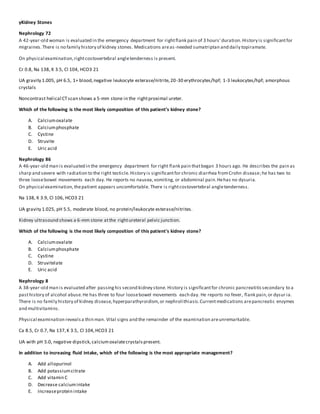
yKidney Stones
Nephrology 72
A 42-year-old woman is evaluated in the emergency department for rightflank pain of 3 hours' duration.History is significantfor
migraines.There is no family history of kidney stones. Medications areas-needed sumatriptan and daily topiramate.
On physical examination,rightcostovertebral angletenderness is present.
Cr 0.8, Na 138, K 3.5, Cl 104, HCO3 21
UA gravity 1.005, pH 6.5, 1+ blood,negative leukocyte esterase/nitrite,20-30 erythrocytes/hpf; 1-3 leukocytes/hpf; amorphous
crystals
Noncontrast helical CTscan shows a 5-mm stone in the rightproximal ureter.
Which of the following is the most likely composition of this patient’s kidney stone?
A. Calciumoxalate
B. Calciumphosphate
C. Cystine
D. Struvite
E. Uric acid
Nephrology 86
A 46-year-old man is evaluated in the emergency department for right flank pain thatbegan 3 hours ago. He describes the pain as
sharp and severe with radiation to the right testicle.History is significantfor chronic diarrhea fromCrohn disease;he has two to
three loosebowel movements each day. He reports no nausea,vomiting, or abdominal pain.Hehas no dysuria.
On physical examination,thepatient appears uncomfortable. There is rightcostovertebral angletenderness.
Na 138, K 3.9, Cl 106, HCO3 21
UA gravity 1.025, pH 5.5, moderate blood, no protein/leukocyte esterase/nitrites.
Kidney ultrasound shows a 6-mm stone atthe rightureteral pelvic junction.
Which of the following is the most likely composition of this patient's kidney stone?
A. Calciumoxalate
B. Calciumphosphate
C. Cystine
D. Struvitelate
E. Uric acid
Nephrology 8
A 38-year-old man is evaluated after passinghis second kidney stone. History is significantfor chronic pancreatitissecondary to a
pasthistory of alcohol abuse.He has three to four loosebowel movements each day. He reports no fever, flank pain,or dysur ia.
There is no family history of kidney disease,hyperparathyroidism,or nephrolithiasis.Currentmedications arepancreatic enzymes
and multivitamins.
Physical examination revealsa thin man. Vital signs and the remainder of the examination areunremarkable.
Ca 8.5, Cr 0.7, Na 137,K 3.5, Cl 104,HCO3 21
UA with pH 5.0, negative dipstick,calciumoxalatecrystalspresent.
In addition to increasing fluid intake, which of the following is the most appropriate management?
A. Add allopurinol
B. Add potassiumcitrate
C. Add vitamin C
D. Decrease calciumintake
E. Increaseprotein intake
Nephrology 30
A 36-year-old man is evaluated in the emergency department for renal colic.He is in otherwise good health and takes no
medications.
Physical examination revealsleftcostovertebral angletenderness. The remainder of the examination is normal.
Noncontrast helical CTscan shows an 11-mm stone at the left ureteral pelvic junction and mild leftcaliectasis.
Analgesics areinitiated.
Which of the following is the most appropriate next step in management?
A. Extracorporeal shock wave lithotripsy
B. Forced diuresis with intravenous normal saline
C. Nifedipine
D. Tamsulosin
Nephrology 36
A 42-year-old man is evaluated duringa follow-up visitfor kidney stones. He had his firststone 4 years ago. Despite increasinghis
water intake, he has had two additional episodes.Stoneanalysishasrevealed only calciumoxalate.He is in otherwise good health.
He has no history of urinary tractinfections.There is no family history of kidney disease,hyperparathyroidism,or nephrolithiasis.
The physical examination and vital signsareunremarkable.The patient weighs 80 kg (176 lb).
Labs: Ca 9.6, Cr 0.9, Na 138,K 4.1, Cl 105,HCO3 25
UA with specific gravity 1.008,pH 5.5, no blood/protein/LE/nitrites
24 hour urine: pH 5.2, Ca 320 (norm: <320), citrate790 (norm: 300-1100),oxalate32 (norm: <40), sodium140 (norm: 40-220),uric
acid 640 (norm: <800).
Noncontrast helical CTscan shows a 4-mm stone in the lower pole of the left kidney and a 3-mm stone in the mid pole of the right
kidney.
Which of the following is the most appropriate next step to decrease this patient’s stone recurrence?
A. Add allopurinol
B. Add hydrochlorothiazide
C. Add potassiumcitrate
D. Increaseurinevolume
E. Recommend a low calciumdiet
Nephrology 49
A 58-year-old woman is evaluated in the emergency department for fever and dysuria of 24 hours' duration.History is significantfor
frequent urinary tractinfections.The patient takes no medications. On physical examination,thepatient appears ill.Temperature is
38.3 °C (101.0 °F), blood pressureis 148/84 mm Hg, pulserate is 98/min,and respiration rateis 18/min.Abdominal exa mination
reveals rightcostovertebral angletenderness. The remainder of the examination is unremarkable.
Labs: Cr 1.1, UA with specific gravity 1.010,pH 8.0, trace blood/protein,2+ LE, 2+ nitrite, 3-4 erythrocytes/hpf, 10-12 leukocytes/hpf,
positivefor bacteria. Abdominal radiograph shows a staghorn calculusin theright kidney. Empiric antibiotic therapy is initiated.
Which of the following is the most appropriate next step in management?
A. Chronic antibiotic suppression
B. Potassiumcitrateadministration
C. Stone removal
D. Urease inhibitor administration
E. Urinary acidification
Answers
Nephrology 72 – B
The most likely composition of this patient's kidney stone is calciumphospate.Approximately 80% of kidney stones contain calcium
oxalate,calciumphosphate,or both. Calciumstones are radiopaqueon plain radiograph.Hypercalciuria,hyperoxaluria,and
hypocitraturia arerisk factorsfor calciumstones.Calciumphosphatestones occur when there is persistently elevated urinepH.
These stones aretherefore commonly associated with distal renal tubular acidosis and hyperparathyroidism.This patientis ta king
topiramate for migraineprophylaxis,a carbonic anhydraseinhibitor thatis associated with calciumphosphatestones. Carbonic
anhydrasepromotes proximal tubulesodium,bicarbonate,and chloridereabsorption.Inhibitorsof carbonicanhydraseproduce
both sodiumchlorideand bicarbonateurinary loss.Theresultantmild metabolic acidosis causes decreased citrateexcretion,and the
persistentalkalineurinefavors the precipitation of calciumphosphate.
Although calciumoxalateis themost common causeof kidney stones, there are no calciumoxalatecrystals noted on this patient's
urinalysis.Themost common crystal formations of calciumoxalatein the urine arethe dumbbell -shaped calciumoxalate
monohydrate crystals and envelope-shaped calciumoxalatedihydratecrystals.Shehas amorphous crystalsin alkalineurine,which
are usually calciumphosphatecrystals.
Cystine stones occur with cystinuria,which is a genetic disease.This patienthas no family history of kidney stones, and the
characteristic hexagonal-shaped crystals arenotseen on urine microscopy. Struvite stones occur in
the presence of urea-splittingbacteria(Proteus, Klebsiella, or, less frequently, Pseudomonas). These bacteria spliturea into
ammonium, which markedly increases urinepHand results in the precipitation of magnesiumammonium phosphate (struvite).The
pH of the urinewill be>7.5.
Struvite stones commonly produce staghorn calculi (stones thatbridge two or more renal calyces) and occur mostfrequently in
older women with chronic urinary infections.Coffin lid–shaped crystalsmay be seen in the urine. This patient does not demonstrate
these findings.
Uric acid stones areuncommon (10% of stones), but the incidenceincreases in hotter, arid climates dueto lowurine volumes. The
main risk factor is lowurinepH, which decreases the solubility of uric acid.Hyperuricosuriais nota consistentfinding.Comorbid risk
factors for uric acid stones includegout, diabetes mellitus,the metabolic syndrome, and chronic diarrhea.Becauseuric acid stones
occur in persistently acidicurine,this is an unlikely diagnosisfor this patient.
Nephrology 86 – A
The most likely composition of this patient's kidney stone is calciumoxalate.This patient has classic symptoms of renal colic,
includingflank pain thatradiates to the groin. Stone movement may resultin pain migration to the genitalia.Nausea,vomiti ng,and
dysuria may also bepresent. Microscopichematuria is usually noted,although its absence does not exclude a stone. Patients with
diarrhea who are volume depleted and have a metabolic acidosis areatincreased risk for developinga kidney stone, particula rly
stones composed of calciumoxalateand uric acid.In this patientwith Crohn diseaseand chronic diarrhea,the most likely
composition of the stone is calciumoxalatebecausethe chronic metabolic acidosis(suggested by the lowserum bicarbonate
concentration and relatively lowurine pH) increases calciumlossfrombone and decreases citrateexcretion. Citrateis the major
inhibitor of calciumcrystallization in theurine. In addition,if there is concomitantfat malabsorption,a common occurrenc ein
inflammatory bowel disease,calciumwill bind to fatin the gut, allowingincreased absorption of oxalate.
Struvite stones are composed of magnesium ammonium phosphate(struvite) and calciumcarbonate-apatiteand occur in the
presence of urea-splittingbacteria,such as Proteus or Klebsiella, in the upper urinary tract.These organisms converturea to
ammonium, which alkalinizes theurine, decreases the solubility of phosphate, and leads to struviteprecipitation.Struvite stones can
rapidly enlargeto fill the entire renal pelvis within weeks to months, takingon a characteristic“staghorn”shape.Calcium phosphate
stones also formin alkalineurine.The low urinepH and absence of signs of infection on urinalysis makethese diagnoses unlikel y.
Cystine stones are caused by cystinuria,a rareautosomal recessivedisorder of proximal tubular transportof dibasic amino acids
such as cystinethat presents at a young age.
The main risk factor for uric acid stones is lowurinepH, usually ≤5.0,which decreases the solubility of uric acid.Hyperur icosuria is
not a consistentfinding.Comorbid risk factors for uric acid stones includegout, diabetes mellitus,the metabolic syndrome, and
chronic diarrhea.
Nephrology 8 – B
In addition to increasingfluid intake,potassiumcitrateis appropriateto prevent future calciumoxalatestones in this patient.
Patients with chronic diarrhea and malabsorption areatincreased risk for formingcalciumoxalatestones for three reasons. First,
because of the diarrhea and concomitantmetabolic acidosis,urinecitrate,an inhibitor of crystallization,is often reduced. In
addition,volume depletion from the diarrhea decreases urinevolume and thus increases the concentration of calciumand oxalatein
the urine. Finally,in malabsorption,especially fatmalabsorption as occursin chronic pancreatitis,enteric calciumbinds to fat as
opposed to oxalate, leavingoxalatefree to be absorbed and excreted in the urine. Although treatment should be based on the
metabolic evaluation in this patient,his lowurine pH and low serum bicarbonatelevel suggest that he has metabolic acidosis.
Decreased systemic pH lowers urine citrateexcretion. Supplementation with citrate as a baseequivalentwill help correctthe
acidosis and increaseurinecitrate,bind urinary calcium,and decreasethe formation of calciumoxalatestones.
If the 24-hour urinemetabolic evaluation showed elevated urineuric acid or if stone analysisrevealed a uric acid nidus,allopurinol
could be considered; however, in the absence of this information,allopurinol should notbe prescribed.
Vitamin C increases urineoxalateexcretion and would not have the desired effect of decreasingcalciumoxalatestone formati on.
Restrictingdietary calciumintakein patients with hypercalciuria may paradoxically increasethe risk of kidney stone formation by
causingdecreased bindingof calciumwith oxalatein the gut with increased absorption and urinary excretion of oxalate;ther efore,
dietary calciumshould notbe limited.
Increased protein intake increases glomerular filtration,and therefore the excretion of calcium,and would not contribute to
decreased kidney stone formation. In addition,high protein diets may exacerbate hypocitraturia.
Nephrology 30 – A
The most appropriatenext step in management is extracorporeal shock wavelithotripsy.Acute management of symptomatic
nephrolithiasisisaimed atpain management and facilitation of stonepassage.Pain can be relieved by NSAIDs and opioids as
needed. Combination NSAID and opioid therapy seems more effective than treatment with either one alone. Stone passage
decreases with increasingsizeof the stone. Only 50% of stones >6 mm will passspontaneously,whereas stones >10 mm are
extremely unlikely to pass spontaneously.Urologic intervention is required in all patients with evidence of infection,acute kidney
injury,intractablenausea or pain,and stones that fail to pass or areunlikely to pass.This patient has an 11 -mm stone that is atthe
ureteral pelvic junction.There is associated dilation of the renal calyces suggestingobstruction to urineoutflow. It is unlikely thata
stone this sizewill pass.Appropriatemanagement therefore is urology consultation.Thepatient will mostlikely require
extracorporeal shock wave lithotripsy or percutaneous nephrolithotomy. Extracorporeal shock wavelithotripsy can beused for
stones in the renal pelvis and proximal ureter,but itis less effective for stones located in the mid/distal ureter or the l ower pole
calyx,larger stones (>15 mm), and hard stones (calciumoxalatemonohydrate or cystine).Potential complicationsof extracorporeal
shock wave lithotripsy includeincompletestone fragmentation, kidney injury,and possibly increased blood pressureor new-onset
hypertension.
Treatment of uncomplicated renal colicwith analgesiaand maintenanceintravenous fluidsis justas efficaciousas with forced
hydration with regard to patient pain perception and opioid use. Moreover, it appears the state of hydration has little impacton
stone passage.This patient may require intravenous fluids to avoid dehydration if pain and nausea prevent adequate oral flui d
intake, but not as a means to expel the kidney stone.
Stones up to 10 mm can be managed conservatively,although the li kelihood of spontaneous passagedecreases with increasingsize.
Medical expulsivetherapy with α-blocker therapy (such as tamsulosin) or a calciumchannel blocker (such as nifedipine) can aid the
passageof small stones (≤10 mm in diameter). This largestone associated with probableobstruction is not a candidatefor medical
expulsivetherapy with either tamsulosin or nifedipine.
Nephrology 36 – B
The most appropriatenext step to decreasethis patient's stone recurrence is to add a thiazidedi uretic such as hydrochlorothiazide.
Hypercalciuria isthemost common metabolic risk factor for calciumoxalatestones.In patients with hypercalcemia,increased
filtered calciumresults in hypercalciuria.However, hypercalciuria isoften idiopathic and commonly familial,occurringwithout
associated hypercalcemia.Hypercalciuria dueto hypercalcemia is treated by addressingthecauseof increased serum calcium. In
patients with other forms of hypercalciuria,thiazidediureticsreducecalciumexcretion in the urine by inducingmild hypovolemia,
triggeringincreased proximal sodiumreabsorption and passivecalciumreabsorption.This effect can be enhanced by the additi on of
sodiumrestriction.
The patient's evaluation reveals a normal uric acid concentration. In calciumstones that form on a uric acid nidus,allopurinol has
been associated with a decrease in stone formation. In this patient, however, stone analysisdid notreveal a uric acid core and thus
would not be the next step in management.
Urinary citrateinhibits stoneformation by bindingcalciumin the tubular lumen, preventing itfrom precipitatingwith oxalate.
Hypocitraturia isseen with diets high in animal protein and metabolic acidosisfromchronic diarrhea,renal tubularacidosis,ureteral
diversion,and carbonic anhydraseinhibitors (includingseizuremedications such astopiramate).The patient's urinecitrate level is in
the high-normal range, and the serum bicarbonatelevel is normal,thus increasingcitratein the urinewould not be benefic ial.
Although increasingurinevolumewill reduce the calciumsaturation,the present urine volume is acceptable.Urinevolume to
prevent stone recurrence should be between 2500 and 3000 mL per day.
Recommending a low calciumdietis inappropriatefor this patient becausereducing calciumin thediet would provide less calcium
in the gastrointestinal tractto bind oxalateand would increaseoxalateabsorption,and thus increaseurineoxalateconcentr ation
and stone formation.
Nephrology 49 – C
In addition to startingantibiotics,stoneremoval should be considered to decreasefuture episodes of urinary tractinfections in this
patient who has a struvitestone. Struvite stones occur most frequently in older women with chronic urinary tractinfections.These
stones occur in the presence of urea-splittingbacteria such as Proteus,Klebsiella, or, less frequently, Pseudomonas. These bacteria
spliturea into ammonium, which markedly increases urinepH(>7.5) and results in the precipitation of magnesium ammonium
phosphate (struvite). Struvite stones can form rapidly and commonly produce staghorn calculi (stones thatbridge two or more renal
calyces).Becausebacteria can livewithin the interstices of the stone, limitingantibioticaccess,theonly intervention that will
decrease recurrent infections is removal of the stone. Although this may be accomplished by means of shock wave lithotripsy,
patients often requirepercutaneous nephrolithotomy and breakup of the stone. A percutaneous nephrostomy tube is often inserted
to allowfor irrigation and to ensure complete removal of all fragments.
Although antibioticsareneeded to treat infection,chronic antibioticsuppression is rarely successful as a primary treatment.
Continued use of antibiotics increases therisk of the development of antibiotic resistance.
Pure struvite stones often occur in women who have upper urinary tractinfections,butoftentimes other components such as
calciumoxalateserveas the initial nidus.Becausethis nidus may not be among the stone fragments submitted for analysis,itmay
be missed.It is therefore important that a metabolic evaluation be performed in all patients.If the evaluation reveals decr eased
levels of urinecitrate, potassiumcitratecan be added. Potassiumcitrateshould not, however, be used empirically.
The urease inhibitor acetohydroxamic acid can decreasestonegrowth; however, it is associated with significantsideeffects (nausea,
vomiting, diarrhea,headache,hallucinations,rash,abdominal discomfort,anemia) and is therefore not recommended as a primary
treatment.
The use of acidifyingagents such as ammonium chloriderarely is ableto achieve acidic urinein patients with urea -splittingbacteria
and therefore is not recommended.

More Related Content

What's hot

high-flux.pptx
high-flux.pptxhigh-flux.pptx
high-flux.pptxayaz9565
 
Renal Replacement therapy in the ICU
Renal Replacement therapy in the ICU Renal Replacement therapy in the ICU
Renal Replacement therapy in the ICU Syed Hussain
 
Hepatorenal syndrome recent advances
Hepatorenal syndrome recent advancesHepatorenal syndrome recent advances
Hepatorenal syndrome recent advancesKushal Dp
 
Peritonitis, exit site care, catheter related infection
Peritonitis, exit site care, catheter related infectionPeritonitis, exit site care, catheter related infection
Peritonitis, exit site care, catheter related infectionMuhammad Mohsin Riaz
 
Is it Hepatorenal Syndrome? - Dr. Gawad
Is it Hepatorenal Syndrome? - Dr. GawadIs it Hepatorenal Syndrome? - Dr. Gawad
Is it Hepatorenal Syndrome? - Dr. GawadNephroTube - Dr.Gawad
 
HTN in Hemodialysis Patients - A Clinical Approach of Management - Dr. Gawad
HTN in Hemodialysis Patients - A Clinical Approach of Management - Dr. GawadHTN in Hemodialysis Patients - A Clinical Approach of Management - Dr. Gawad
HTN in Hemodialysis Patients - A Clinical Approach of Management - Dr. GawadNephroTube - Dr.Gawad
 
renal replacement therapies
renal replacement therapiesrenal replacement therapies
renal replacement therapiesRia Saira
 
Renal replacement therapy_
Renal replacement therapy_Renal replacement therapy_
Renal replacement therapy_shashank agrawal
 
Intradialytic hypotension & Its Managemnet
 Intradialytic hypotension & Its Managemnet Intradialytic hypotension & Its Managemnet
Intradialytic hypotension & Its ManagemnetDr Ashutosh Ojha
 
Evaluation of adult kidney transplant candidates
Evaluation of adult kidney transplant candidatesEvaluation of adult kidney transplant candidates
Evaluation of adult kidney transplant candidatesSunil Kumar Prajapati
 
Gastrocon 2016 - Hepatorenal Syndrome
Gastrocon 2016 - Hepatorenal SyndromeGastrocon 2016 - Hepatorenal Syndrome
Gastrocon 2016 - Hepatorenal SyndromeApolloGleaneagls
 
Chronic renal failure
Chronic renal failureChronic renal failure
Chronic renal failureSUBIN S
 
Dialysis various modalities and indices used
Dialysis various modalities and indices usedDialysis various modalities and indices used
Dialysis various modalities and indices usedAbhay Mange
 
Hepatorenal syndrome
Hepatorenal syndromeHepatorenal syndrome
Hepatorenal syndromeheyraghul
 
Hemodialysis anticoagulation
Hemodialysis anticoagulationHemodialysis anticoagulation
Hemodialysis anticoagulationAbdullah Ansari
 
CRRT workshop (Therapy overview I)
CRRT workshop (Therapy overview I)CRRT workshop (Therapy overview I)
CRRT workshop (Therapy overview I)Dr.Mahmoud Abbas
 

What's hot (20)

high-flux.pptx
high-flux.pptxhigh-flux.pptx
high-flux.pptx
 
Renal Replacement therapy in the ICU
Renal Replacement therapy in the ICU Renal Replacement therapy in the ICU
Renal Replacement therapy in the ICU
 
Renal replacement therapy
Renal replacement therapyRenal replacement therapy
Renal replacement therapy
 
Adequacy of haemodialysis
Adequacy of haemodialysisAdequacy of haemodialysis
Adequacy of haemodialysis
 
Hepatorenal syndrome recent advances
Hepatorenal syndrome recent advancesHepatorenal syndrome recent advances
Hepatorenal syndrome recent advances
 
Approach to aki
Approach to akiApproach to aki
Approach to aki
 
Peritonitis, exit site care, catheter related infection
Peritonitis, exit site care, catheter related infectionPeritonitis, exit site care, catheter related infection
Peritonitis, exit site care, catheter related infection
 
Is it Hepatorenal Syndrome? - Dr. Gawad
Is it Hepatorenal Syndrome? - Dr. GawadIs it Hepatorenal Syndrome? - Dr. Gawad
Is it Hepatorenal Syndrome? - Dr. Gawad
 
HTN in Hemodialysis Patients - A Clinical Approach of Management - Dr. Gawad
HTN in Hemodialysis Patients - A Clinical Approach of Management - Dr. GawadHTN in Hemodialysis Patients - A Clinical Approach of Management - Dr. Gawad
HTN in Hemodialysis Patients - A Clinical Approach of Management - Dr. Gawad
 
renal replacement therapies
renal replacement therapiesrenal replacement therapies
renal replacement therapies
 
Renal replacement therapy_
Renal replacement therapy_Renal replacement therapy_
Renal replacement therapy_
 
Intradialytic hypotension & Its Managemnet
 Intradialytic hypotension & Its Managemnet Intradialytic hypotension & Its Managemnet
Intradialytic hypotension & Its Managemnet
 
Evaluation of adult kidney transplant candidates
Evaluation of adult kidney transplant candidatesEvaluation of adult kidney transplant candidates
Evaluation of adult kidney transplant candidates
 
Hemodialysis And CRRT
Hemodialysis And CRRTHemodialysis And CRRT
Hemodialysis And CRRT
 
Gastrocon 2016 - Hepatorenal Syndrome
Gastrocon 2016 - Hepatorenal SyndromeGastrocon 2016 - Hepatorenal Syndrome
Gastrocon 2016 - Hepatorenal Syndrome
 
Chronic renal failure
Chronic renal failureChronic renal failure
Chronic renal failure
 
Dialysis various modalities and indices used
Dialysis various modalities and indices usedDialysis various modalities and indices used
Dialysis various modalities and indices used
 
Hepatorenal syndrome
Hepatorenal syndromeHepatorenal syndrome
Hepatorenal syndrome
 
Hemodialysis anticoagulation
Hemodialysis anticoagulationHemodialysis anticoagulation
Hemodialysis anticoagulation
 
CRRT workshop (Therapy overview I)
CRRT workshop (Therapy overview I)CRRT workshop (Therapy overview I)
CRRT workshop (Therapy overview I)
 

Similar to Mksap review 2 26-19 kidney stones

urolithasis 2.pptx
urolithasis 2.pptxurolithasis 2.pptx
urolithasis 2.pptxSuburHantoro
 
Urolithiasis by prof dr ahmed ragab
Urolithiasis by prof dr ahmed ragabUrolithiasis by prof dr ahmed ragab
Urolithiasis by prof dr ahmed ragabAhmed ragab
 
urolithasis.pptx for medical purposes...
urolithasis.pptx for medical purposes...urolithasis.pptx for medical purposes...
urolithasis.pptx for medical purposes...GokulnathMbbs
 
Notes on urinary disorders 2
Notes on urinary disorders   2Notes on urinary disorders   2
Notes on urinary disorders 2Babitha Devu
 
Aetiopathogenesis and treatment of urolithiasis
Aetiopathogenesis and treatment of urolithiasisAetiopathogenesis and treatment of urolithiasis
Aetiopathogenesis and treatment of urolithiasisAbdullahi Sanusi
 
CHOLELITHIASIS, NEPHROLITHIASIS SECONDARY HYPERTENSION, DM TYPE 2, HYDRONEPH...
CHOLELITHIASIS, NEPHROLITHIASIS  SECONDARY HYPERTENSION, DM TYPE 2, HYDRONEPH...CHOLELITHIASIS, NEPHROLITHIASIS  SECONDARY HYPERTENSION, DM TYPE 2, HYDRONEPH...
CHOLELITHIASIS, NEPHROLITHIASIS SECONDARY HYPERTENSION, DM TYPE 2, HYDRONEPH...Jack Frost
 
Renal calculi (nursing ppt)
Renal calculi (nursing ppt)Renal calculi (nursing ppt)
Renal calculi (nursing ppt)obieda mansour
 
Management of urolithiasis
Management of urolithiasisManagement of urolithiasis
Management of urolithiasisyakubuahmed1
 
Nephrolithiasis and Pyelonephritis
Nephrolithiasis and PyelonephritisNephrolithiasis and Pyelonephritis
Nephrolithiasis and Pyelonephritisrod prasad
 
09. RENAL CALCULI.ppt
09. RENAL CALCULI.ppt09. RENAL CALCULI.ppt
09. RENAL CALCULI.pptClementPeter4
 

Similar to Mksap review 2 26-19 kidney stones (20)

Urolithiasis: Renal calculi rsd
Urolithiasis: Renal calculi rsdUrolithiasis: Renal calculi rsd
Urolithiasis: Renal calculi rsd
 
urolithasis 2.pptx
urolithasis 2.pptxurolithasis 2.pptx
urolithasis 2.pptx
 
Urolithiasis by prof dr ahmed ragab
Urolithiasis by prof dr ahmed ragabUrolithiasis by prof dr ahmed ragab
Urolithiasis by prof dr ahmed ragab
 
urolithasis.pptx for medical purposes...
urolithasis.pptx for medical purposes...urolithasis.pptx for medical purposes...
urolithasis.pptx for medical purposes...
 
Nephrolithiasis
NephrolithiasisNephrolithiasis
Nephrolithiasis
 
Notes on urinary disorders 2
Notes on urinary disorders   2Notes on urinary disorders   2
Notes on urinary disorders 2
 
Renal calculi
Renal calculiRenal calculi
Renal calculi
 
renal and bladder calculi
renal and bladder calculirenal and bladder calculi
renal and bladder calculi
 
Renal calculi
Renal calculiRenal calculi
Renal calculi
 
Aetiopathogenesis and treatment of urolithiasis
Aetiopathogenesis and treatment of urolithiasisAetiopathogenesis and treatment of urolithiasis
Aetiopathogenesis and treatment of urolithiasis
 
CHOLELITHIASIS, NEPHROLITHIASIS SECONDARY HYPERTENSION, DM TYPE 2, HYDRONEPH...
CHOLELITHIASIS, NEPHROLITHIASIS  SECONDARY HYPERTENSION, DM TYPE 2, HYDRONEPH...CHOLELITHIASIS, NEPHROLITHIASIS  SECONDARY HYPERTENSION, DM TYPE 2, HYDRONEPH...
CHOLELITHIASIS, NEPHROLITHIASIS SECONDARY HYPERTENSION, DM TYPE 2, HYDRONEPH...
 
Renal calculi (nursing ppt)
Renal calculi (nursing ppt)Renal calculi (nursing ppt)
Renal calculi (nursing ppt)
 
Management of urolithiasis
Management of urolithiasisManagement of urolithiasis
Management of urolithiasis
 
Renal calculi
Renal calculiRenal calculi
Renal calculi
 
Staghorn calculi
Staghorn calculiStaghorn calculi
Staghorn calculi
 
Renal Calculi
Renal CalculiRenal Calculi
Renal Calculi
 
Ppt by Basheer Oudah urolithiasis imaging
Ppt by Basheer Oudah    urolithiasis imaging Ppt by Basheer Oudah    urolithiasis imaging
Ppt by Basheer Oudah urolithiasis imaging
 
Obstructive jaundice
Obstructive  jaundiceObstructive  jaundice
Obstructive jaundice
 
Nephrolithiasis and Pyelonephritis
Nephrolithiasis and PyelonephritisNephrolithiasis and Pyelonephritis
Nephrolithiasis and Pyelonephritis
 
09. RENAL CALCULI.ppt
09. RENAL CALCULI.ppt09. RENAL CALCULI.ppt
09. RENAL CALCULI.ppt
 

More from Virginia Mason Internal Medicine Residency

More from Virginia Mason Internal Medicine Residency (20)

Noon conference specialty talk ccu 5-7-19
Noon conference specialty talk   ccu 5-7-19Noon conference specialty talk   ccu 5-7-19
Noon conference specialty talk ccu 5-7-19
 
Jgk noon conference 5.7.19
Jgk noon conference 5.7.19Jgk noon conference 5.7.19
Jgk noon conference 5.7.19
 
Organism potpourri 5 6-2019
Organism potpourri 5 6-2019Organism potpourri 5 6-2019
Organism potpourri 5 6-2019
 
Noon conference 2 caballero
Noon conference 2 caballeroNoon conference 2 caballero
Noon conference 2 caballero
 
Clinical osa evaluation (residents)
Clinical osa evaluation (residents)Clinical osa evaluation (residents)
Clinical osa evaluation (residents)
 
Noon conference opheim 050219
Noon conference opheim 050219Noon conference opheim 050219
Noon conference opheim 050219
 
Tb answer sheet
Tb answer sheetTb answer sheet
Tb answer sheet
 
Latent tb worksheet
Latent tb worksheetLatent tb worksheet
Latent tb worksheet
 
Intro to ct head prr
Intro to ct head   prrIntro to ct head   prr
Intro to ct head prr
 
2019 04-30 noon conference [stephen slade]
2019 04-30 noon conference [stephen slade]2019 04-30 noon conference [stephen slade]
2019 04-30 noon conference [stephen slade]
 
Noon conference banta
Noon conference bantaNoon conference banta
Noon conference banta
 
Mm 4 29-19
Mm 4 29-19Mm 4 29-19
Mm 4 29-19
 
Migraine headache presentation resident
Migraine headache presentation residentMigraine headache presentation resident
Migraine headache presentation resident
 
Noon conference Lobaton
Noon conference LobatonNoon conference Lobaton
Noon conference Lobaton
 
Noon conference kaylee park
Noon conference kaylee parkNoon conference kaylee park
Noon conference kaylee park
 
Uri presentation 4 23-19
Uri presentation 4 23-19Uri presentation 4 23-19
Uri presentation 4 23-19
 
Case report 4 23-19
Case report 4 23-19Case report 4 23-19
Case report 4 23-19
 
Crc talk for residents 2019
Crc talk for residents 2019Crc talk for residents 2019
Crc talk for residents 2019
 
Noon conference mgus
Noon conference   mgusNoon conference   mgus
Noon conference mgus
 
19 im resident future of rectal cancer
19 im resident future of rectal cancer19 im resident future of rectal cancer
19 im resident future of rectal cancer
 

Recently uploaded

Final CAPNOCYTOPHAGA INFECTION by Gauri Gawande.pptx
Final CAPNOCYTOPHAGA INFECTION by Gauri Gawande.pptxFinal CAPNOCYTOPHAGA INFECTION by Gauri Gawande.pptx
Final CAPNOCYTOPHAGA INFECTION by Gauri Gawande.pptxgauripg8
 
Hemodialysis: Chapter 3, Dialysis Water Unit - Dr.Gawad
Hemodialysis: Chapter 3, Dialysis Water Unit - Dr.GawadHemodialysis: Chapter 3, Dialysis Water Unit - Dr.Gawad
Hemodialysis: Chapter 3, Dialysis Water Unit - Dr.GawadNephroTube - Dr.Gawad
 
Antiplatelets in IHD, Dose Duration, DAPT vs SAPT
Antiplatelets in IHD, Dose Duration, DAPT vs SAPTAntiplatelets in IHD, Dose Duration, DAPT vs SAPT
Antiplatelets in IHD, Dose Duration, DAPT vs SAPTAkashGanganePatil1
 
CURRENT HEALTH PROBLEMS AND ITS SOLUTION BY AYURVEDA.pptx
CURRENT HEALTH PROBLEMS AND ITS SOLUTION BY AYURVEDA.pptxCURRENT HEALTH PROBLEMS AND ITS SOLUTION BY AYURVEDA.pptx
CURRENT HEALTH PROBLEMS AND ITS SOLUTION BY AYURVEDA.pptxDr KHALID B.M
 
How to Give Better Lectures: Some Tips for Doctors
How to Give Better Lectures: Some Tips for DoctorsHow to Give Better Lectures: Some Tips for Doctors
How to Give Better Lectures: Some Tips for DoctorsLanceCatedral
 
Temporal, Infratemporal & Pterygopalatine BY Dr.RIG.pptx
Temporal, Infratemporal & Pterygopalatine BY Dr.RIG.pptxTemporal, Infratemporal & Pterygopalatine BY Dr.RIG.pptx
Temporal, Infratemporal & Pterygopalatine BY Dr.RIG.pptxDr. Rabia Inam Gandapore
 
Scientificity and feasibility study of non-invasive central arterial pressure...
Scientificity and feasibility study of non-invasive central arterial pressure...Scientificity and feasibility study of non-invasive central arterial pressure...
Scientificity and feasibility study of non-invasive central arterial pressure...Catherine Liao
 
Mastering Wealth: A Path to Financial Freedom
Mastering Wealth: A Path to Financial FreedomMastering Wealth: A Path to Financial Freedom
Mastering Wealth: A Path to Financial FreedomFatimaMary4
 
TEST BANK For Timby's Introductory Medical-Surgical Nursing, 13th American Ed...
TEST BANK For Timby's Introductory Medical-Surgical Nursing, 13th American Ed...TEST BANK For Timby's Introductory Medical-Surgical Nursing, 13th American Ed...
TEST BANK For Timby's Introductory Medical-Surgical Nursing, 13th American Ed...kevinkariuki227
 
HOT NEW PRODUCT! BIG SALES FAST SHIPPING NOW FROM CHINA!! EU KU DB BK substit...
HOT NEW PRODUCT! BIG SALES FAST SHIPPING NOW FROM CHINA!! EU KU DB BK substit...HOT NEW PRODUCT! BIG SALES FAST SHIPPING NOW FROM CHINA!! EU KU DB BK substit...
HOT NEW PRODUCT! BIG SALES FAST SHIPPING NOW FROM CHINA!! EU KU DB BK substit...GL Anaacs
 
Surat @ℂall @Girls ꧁❤8527049040❤꧂@ℂall @Girls Service Vip Top Model Safe
Surat @ℂall @Girls ꧁❤8527049040❤꧂@ℂall @Girls Service Vip Top Model SafeSurat @ℂall @Girls ꧁❤8527049040❤꧂@ℂall @Girls Service Vip Top Model Safe
Surat @ℂall @Girls ꧁❤8527049040❤꧂@ℂall @Girls Service Vip Top Model SafeSavita Shen $i11
 
Aptopadesha Pramana / Pariksha: The Verbal Testimony
Aptopadesha Pramana / Pariksha: The Verbal TestimonyAptopadesha Pramana / Pariksha: The Verbal Testimony
Aptopadesha Pramana / Pariksha: The Verbal TestimonyDr KHALID B.M
 
Fundamental of Radiobiology -SABBU.pptx
Fundamental of Radiobiology  -SABBU.pptxFundamental of Radiobiology  -SABBU.pptx
Fundamental of Radiobiology -SABBU.pptxSabbu Khatoon
 
Anuman- An inference for helpful in diagnosis and treatment
Anuman- An inference for helpful in diagnosis and treatmentAnuman- An inference for helpful in diagnosis and treatment
Anuman- An inference for helpful in diagnosis and treatmentabdeli bhadarva
 
Alcohol_Dr. Jeenal Mistry MD Pharmacology.pdf
Alcohol_Dr. Jeenal Mistry MD Pharmacology.pdfAlcohol_Dr. Jeenal Mistry MD Pharmacology.pdf
Alcohol_Dr. Jeenal Mistry MD Pharmacology.pdfDr Jeenal Mistry
 
Retinal consideration in cataract surgery
Retinal consideration in cataract surgeryRetinal consideration in cataract surgery
Retinal consideration in cataract surgeryKafrELShiekh University
 
Impact of cancers therapies on the loss in cardiac function, myocardial fffic...
Impact of cancers therapies on the loss in cardiac function, myocardial fffic...Impact of cancers therapies on the loss in cardiac function, myocardial fffic...
Impact of cancers therapies on the loss in cardiac function, myocardial fffic...Catherine Liao
 
PT MANAGEMENT OF URINARY INCONTINENCE.pptx
PT MANAGEMENT OF URINARY INCONTINENCE.pptxPT MANAGEMENT OF URINARY INCONTINENCE.pptx
PT MANAGEMENT OF URINARY INCONTINENCE.pptxdrtabassum4
 
New Directions in Targeted Therapeutic Approaches for Older Adults With Mantl...
New Directions in Targeted Therapeutic Approaches for Older Adults With Mantl...New Directions in Targeted Therapeutic Approaches for Older Adults With Mantl...
New Directions in Targeted Therapeutic Approaches for Older Adults With Mantl...i3 Health
 
Lung Cancer: Artificial Intelligence, Synergetics, Complex System Analysis, S...
Lung Cancer: Artificial Intelligence, Synergetics, Complex System Analysis, S...Lung Cancer: Artificial Intelligence, Synergetics, Complex System Analysis, S...
Lung Cancer: Artificial Intelligence, Synergetics, Complex System Analysis, S...Oleg Kshivets
 

Recently uploaded (20)

Final CAPNOCYTOPHAGA INFECTION by Gauri Gawande.pptx
Final CAPNOCYTOPHAGA INFECTION by Gauri Gawande.pptxFinal CAPNOCYTOPHAGA INFECTION by Gauri Gawande.pptx
Final CAPNOCYTOPHAGA INFECTION by Gauri Gawande.pptx
 
Hemodialysis: Chapter 3, Dialysis Water Unit - Dr.Gawad
Hemodialysis: Chapter 3, Dialysis Water Unit - Dr.GawadHemodialysis: Chapter 3, Dialysis Water Unit - Dr.Gawad
Hemodialysis: Chapter 3, Dialysis Water Unit - Dr.Gawad
 
Antiplatelets in IHD, Dose Duration, DAPT vs SAPT
Antiplatelets in IHD, Dose Duration, DAPT vs SAPTAntiplatelets in IHD, Dose Duration, DAPT vs SAPT
Antiplatelets in IHD, Dose Duration, DAPT vs SAPT
 
CURRENT HEALTH PROBLEMS AND ITS SOLUTION BY AYURVEDA.pptx
CURRENT HEALTH PROBLEMS AND ITS SOLUTION BY AYURVEDA.pptxCURRENT HEALTH PROBLEMS AND ITS SOLUTION BY AYURVEDA.pptx
CURRENT HEALTH PROBLEMS AND ITS SOLUTION BY AYURVEDA.pptx
 
How to Give Better Lectures: Some Tips for Doctors
How to Give Better Lectures: Some Tips for DoctorsHow to Give Better Lectures: Some Tips for Doctors
How to Give Better Lectures: Some Tips for Doctors
 
Temporal, Infratemporal & Pterygopalatine BY Dr.RIG.pptx
Temporal, Infratemporal & Pterygopalatine BY Dr.RIG.pptxTemporal, Infratemporal & Pterygopalatine BY Dr.RIG.pptx
Temporal, Infratemporal & Pterygopalatine BY Dr.RIG.pptx
 
Scientificity and feasibility study of non-invasive central arterial pressure...
Scientificity and feasibility study of non-invasive central arterial pressure...Scientificity and feasibility study of non-invasive central arterial pressure...
Scientificity and feasibility study of non-invasive central arterial pressure...
 
Mastering Wealth: A Path to Financial Freedom
Mastering Wealth: A Path to Financial FreedomMastering Wealth: A Path to Financial Freedom
Mastering Wealth: A Path to Financial Freedom
 
TEST BANK For Timby's Introductory Medical-Surgical Nursing, 13th American Ed...
TEST BANK For Timby's Introductory Medical-Surgical Nursing, 13th American Ed...TEST BANK For Timby's Introductory Medical-Surgical Nursing, 13th American Ed...
TEST BANK For Timby's Introductory Medical-Surgical Nursing, 13th American Ed...
 
HOT NEW PRODUCT! BIG SALES FAST SHIPPING NOW FROM CHINA!! EU KU DB BK substit...
HOT NEW PRODUCT! BIG SALES FAST SHIPPING NOW FROM CHINA!! EU KU DB BK substit...HOT NEW PRODUCT! BIG SALES FAST SHIPPING NOW FROM CHINA!! EU KU DB BK substit...
HOT NEW PRODUCT! BIG SALES FAST SHIPPING NOW FROM CHINA!! EU KU DB BK substit...
 
Surat @ℂall @Girls ꧁❤8527049040❤꧂@ℂall @Girls Service Vip Top Model Safe
Surat @ℂall @Girls ꧁❤8527049040❤꧂@ℂall @Girls Service Vip Top Model SafeSurat @ℂall @Girls ꧁❤8527049040❤꧂@ℂall @Girls Service Vip Top Model Safe
Surat @ℂall @Girls ꧁❤8527049040❤꧂@ℂall @Girls Service Vip Top Model Safe
 
Aptopadesha Pramana / Pariksha: The Verbal Testimony
Aptopadesha Pramana / Pariksha: The Verbal TestimonyAptopadesha Pramana / Pariksha: The Verbal Testimony
Aptopadesha Pramana / Pariksha: The Verbal Testimony
 
Fundamental of Radiobiology -SABBU.pptx
Fundamental of Radiobiology  -SABBU.pptxFundamental of Radiobiology  -SABBU.pptx
Fundamental of Radiobiology -SABBU.pptx
 
Anuman- An inference for helpful in diagnosis and treatment
Anuman- An inference for helpful in diagnosis and treatmentAnuman- An inference for helpful in diagnosis and treatment
Anuman- An inference for helpful in diagnosis and treatment
 
Alcohol_Dr. Jeenal Mistry MD Pharmacology.pdf
Alcohol_Dr. Jeenal Mistry MD Pharmacology.pdfAlcohol_Dr. Jeenal Mistry MD Pharmacology.pdf
Alcohol_Dr. Jeenal Mistry MD Pharmacology.pdf
 
Retinal consideration in cataract surgery
Retinal consideration in cataract surgeryRetinal consideration in cataract surgery
Retinal consideration in cataract surgery
 
Impact of cancers therapies on the loss in cardiac function, myocardial fffic...
Impact of cancers therapies on the loss in cardiac function, myocardial fffic...Impact of cancers therapies on the loss in cardiac function, myocardial fffic...
Impact of cancers therapies on the loss in cardiac function, myocardial fffic...
 
PT MANAGEMENT OF URINARY INCONTINENCE.pptx
PT MANAGEMENT OF URINARY INCONTINENCE.pptxPT MANAGEMENT OF URINARY INCONTINENCE.pptx
PT MANAGEMENT OF URINARY INCONTINENCE.pptx
 
New Directions in Targeted Therapeutic Approaches for Older Adults With Mantl...
New Directions in Targeted Therapeutic Approaches for Older Adults With Mantl...New Directions in Targeted Therapeutic Approaches for Older Adults With Mantl...
New Directions in Targeted Therapeutic Approaches for Older Adults With Mantl...
 
Lung Cancer: Artificial Intelligence, Synergetics, Complex System Analysis, S...
Lung Cancer: Artificial Intelligence, Synergetics, Complex System Analysis, S...Lung Cancer: Artificial Intelligence, Synergetics, Complex System Analysis, S...
Lung Cancer: Artificial Intelligence, Synergetics, Complex System Analysis, S...
 

Mksap review 2 26-19 kidney stones

  • 1. yKidney Stones Nephrology 72 A 42-year-old woman is evaluated in the emergency department for rightflank pain of 3 hours' duration.History is significantfor migraines.There is no family history of kidney stones. Medications areas-needed sumatriptan and daily topiramate. On physical examination,rightcostovertebral angletenderness is present. Cr 0.8, Na 138, K 3.5, Cl 104, HCO3 21 UA gravity 1.005, pH 6.5, 1+ blood,negative leukocyte esterase/nitrite,20-30 erythrocytes/hpf; 1-3 leukocytes/hpf; amorphous crystals Noncontrast helical CTscan shows a 5-mm stone in the rightproximal ureter. Which of the following is the most likely composition of this patient’s kidney stone? A. Calciumoxalate B. Calciumphosphate C. Cystine D. Struvite E. Uric acid Nephrology 86 A 46-year-old man is evaluated in the emergency department for right flank pain thatbegan 3 hours ago. He describes the pain as sharp and severe with radiation to the right testicle.History is significantfor chronic diarrhea fromCrohn disease;he has two to three loosebowel movements each day. He reports no nausea,vomiting, or abdominal pain.Hehas no dysuria. On physical examination,thepatient appears uncomfortable. There is rightcostovertebral angletenderness. Na 138, K 3.9, Cl 106, HCO3 21 UA gravity 1.025, pH 5.5, moderate blood, no protein/leukocyte esterase/nitrites. Kidney ultrasound shows a 6-mm stone atthe rightureteral pelvic junction. Which of the following is the most likely composition of this patient's kidney stone? A. Calciumoxalate B. Calciumphosphate C. Cystine D. Struvitelate E. Uric acid Nephrology 8 A 38-year-old man is evaluated after passinghis second kidney stone. History is significantfor chronic pancreatitissecondary to a pasthistory of alcohol abuse.He has three to four loosebowel movements each day. He reports no fever, flank pain,or dysur ia. There is no family history of kidney disease,hyperparathyroidism,or nephrolithiasis.Currentmedications arepancreatic enzymes and multivitamins. Physical examination revealsa thin man. Vital signs and the remainder of the examination areunremarkable. Ca 8.5, Cr 0.7, Na 137,K 3.5, Cl 104,HCO3 21 UA with pH 5.0, negative dipstick,calciumoxalatecrystalspresent. In addition to increasing fluid intake, which of the following is the most appropriate management? A. Add allopurinol B. Add potassiumcitrate C. Add vitamin C D. Decrease calciumintake E. Increaseprotein intake
  • 2. Nephrology 30 A 36-year-old man is evaluated in the emergency department for renal colic.He is in otherwise good health and takes no medications. Physical examination revealsleftcostovertebral angletenderness. The remainder of the examination is normal. Noncontrast helical CTscan shows an 11-mm stone at the left ureteral pelvic junction and mild leftcaliectasis. Analgesics areinitiated. Which of the following is the most appropriate next step in management? A. Extracorporeal shock wave lithotripsy B. Forced diuresis with intravenous normal saline C. Nifedipine D. Tamsulosin Nephrology 36 A 42-year-old man is evaluated duringa follow-up visitfor kidney stones. He had his firststone 4 years ago. Despite increasinghis water intake, he has had two additional episodes.Stoneanalysishasrevealed only calciumoxalate.He is in otherwise good health. He has no history of urinary tractinfections.There is no family history of kidney disease,hyperparathyroidism,or nephrolithiasis. The physical examination and vital signsareunremarkable.The patient weighs 80 kg (176 lb). Labs: Ca 9.6, Cr 0.9, Na 138,K 4.1, Cl 105,HCO3 25 UA with specific gravity 1.008,pH 5.5, no blood/protein/LE/nitrites 24 hour urine: pH 5.2, Ca 320 (norm: <320), citrate790 (norm: 300-1100),oxalate32 (norm: <40), sodium140 (norm: 40-220),uric acid 640 (norm: <800). Noncontrast helical CTscan shows a 4-mm stone in the lower pole of the left kidney and a 3-mm stone in the mid pole of the right kidney. Which of the following is the most appropriate next step to decrease this patient’s stone recurrence? A. Add allopurinol B. Add hydrochlorothiazide C. Add potassiumcitrate D. Increaseurinevolume E. Recommend a low calciumdiet Nephrology 49 A 58-year-old woman is evaluated in the emergency department for fever and dysuria of 24 hours' duration.History is significantfor frequent urinary tractinfections.The patient takes no medications. On physical examination,thepatient appears ill.Temperature is 38.3 °C (101.0 °F), blood pressureis 148/84 mm Hg, pulserate is 98/min,and respiration rateis 18/min.Abdominal exa mination reveals rightcostovertebral angletenderness. The remainder of the examination is unremarkable. Labs: Cr 1.1, UA with specific gravity 1.010,pH 8.0, trace blood/protein,2+ LE, 2+ nitrite, 3-4 erythrocytes/hpf, 10-12 leukocytes/hpf, positivefor bacteria. Abdominal radiograph shows a staghorn calculusin theright kidney. Empiric antibiotic therapy is initiated. Which of the following is the most appropriate next step in management? A. Chronic antibiotic suppression B. Potassiumcitrateadministration C. Stone removal D. Urease inhibitor administration E. Urinary acidification
  • 3. Answers Nephrology 72 – B The most likely composition of this patient's kidney stone is calciumphospate.Approximately 80% of kidney stones contain calcium oxalate,calciumphosphate,or both. Calciumstones are radiopaqueon plain radiograph.Hypercalciuria,hyperoxaluria,and hypocitraturia arerisk factorsfor calciumstones.Calciumphosphatestones occur when there is persistently elevated urinepH. These stones aretherefore commonly associated with distal renal tubular acidosis and hyperparathyroidism.This patientis ta king topiramate for migraineprophylaxis,a carbonic anhydraseinhibitor thatis associated with calciumphosphatestones. Carbonic anhydrasepromotes proximal tubulesodium,bicarbonate,and chloridereabsorption.Inhibitorsof carbonicanhydraseproduce both sodiumchlorideand bicarbonateurinary loss.Theresultantmild metabolic acidosis causes decreased citrateexcretion,and the persistentalkalineurinefavors the precipitation of calciumphosphate. Although calciumoxalateis themost common causeof kidney stones, there are no calciumoxalatecrystals noted on this patient's urinalysis.Themost common crystal formations of calciumoxalatein the urine arethe dumbbell -shaped calciumoxalate monohydrate crystals and envelope-shaped calciumoxalatedihydratecrystals.Shehas amorphous crystalsin alkalineurine,which are usually calciumphosphatecrystals. Cystine stones occur with cystinuria,which is a genetic disease.This patienthas no family history of kidney stones, and the characteristic hexagonal-shaped crystals arenotseen on urine microscopy. Struvite stones occur in the presence of urea-splittingbacteria(Proteus, Klebsiella, or, less frequently, Pseudomonas). These bacteria spliturea into ammonium, which markedly increases urinepHand results in the precipitation of magnesiumammonium phosphate (struvite).The pH of the urinewill be>7.5. Struvite stones commonly produce staghorn calculi (stones thatbridge two or more renal calyces) and occur mostfrequently in older women with chronic urinary infections.Coffin lid–shaped crystalsmay be seen in the urine. This patient does not demonstrate these findings. Uric acid stones areuncommon (10% of stones), but the incidenceincreases in hotter, arid climates dueto lowurine volumes. The main risk factor is lowurinepH, which decreases the solubility of uric acid.Hyperuricosuriais nota consistentfinding.Comorbid risk factors for uric acid stones includegout, diabetes mellitus,the metabolic syndrome, and chronic diarrhea.Becauseuric acid stones occur in persistently acidicurine,this is an unlikely diagnosisfor this patient. Nephrology 86 – A The most likely composition of this patient's kidney stone is calciumoxalate.This patient has classic symptoms of renal colic, includingflank pain thatradiates to the groin. Stone movement may resultin pain migration to the genitalia.Nausea,vomiti ng,and dysuria may also bepresent. Microscopichematuria is usually noted,although its absence does not exclude a stone. Patients with diarrhea who are volume depleted and have a metabolic acidosis areatincreased risk for developinga kidney stone, particula rly stones composed of calciumoxalateand uric acid.In this patientwith Crohn diseaseand chronic diarrhea,the most likely composition of the stone is calciumoxalatebecausethe chronic metabolic acidosis(suggested by the lowserum bicarbonate concentration and relatively lowurine pH) increases calciumlossfrombone and decreases citrateexcretion. Citrateis the major inhibitor of calciumcrystallization in theurine. In addition,if there is concomitantfat malabsorption,a common occurrenc ein inflammatory bowel disease,calciumwill bind to fatin the gut, allowingincreased absorption of oxalate. Struvite stones are composed of magnesium ammonium phosphate(struvite) and calciumcarbonate-apatiteand occur in the presence of urea-splittingbacteria,such as Proteus or Klebsiella, in the upper urinary tract.These organisms converturea to ammonium, which alkalinizes theurine, decreases the solubility of phosphate, and leads to struviteprecipitation.Struvite stones can rapidly enlargeto fill the entire renal pelvis within weeks to months, takingon a characteristic“staghorn”shape.Calcium phosphate stones also formin alkalineurine.The low urinepH and absence of signs of infection on urinalysis makethese diagnoses unlikel y. Cystine stones are caused by cystinuria,a rareautosomal recessivedisorder of proximal tubular transportof dibasic amino acids such as cystinethat presents at a young age. The main risk factor for uric acid stones is lowurinepH, usually ≤5.0,which decreases the solubility of uric acid.Hyperur icosuria is not a consistentfinding.Comorbid risk factors for uric acid stones includegout, diabetes mellitus,the metabolic syndrome, and chronic diarrhea.
  • 4. Nephrology 8 – B In addition to increasingfluid intake,potassiumcitrateis appropriateto prevent future calciumoxalatestones in this patient. Patients with chronic diarrhea and malabsorption areatincreased risk for formingcalciumoxalatestones for three reasons. First, because of the diarrhea and concomitantmetabolic acidosis,urinecitrate,an inhibitor of crystallization,is often reduced. In addition,volume depletion from the diarrhea decreases urinevolume and thus increases the concentration of calciumand oxalatein the urine. Finally,in malabsorption,especially fatmalabsorption as occursin chronic pancreatitis,enteric calciumbinds to fat as opposed to oxalate, leavingoxalatefree to be absorbed and excreted in the urine. Although treatment should be based on the metabolic evaluation in this patient,his lowurine pH and low serum bicarbonatelevel suggest that he has metabolic acidosis. Decreased systemic pH lowers urine citrateexcretion. Supplementation with citrate as a baseequivalentwill help correctthe acidosis and increaseurinecitrate,bind urinary calcium,and decreasethe formation of calciumoxalatestones. If the 24-hour urinemetabolic evaluation showed elevated urineuric acid or if stone analysisrevealed a uric acid nidus,allopurinol could be considered; however, in the absence of this information,allopurinol should notbe prescribed. Vitamin C increases urineoxalateexcretion and would not have the desired effect of decreasingcalciumoxalatestone formati on. Restrictingdietary calciumintakein patients with hypercalciuria may paradoxically increasethe risk of kidney stone formation by causingdecreased bindingof calciumwith oxalatein the gut with increased absorption and urinary excretion of oxalate;ther efore, dietary calciumshould notbe limited. Increased protein intake increases glomerular filtration,and therefore the excretion of calcium,and would not contribute to decreased kidney stone formation. In addition,high protein diets may exacerbate hypocitraturia. Nephrology 30 – A The most appropriatenext step in management is extracorporeal shock wavelithotripsy.Acute management of symptomatic nephrolithiasisisaimed atpain management and facilitation of stonepassage.Pain can be relieved by NSAIDs and opioids as needed. Combination NSAID and opioid therapy seems more effective than treatment with either one alone. Stone passage decreases with increasingsizeof the stone. Only 50% of stones >6 mm will passspontaneously,whereas stones >10 mm are extremely unlikely to pass spontaneously.Urologic intervention is required in all patients with evidence of infection,acute kidney injury,intractablenausea or pain,and stones that fail to pass or areunlikely to pass.This patient has an 11 -mm stone that is atthe ureteral pelvic junction.There is associated dilation of the renal calyces suggestingobstruction to urineoutflow. It is unlikely thata stone this sizewill pass.Appropriatemanagement therefore is urology consultation.Thepatient will mostlikely require extracorporeal shock wave lithotripsy or percutaneous nephrolithotomy. Extracorporeal shock wavelithotripsy can beused for stones in the renal pelvis and proximal ureter,but itis less effective for stones located in the mid/distal ureter or the l ower pole calyx,larger stones (>15 mm), and hard stones (calciumoxalatemonohydrate or cystine).Potential complicationsof extracorporeal shock wave lithotripsy includeincompletestone fragmentation, kidney injury,and possibly increased blood pressureor new-onset hypertension. Treatment of uncomplicated renal colicwith analgesiaand maintenanceintravenous fluidsis justas efficaciousas with forced hydration with regard to patient pain perception and opioid use. Moreover, it appears the state of hydration has little impacton stone passage.This patient may require intravenous fluids to avoid dehydration if pain and nausea prevent adequate oral flui d intake, but not as a means to expel the kidney stone. Stones up to 10 mm can be managed conservatively,although the li kelihood of spontaneous passagedecreases with increasingsize. Medical expulsivetherapy with α-blocker therapy (such as tamsulosin) or a calciumchannel blocker (such as nifedipine) can aid the passageof small stones (≤10 mm in diameter). This largestone associated with probableobstruction is not a candidatefor medical expulsivetherapy with either tamsulosin or nifedipine. Nephrology 36 – B The most appropriatenext step to decreasethis patient's stone recurrence is to add a thiazidedi uretic such as hydrochlorothiazide. Hypercalciuria isthemost common metabolic risk factor for calciumoxalatestones.In patients with hypercalcemia,increased filtered calciumresults in hypercalciuria.However, hypercalciuria isoften idiopathic and commonly familial,occurringwithout associated hypercalcemia.Hypercalciuria dueto hypercalcemia is treated by addressingthecauseof increased serum calcium. In patients with other forms of hypercalciuria,thiazidediureticsreducecalciumexcretion in the urine by inducingmild hypovolemia, triggeringincreased proximal sodiumreabsorption and passivecalciumreabsorption.This effect can be enhanced by the additi on of sodiumrestriction.
  • 5. The patient's evaluation reveals a normal uric acid concentration. In calciumstones that form on a uric acid nidus,allopurinol has been associated with a decrease in stone formation. In this patient, however, stone analysisdid notreveal a uric acid core and thus would not be the next step in management. Urinary citrateinhibits stoneformation by bindingcalciumin the tubular lumen, preventing itfrom precipitatingwith oxalate. Hypocitraturia isseen with diets high in animal protein and metabolic acidosisfromchronic diarrhea,renal tubularacidosis,ureteral diversion,and carbonic anhydraseinhibitors (includingseizuremedications such astopiramate).The patient's urinecitrate level is in the high-normal range, and the serum bicarbonatelevel is normal,thus increasingcitratein the urinewould not be benefic ial. Although increasingurinevolumewill reduce the calciumsaturation,the present urine volume is acceptable.Urinevolume to prevent stone recurrence should be between 2500 and 3000 mL per day. Recommending a low calciumdietis inappropriatefor this patient becausereducing calciumin thediet would provide less calcium in the gastrointestinal tractto bind oxalateand would increaseoxalateabsorption,and thus increaseurineoxalateconcentr ation and stone formation. Nephrology 49 – C In addition to startingantibiotics,stoneremoval should be considered to decreasefuture episodes of urinary tractinfections in this patient who has a struvitestone. Struvite stones occur most frequently in older women with chronic urinary tractinfections.These stones occur in the presence of urea-splittingbacteria such as Proteus,Klebsiella, or, less frequently, Pseudomonas. These bacteria spliturea into ammonium, which markedly increases urinepH(>7.5) and results in the precipitation of magnesium ammonium phosphate (struvite). Struvite stones can form rapidly and commonly produce staghorn calculi (stones thatbridge two or more renal calyces).Becausebacteria can livewithin the interstices of the stone, limitingantibioticaccess,theonly intervention that will decrease recurrent infections is removal of the stone. Although this may be accomplished by means of shock wave lithotripsy, patients often requirepercutaneous nephrolithotomy and breakup of the stone. A percutaneous nephrostomy tube is often inserted to allowfor irrigation and to ensure complete removal of all fragments. Although antibioticsareneeded to treat infection,chronic antibioticsuppression is rarely successful as a primary treatment. Continued use of antibiotics increases therisk of the development of antibiotic resistance. Pure struvite stones often occur in women who have upper urinary tractinfections,butoftentimes other components such as calciumoxalateserveas the initial nidus.Becausethis nidus may not be among the stone fragments submitted for analysis,itmay be missed.It is therefore important that a metabolic evaluation be performed in all patients.If the evaluation reveals decr eased levels of urinecitrate, potassiumcitratecan be added. Potassiumcitrateshould not, however, be used empirically. The urease inhibitor acetohydroxamic acid can decreasestonegrowth; however, it is associated with significantsideeffects (nausea, vomiting, diarrhea,headache,hallucinations,rash,abdominal discomfort,anemia) and is therefore not recommended as a primary treatment. The use of acidifyingagents such as ammonium chloriderarely is ableto achieve acidic urinein patients with urea -splittingbacteria and therefore is not recommended.