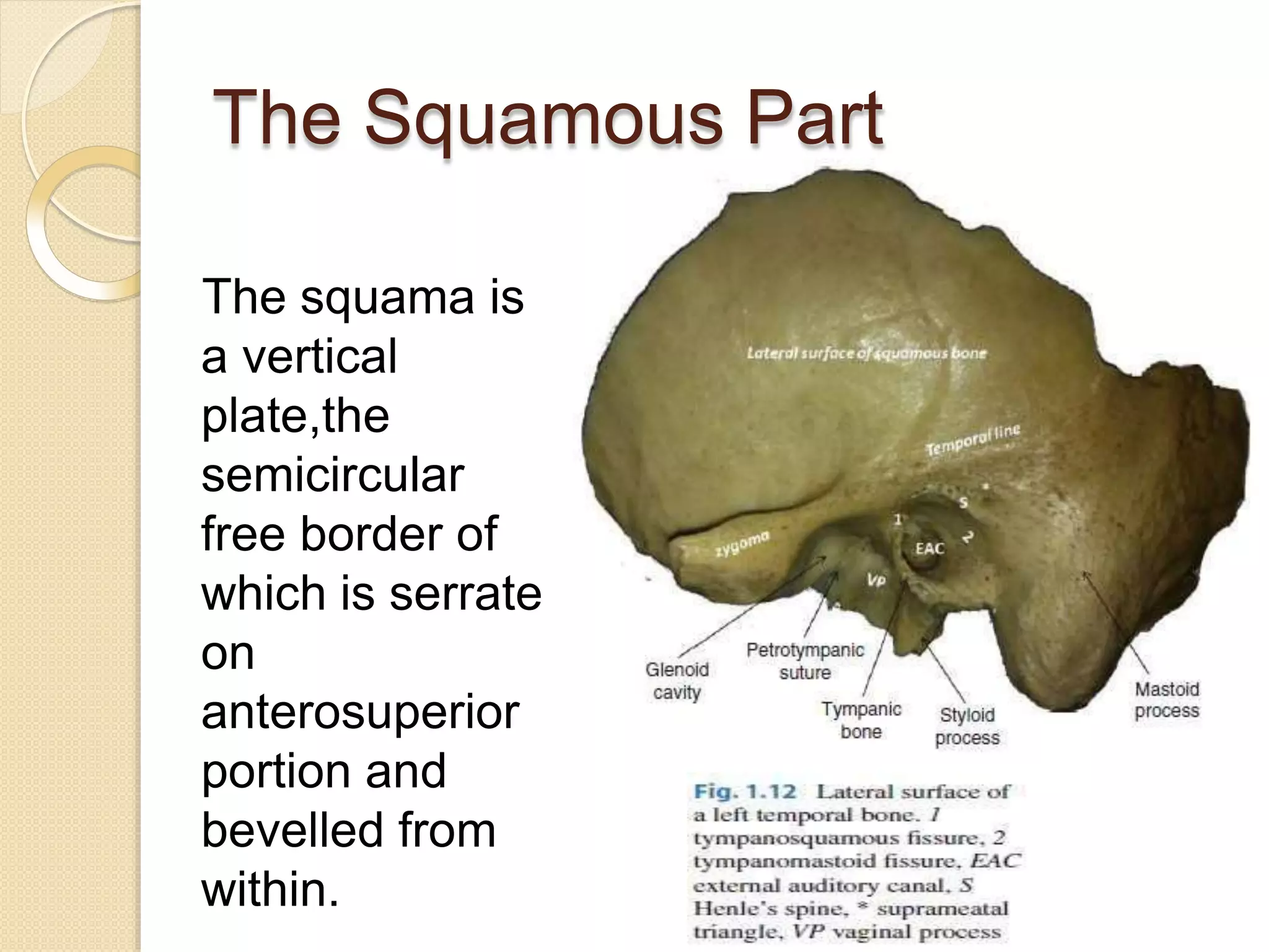
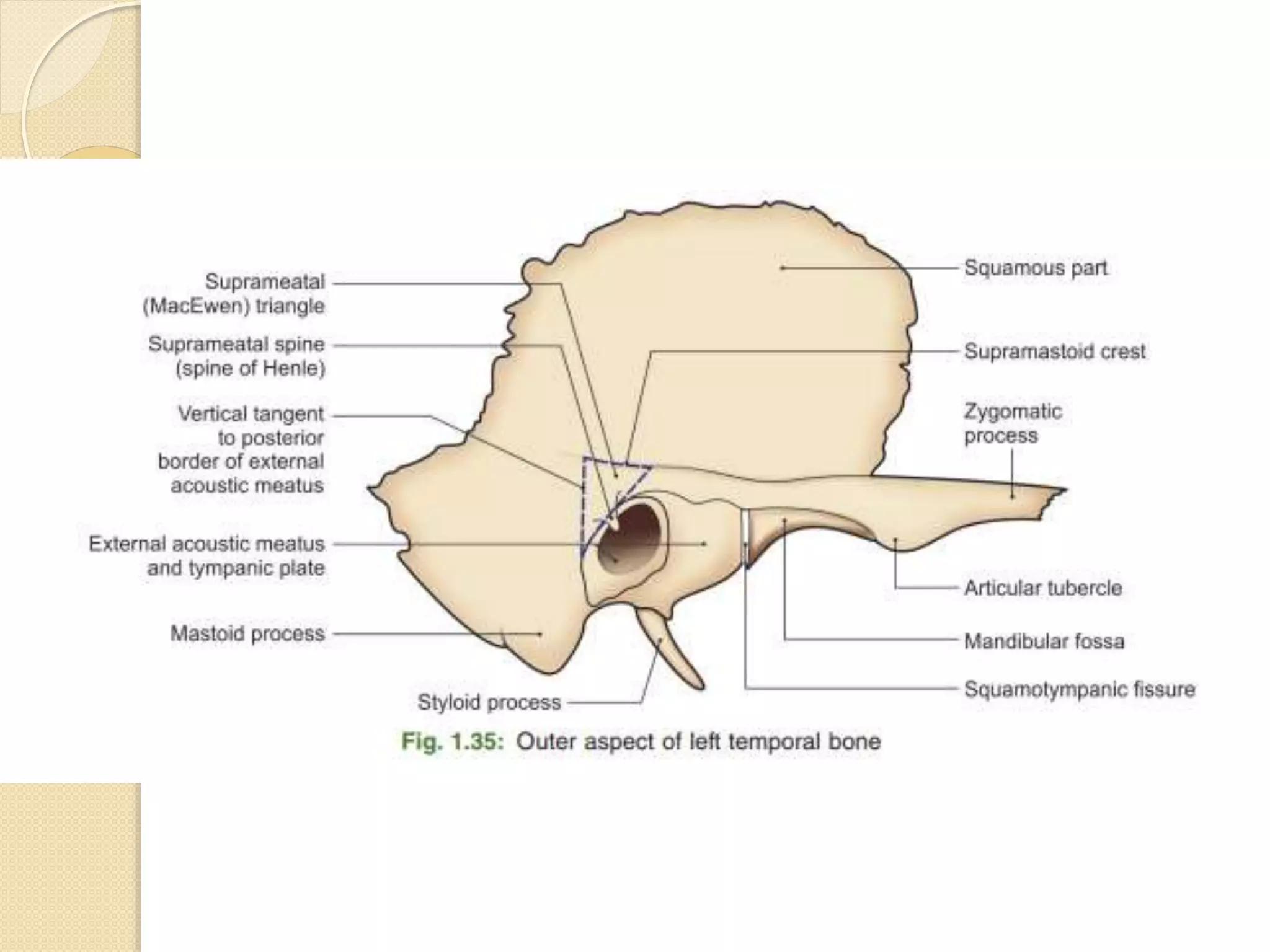
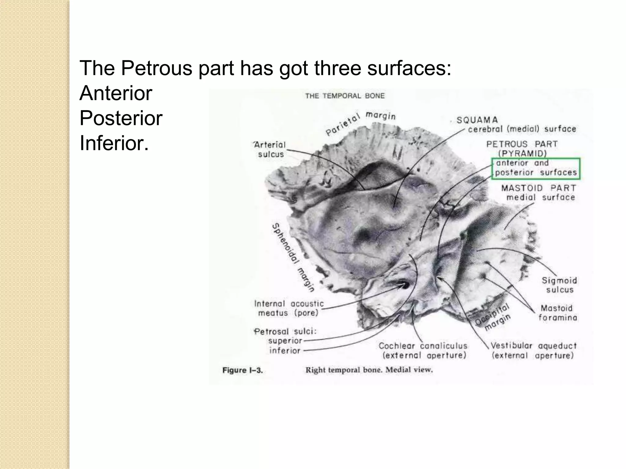
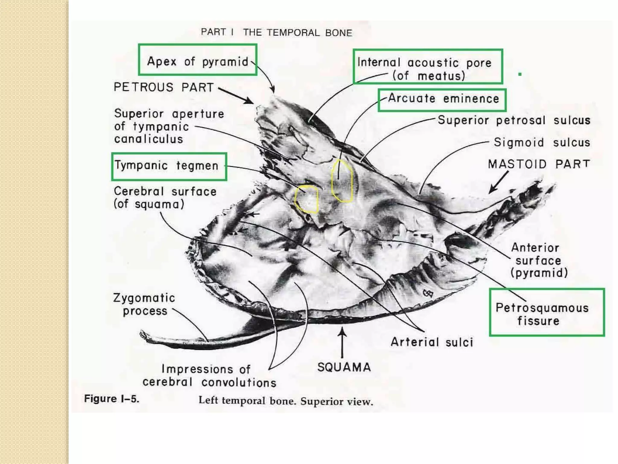
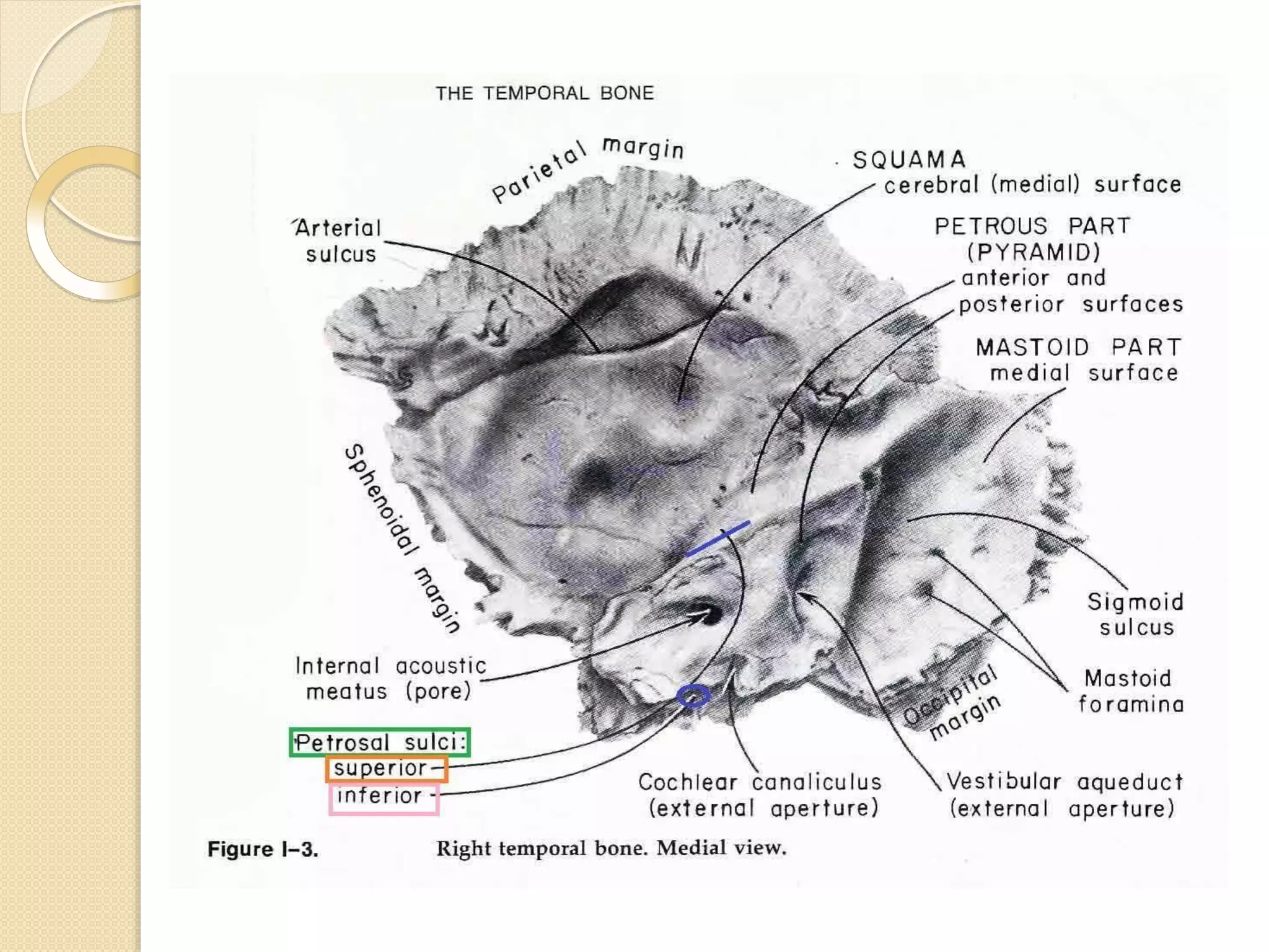
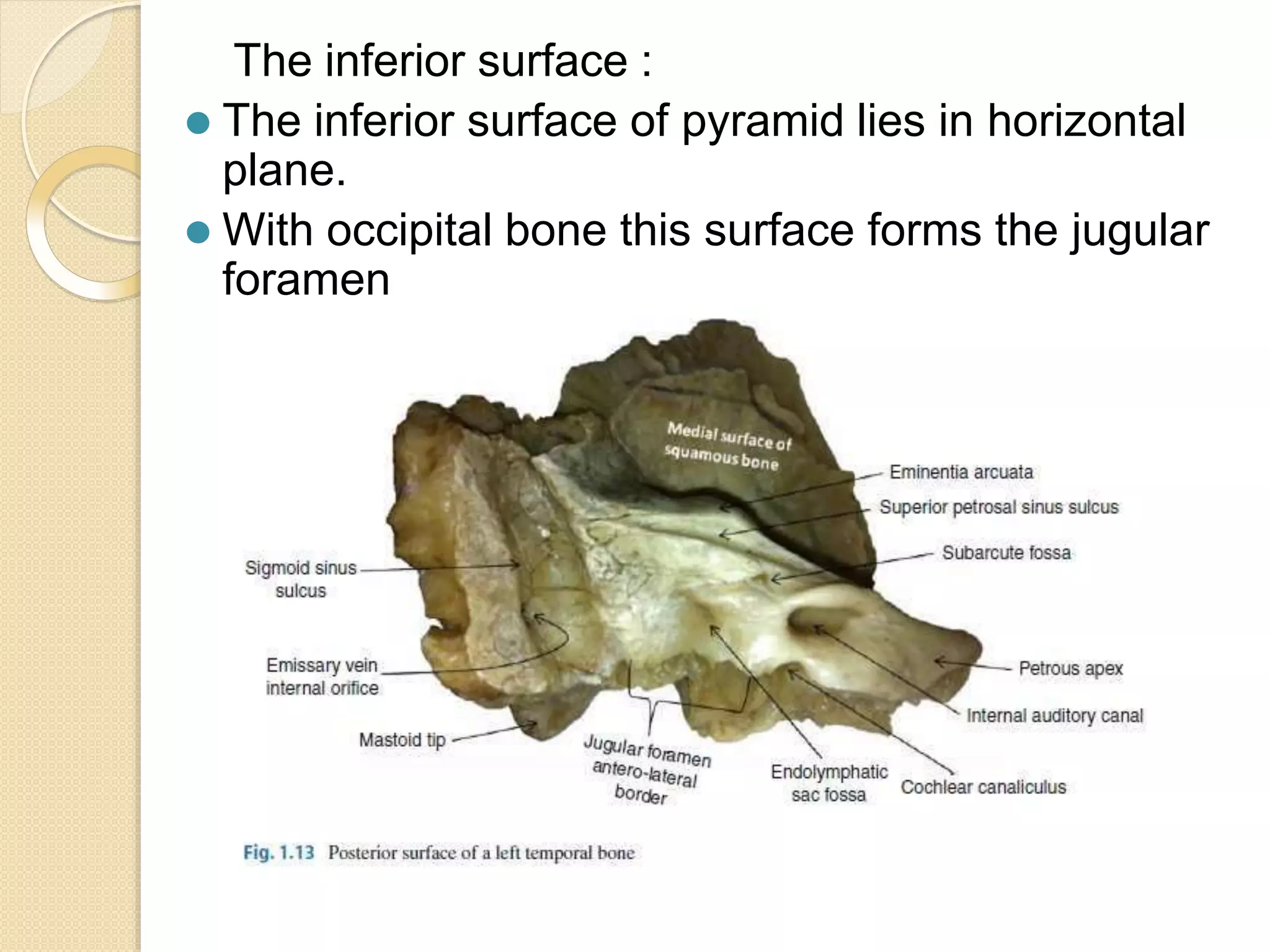
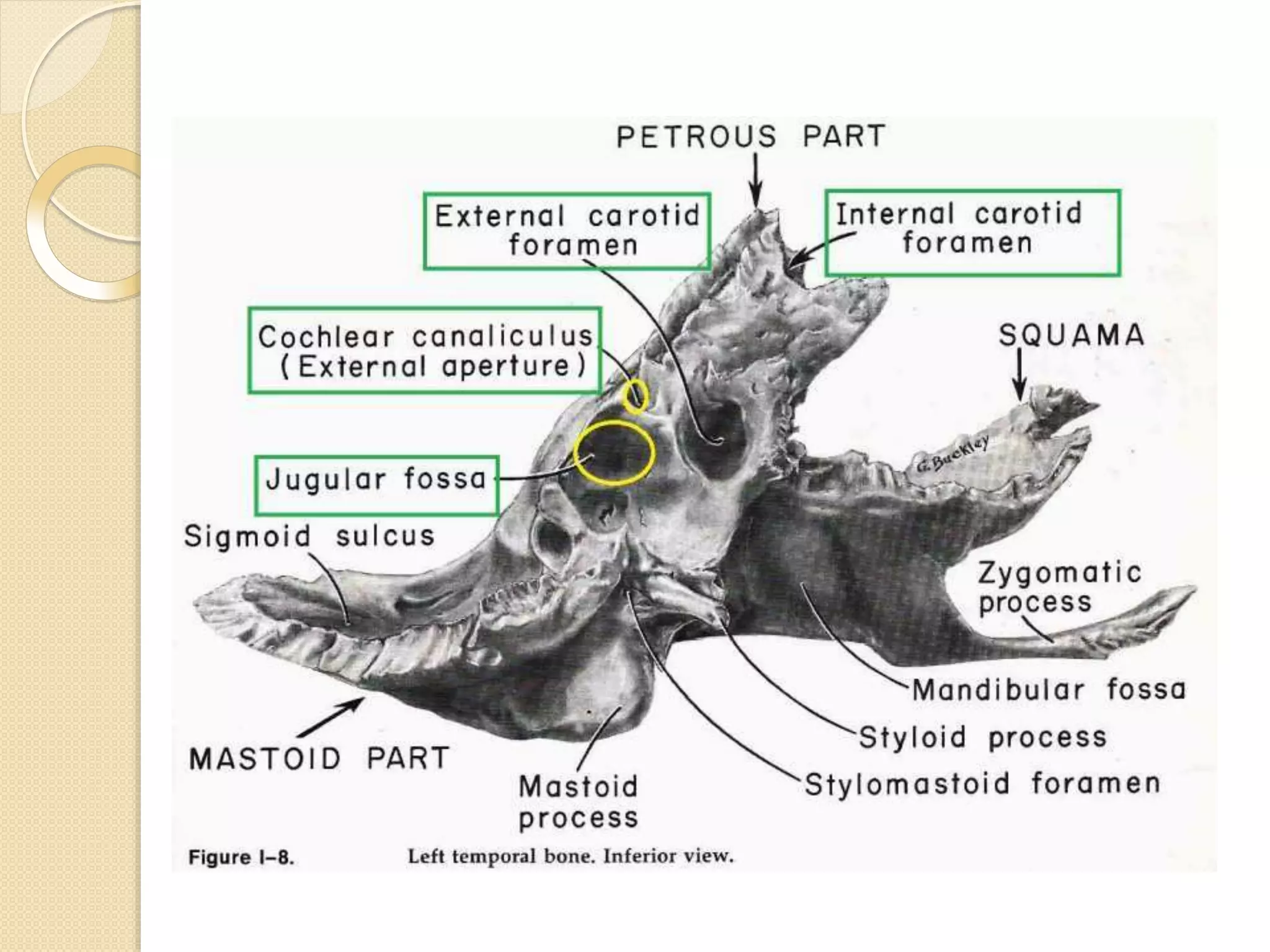
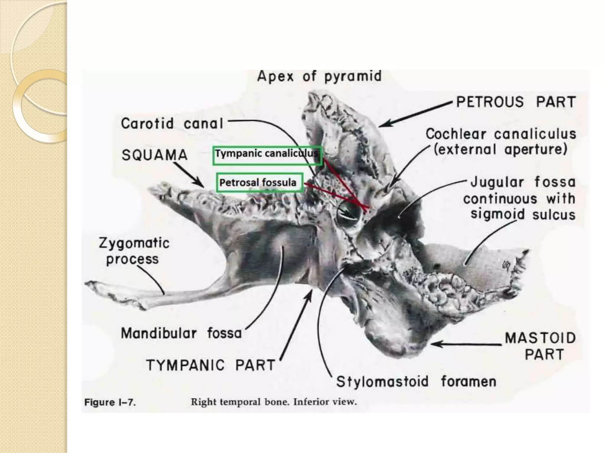
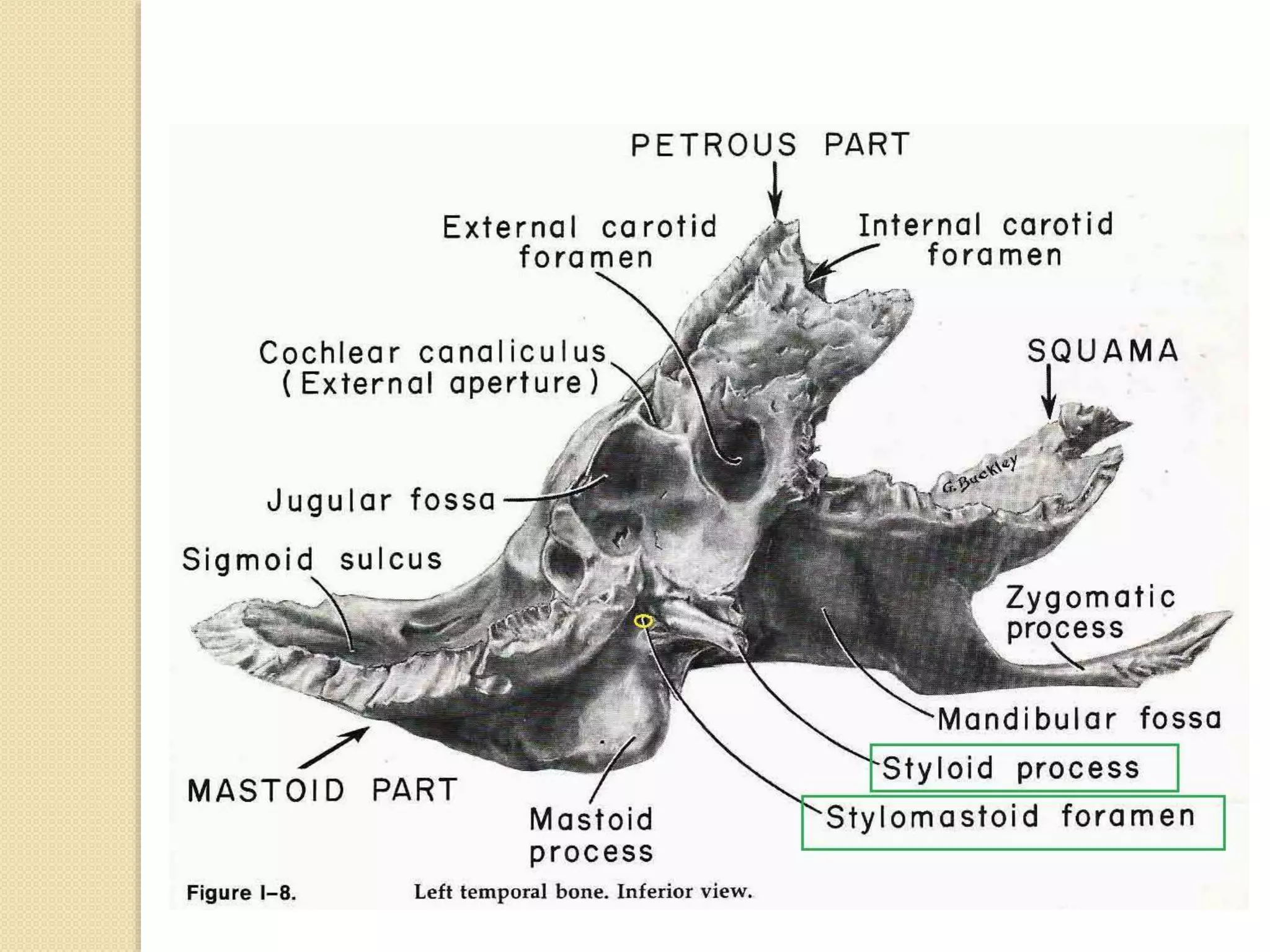
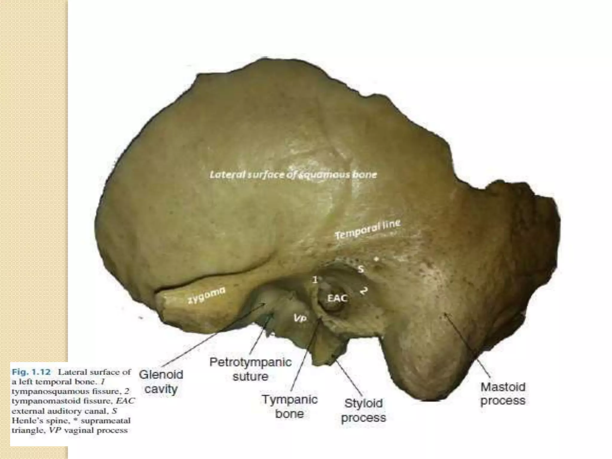
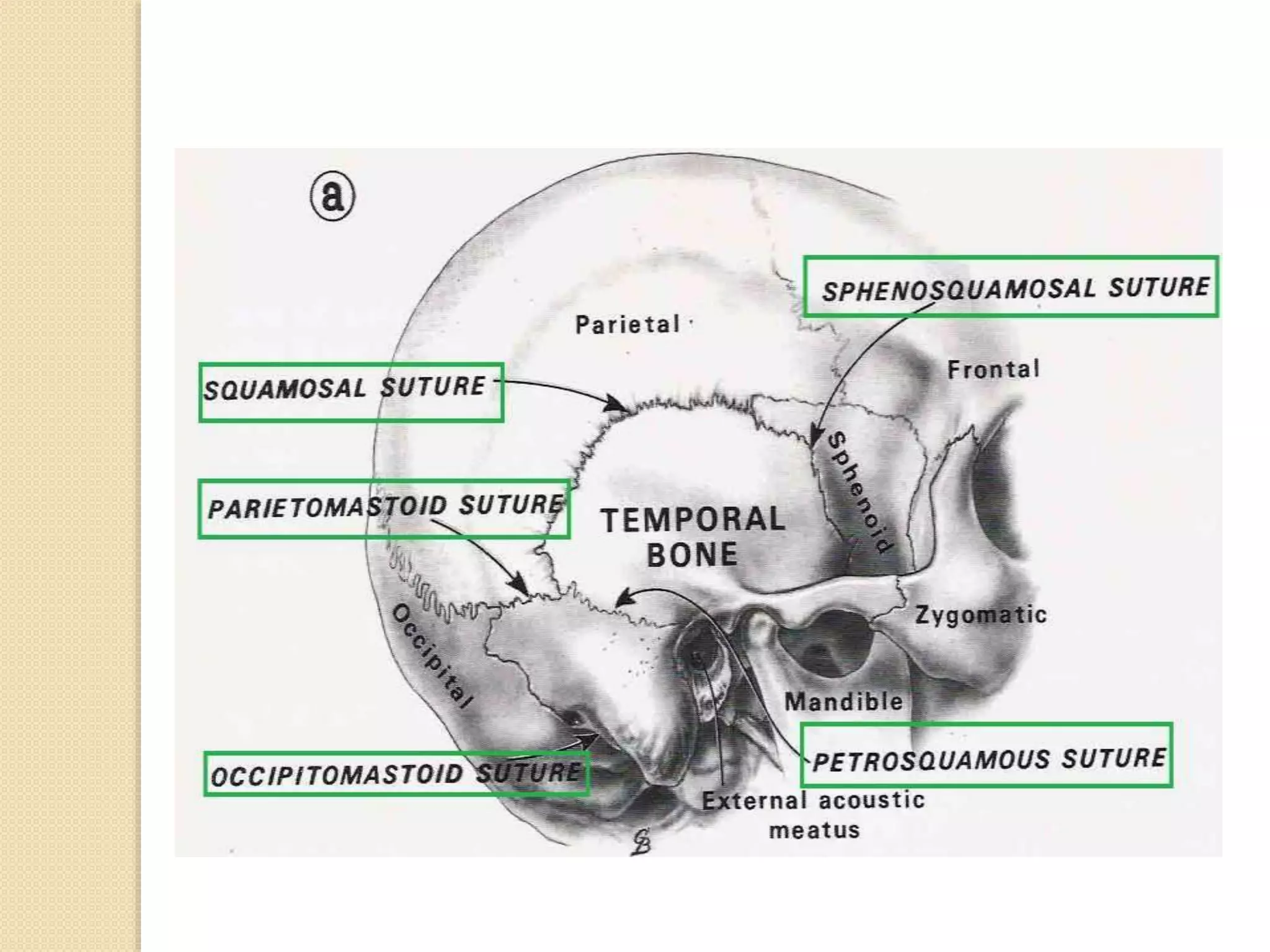
SlideShare a Scribd company logo
ANATOMY OF TEMPORAL BONE
BY Dr. VIJAY KUMAR
⚫ The paired
temporal bones
contribute to both
the base and the
lateral wall of the
skull.
⚫ For cranial cavity
each forms part of
the middle and
posterior cranial
fossae.
⚫ Each bone is divisible
into the following four
parts:
1.Squamous
2.Mastoid
3.Petrous
4.Tympanic
When the pores of the
external acoustic
meatus is taken as a
point of topographical
referrence,the
squamous part is
directed upward
the mastoid part
backward
the tympanic forward
and downward
the petrous portion
medially.
The temporal bone articulates with :
 Sphenoid bone
 Parietal bone
 Occipital bone
 Zygomatic bone.
The Squamous Part
The squama is
a vertical
plate,the
semicircular
free border of
which is serrate
on
anterosuperior
portion and
bevelled from
within.
⚫Outerortemporal
Itissmoothandformsapartoftemporalfossa.Above
externalacousticmeatus,thereisagrooveformiddle
temporalartery.Itsposteriorpartpresents
supramastoidcrest.Belowtheanteriorendof
supramastoidcrestandposterosuperiortoexternal
acousticmeatus,thereis suprameataltriangle.
Zygomaticprocessspringsforwardsfromtheouter
surfaceofsquamouspart.Itsposteriorpartcomprises
superiorandinferiorsurfaces.Theinferiorsurfaceis
boundedbytwo rootswhich convergeatthetubercleof
rootofthezygoma.
Anteriorrootprojectsasthearticulartubercleinfront
ofmandibularfossa.Posteriorrootbeginsabovethe
externalacousticmeatus.Mandibularfossaliesbehind
articulartubercleandconsistsofanteriorarticular
partformedbysquamouspartoftemporalboneanda
posteriornon-articularportionformedbytympanic
plate
• Innerorcerebral:
Itisconcaveandshowsgroovesforthemiddle
meningealvessels.
Itssuperiorborderarticulateswiththelowerborderof
parietalbone.
Itsanteroinferiorborderarticulateswiththegreater
wing ofsphenoid.
The Mastoid Part
⚫ Thisisathick conical
projection fromthe
squamousand petrous
partsof thetemporal
boneanditcomesinto
appostionwith occipital
and parietalbones
•Twosurfaces—outerandinner
•Twoborders—superiorand
posterior,andenclosethemastoid
aircells.
Theoutersurfaceformsaconical
process,themastoidprocess.
⚫ The lateral
surface: is
rough and
gives
attachment
to posterior
auricular
and
occipital
muscles.
Mastoid Part
• OuterSurface:
Theoutersurfacegivesattachmenttooccipitalismuscle.
Mastoidforamenopensnearitsposteriorborderand
transmitsanemissaryveinandabranch ofoccipital
artery.Mastoidprocessappearsattheendof2ndyear.
Lateralsurfacegivesattachmenttosternocleidomastoid,
spleniuscapitis,andlongissimuscapitis).Medialsurface
oftheprocessshowsadeepmastoidnotchfortheoriginof
posteriorbellyofdigastric.Medialtothisnotchisagroove
fortheoccipitalartery.
• InnerSurface:
Theinnersurfaceismarkedbyadeepsigmoidsulcus
Borders:
Superiorborder:Articulateswithparietalboneat
asterion.
Posteriorborder:Articulateswithoccipitalboneat
occipitomastoidsuture.
⚫ The medial
surface:
presents a
deep groove
,the sigmoid
sulcus for
lodgement of
the sigmoid
venous sinus
of the dura
mater.
The petrous part
⚫This part of the temporal bone is like a
three sided pyramid.
⚫The base united with the mastoid part
is inserted into the angle between
occipital and sphenoid bones.
⚫The apex is directed medially and
forward.
The foramen lacerum intervenes between the
apex and the sphenoid bone .
Structure passing whole length:
Meningeal branch of ascending pharyngeal
artery
Emissary veins
Other structure partialy traversing is :
Internal carotid artery with venous and
sympathetic plexus around it.
Greater petrosal nerve unite with dep petrosal
nerve to form nerve to pterygoid canal
The petrous part…
The Petrous part has got three surfaces:
Anterior
Posterior
Inferior.
⚫The anterior surface:
It is smooth , forms part of the middle
cranial fossa and inclines forward and
downward.
⚫Laterally it is fused with squama at the
petrosquamous suture.
The petrous part…
The petrous part…
⚫ The anterior margin(termed the anterior
angle) is free & roughened , with the
greater wing of the sphenoid bone bounds
an opening : the musculotubal canal
⚫ Canal is subdivided by leaflet of bone into
smaller upper part - the semicanal for the
tensor tympani muscle
and
a large lower portion the semicanal of
auditory(eustachian ) tube
⚫ Near the middle of the anterior surface is
the Arcuate eminence ,caused by
underlying superior semicircular canal.
⚫ Anterior and lateral to this eminence is the
tympanic tegmen,which forms the roof of
the tympanic cavity.
⚫ In the anterior direction near the apex is
a medial opening, hiatus of the facial
canal
(it transmits the superficial petrosal
branch of the middle meningeal artery
and the greater superficial petrosal
nerve)
and
a lateral smaller opening, superior
aperture of the tympanic canaliculus(it
transmits the superior tympanic artery
and the lesser superficial petrosal nerve)
The posterior surface:
⚫ It lies in an almost vertical plane and it
faces the posterior cranial fossa.
⚫ It is bounded above at the superior angle
by the sulcus for superior petrosal sinus,
⚫ below at the posterior angle ,the pyramid
unites with the occipital bone ,along the
line of fusion accommodating the sulcus
for inferior petrosal sinus.
⚫ Midway between the base and apex is the
opening of internal acoustic meatus(short
canal for acoustic and facial nerves and the
internal auditory blood vessels)
⚫ Behind and above this is the subarcuate
fossa (it carries blood vessels to otic
capsule during fetal life.) it is of pin point
caliber in adults and may transmit small
veins to dura mater.
⚫ Further laterally and downward is the
vestibular aqueduct (for transmission of
endolymphatic duct and sac)
The inferior surface :
⚫ The inferior surface of pyramid lies in horizontal
plane.
⚫ With occipital bone this surface forms the jugular
foramen
⚫ The lateral
part of the
foramen
contains the
junction of
sigmoid sinus
and the
internal
jugular vein
⚫ Medial part
contains
inferior
petrosal sinus
and middle
portion
contains
cranial
nerves IX,X
and XI.
⚫ In front of the lateral compartment of the
foramen is jugular fossa (for the bulb of
jugular vein).
⚫ Medial to fossa is the funnel-shaped
external aperture of the cochlear canaliculus
(containing perilymphatic duct).
⚫ In front of the fossa is the external carotid
foramen(entrance to the canal for the
internal carotid artery and its plexus of veins
and sympathetic nerves.)
⚫ Near the external carotid foramen small
openings ,the caroticotympanic canaliculi
are present which transmit the
caroticotympanic artery and nerves into the
middle ear.
⚫ The external carotid foramen is separated
from the jugular fossa by the carotid ridge.
On the edge of the carotid ridge is petrosal
fossula for lodgement of petrosal ganglion
of the glossopharyngeal nerve.
At the bottom of this fossula ,tympanic
canaliculus is situated ,which transmits
tympanic branch of glossopharyngeal nerve
(Jacbson’s nerve) and tympanic branch of
ascending pharyngeal artery.
⚫ Rough jugular surface behind the jugular
fossa articulates with the jugular process of
the occipital bone.
⚫ Lateral to this surface,a downward directed
cylindrical spur ,the styloid process is
present.
⚫ The stylomastoid foramen is present at its
base on the posterior aspect.
⚫ This is the external orifice of the facial canal
and transmits the facial nerve ,the
stylomastoid artery and in some cases the
auricular branch of the vagus nerve.
⚫ Mastoid
incisure/notch (for
the attachment of
the digastric
muscle) and the
temoral/occipital
groove(for the
occipital artery) is
present in the
posterior direction.
The Tympanic Part
⚫ The tympanic bone is thin and roughly
quadrilateral bone.
⚫ It is hollow above and concave in front
and below.
⚫ It forms all the anterior and inferior wall
and part of posterior wall of the external
acoustic meatus.
⚫ The posterosuperior surface faces the
external acoustic meatus and the
tympanic cavity.
⚫ The middle of anteroinferior surface is thin &
sometimes presenting a small Foramen of
Huschke.
⚫ It represents a nonossified portion of the
plate.
⚫ At the medial end a groove called tympanic
sulcus is present whichh is deficient
superiorly ,the tympanic membrane is present
in this sulcus.
⚫ The inferior surface is prolonged into a
vaginal process,which encircles the lateral
aspect of the base of the styloid process.
Sutures and Articulations
Articulations:
The temporal bone articulates with:
⚫ occipital
⚫ parietal
⚫ Sphenoid
⚫ zygomatic bones and – by a movable
joint- with the mandible.
⚫ Articulations are formed also with the
auditory ossicles and the hyoid bone
through the ligaments.
Embryology and Ossification
⚫The skull is developed from the
mesenchyme surrounding the
developing brain.
⚫Some of the bones of skull are formed in
membrane, some in cartilage and some
partly in membrane and partly in
cartilage.
⚫The squamous and tympanic parts of the
temporal bone develop in membrane
while the petrous part in the cartilage.
⚫The squamous portion of the subsequent
temporal bone is formed from one(or
two) ossification centers which arise in
membrane in the second month of
⚫ The tympanic portion has three ossification
centers which appear by ninth to tenth fetal
week.
⚫ The tympanic and squamous part unite by ninth
month of fetal life.
⚫ The petrous bone by the fifth month is
represented by the otic capsule. This bone later
ossifies in 14 centers. (petrous part is formed in
cartilage.)
⚫ squamous and petrous part fuse immediately
after birth.
⚫ The styloid process (formed in cartilage) ossifies
from six months onwards.
⚫ Zygomatic bone arises as a thin bony plate by
ossification in membrane at the end of second
month of embryonic life.
Applied anatomy and important
relations
⚫ Glossopharyngeal nerve lies in close relation to
styloid process and Elongated styloid process or
calcified stylohoid ligament may lead to Eagle’s
syndrome or styalgia.(characterised by pain in
tonsillar fossa and upper neck , radiating to
ipsilateral ear and aggravated on swallowing)
⚫ Mac Ewen’s triangle is a landmark for mastoid
antrum and is formed by posterosuperior wall of
external acoustic meatus ,the posterior extension
of the root of zygomatic arch and a tangent to it
joining external auditory canal.
⚫ The position of sigmoid sinus in the sigmoid
sulcus just behind and deep to the mastoid
antrum makes it vulnerable to damage in mastoid
surgeries.
⚫ The anterior relation of temporomandibular joint to
external acoustic meatus is important as over
enthusiastic attempts at straightening the anterior canal
wall can lead to sagging of head of mandible in external
auditory canal.
⚫ And for the same reason a backward directed force on
the mandible can cause trauma and bleeding in
external auditory canal(either unilateral or bilateral)
⚫ The intratemporal course of the facial nerve from the
entrance of facial canal at the fundus of internal
acoustic meatus to the stylomastoid foramen is
extremely important in ear surgeries.
⚫ The horizontal semicircular canal ,the processus
cochleariformis , the oval window , the pyramid , the
aditus , digastric ridge and the short process of incus
are few important landmarks which help to identify the
position and course of facial nerve in the temporal
bone.
THANK YOU

More Related Content

What's hot

Auditory physiology of Inner Ear
Auditory physiology of Inner EarAuditory physiology of Inner Ear
Auditory physiology of Inner Ear
Diptiman Baliarsingh
 
Hadad.bassagasteguy flap
Hadad.bassagasteguy flap Hadad.bassagasteguy flap
Hadad.bassagasteguy flap
Chandra Veer Suryavanshi
 
The pneumatic system of the temporal bone
The pneumatic system of the temporal boneThe pneumatic system of the temporal bone
The pneumatic system of the temporal bone
Surbhi narayan
 
Anatomy facial nerve
Anatomy facial nerveAnatomy facial nerve
Anatomy facial nerve
محمد حسين خميس
 
Infratemporal fossa a systematic approach
Infratemporal fossa a systematic approachInfratemporal fossa a systematic approach
Infratemporal fossa a systematic approach
Augustine raj
 
Middle ear ventilatory pathway and Mucosal folds.pptx
Middle ear ventilatory pathway and Mucosal folds.pptxMiddle ear ventilatory pathway and Mucosal folds.pptx
Middle ear ventilatory pathway and Mucosal folds.pptx
SaneeshDamodaran
 
Endoscopic anatomy of lateral wall of sphenoid sinus
Endoscopic anatomy of lateral wall of sphenoid sinusEndoscopic anatomy of lateral wall of sphenoid sinus
Endoscopic anatomy of lateral wall of sphenoid sinus
Ankit Choudhary
 
surgical approaches to frontal sinus ppt
surgical approaches to frontal sinus pptsurgical approaches to frontal sinus ppt
surgical approaches to frontal sinus ppt
Vaibhav Lahane
 
Anatomy temporal bone
Anatomy temporal bone Anatomy temporal bone
Anatomy temporal bone
KevinMungasia
 
Surgical anatomy of nose
Surgical anatomy of noseSurgical anatomy of nose
Surgical anatomy of nose
Augustine raj
 
Facial nerve decompression
Facial nerve decompressionFacial nerve decompression
Facial nerve decompression
Mamoon Ameen
 
Endoscopic anatomy of nose and sphenoid sinus
Endoscopic anatomy of nose and sphenoid sinusEndoscopic anatomy of nose and sphenoid sinus
Endoscopic anatomy of nose and sphenoid sinus
Dr Zeeshan Ahmad
 
Pterygopalatine fossa
Pterygopalatine fossaPterygopalatine fossa
Pterygopalatine fossa
Chitransha03
 
Mucosal folds of the middle ear
Mucosal folds of the middle earMucosal folds of the middle ear
Mucosal folds of the middle ear
Dʀ Smruti Ranjan Samal
 
anatomy of inner ear by dr. ravindra daggupati
anatomy of inner ear by dr. ravindra daggupatianatomy of inner ear by dr. ravindra daggupati
anatomy of inner ear by dr. ravindra daggupati
Ravindra Daggupati
 
Anatomy of temporal bone
Anatomy of temporal boneAnatomy of temporal bone
Anatomy of temporal bone
praneeth koduru
 
Jugular foramen anatomy and approaches
Jugular foramen anatomy and approachesJugular foramen anatomy and approaches
Jugular foramen anatomy and approaches
Dikpal Singh
 
Anatomy of Larynx
Anatomy of LarynxAnatomy of Larynx
Anatomy of Larynx
Diptiman Baliarsingh
 
Fisch approaches Dr Zeeshan Ahmad
Fisch approaches Dr Zeeshan AhmadFisch approaches Dr Zeeshan Ahmad
Fisch approaches Dr Zeeshan Ahmad
Dr Zeeshan Ahmad
 

What's hot (20)

Auditory physiology of Inner Ear
Auditory physiology of Inner EarAuditory physiology of Inner Ear
Auditory physiology of Inner Ear
 
Hadad.bassagasteguy flap
Hadad.bassagasteguy flap Hadad.bassagasteguy flap
Hadad.bassagasteguy flap
 
The pneumatic system of the temporal bone
The pneumatic system of the temporal boneThe pneumatic system of the temporal bone
The pneumatic system of the temporal bone
 
Anatomy facial nerve
Anatomy facial nerveAnatomy facial nerve
Anatomy facial nerve
 
Infratemporal fossa a systematic approach
Infratemporal fossa a systematic approachInfratemporal fossa a systematic approach
Infratemporal fossa a systematic approach
 
Middle ear ventilatory pathway and Mucosal folds.pptx
Middle ear ventilatory pathway and Mucosal folds.pptxMiddle ear ventilatory pathway and Mucosal folds.pptx
Middle ear ventilatory pathway and Mucosal folds.pptx
 
Endoscopic anatomy of lateral wall of sphenoid sinus
Endoscopic anatomy of lateral wall of sphenoid sinusEndoscopic anatomy of lateral wall of sphenoid sinus
Endoscopic anatomy of lateral wall of sphenoid sinus
 
surgical approaches to frontal sinus ppt
surgical approaches to frontal sinus pptsurgical approaches to frontal sinus ppt
surgical approaches to frontal sinus ppt
 
Anatomy temporal bone
Anatomy temporal bone Anatomy temporal bone
Anatomy temporal bone
 
Surgical anatomy of nose
Surgical anatomy of noseSurgical anatomy of nose
Surgical anatomy of nose
 
Facial nerve decompression
Facial nerve decompressionFacial nerve decompression
Facial nerve decompression
 
Endoscopic anatomy of nose and sphenoid sinus
Endoscopic anatomy of nose and sphenoid sinusEndoscopic anatomy of nose and sphenoid sinus
Endoscopic anatomy of nose and sphenoid sinus
 
Pterygopalatine fossa
Pterygopalatine fossaPterygopalatine fossa
Pterygopalatine fossa
 
Mucosal folds of the middle ear
Mucosal folds of the middle earMucosal folds of the middle ear
Mucosal folds of the middle ear
 
IAC 360°
IAC 360°IAC 360°
IAC 360°
 
anatomy of inner ear by dr. ravindra daggupati
anatomy of inner ear by dr. ravindra daggupatianatomy of inner ear by dr. ravindra daggupati
anatomy of inner ear by dr. ravindra daggupati
 
Anatomy of temporal bone
Anatomy of temporal boneAnatomy of temporal bone
Anatomy of temporal bone
 
Jugular foramen anatomy and approaches
Jugular foramen anatomy and approachesJugular foramen anatomy and approaches
Jugular foramen anatomy and approaches
 
Anatomy of Larynx
Anatomy of LarynxAnatomy of Larynx
Anatomy of Larynx
 
Fisch approaches Dr Zeeshan Ahmad
Fisch approaches Dr Zeeshan AhmadFisch approaches Dr Zeeshan Ahmad
Fisch approaches Dr Zeeshan Ahmad
 

Similar to Temporal Bone anatomy ppt.pptx

Anatomy of temporal bone By Dr.Vijay kumar , AMU
Anatomy of temporal bone By Dr.Vijay kumar , AMUAnatomy of temporal bone By Dr.Vijay kumar , AMU
Anatomy of temporal bone By Dr.Vijay kumar , AMU
vijaymgims
 
TEMPORAL BONE.pptx
TEMPORAL BONE.pptxTEMPORAL BONE.pptx
TEMPORAL BONE.pptx
DrRashidKaziMahbubur
 
Anatomy of skull base
Anatomy of skull baseAnatomy of skull base
Anatomy of skull base
Dr. Shilpa M J
 
Radiological anatomy of_temporal_bone[1]
Radiological anatomy of_temporal_bone[1]Radiological anatomy of_temporal_bone[1]
Radiological anatomy of_temporal_bone[1]
suriyaprakash nagarajan
 
Skull base anatomy by Dr. Aditya Tiwari
Skull base anatomy by Dr. Aditya TiwariSkull base anatomy by Dr. Aditya Tiwari
Skull base anatomy by Dr. Aditya Tiwari
Aditya Tiwari
 
Lec7
Lec7Lec7
Anatomy of Skullbase
Anatomy of SkullbaseAnatomy of Skullbase
Anatomy of Skullbase
Jinu Iype
 
Basic Skull Anatomy
Basic Skull AnatomyBasic Skull Anatomy
Basic Skull Anatomy
Hadi Munib
 
Individual skull bones
Individual skull bonesIndividual skull bones
Individual skull bones
Idris Siddiqui
 
Anatomy skull anatomy dr.mohammed
Anatomy skull anatomy dr.mohammedAnatomy skull anatomy dr.mohammed
Anatomy skull anatomy dr.mohammed
mohammed muneer
 
Skull inside and some separate bones
Skull   inside and some separate bonesSkull   inside and some separate bones
Skull inside and some separate bones
Akram Jaffar
 
Head & Neck Anatomy the EYEBALL lec 7.pdf
Head & Neck Anatomy the  EYEBALL lec 7.pdfHead & Neck Anatomy the  EYEBALL lec 7.pdf
Head & Neck Anatomy the EYEBALL lec 7.pdf
ssuser386649
 
The Skull and Vertebral column
The Skull and Vertebral columnThe Skull and Vertebral column
The Skull and Vertebral column
Seddie Chitamu
 
1 Skull
1   Skull1   Skull
1 Skull
mehrano
 
Anatomy of orbital cavity
Anatomy of orbital cavityAnatomy of orbital cavity
Anatomy of orbital cavity
Othman Al-Abbadi
 
Anatomy of middle ear
Anatomy of middle earAnatomy of middle ear
Anatomy of middle ear
Dr Soumya Singh
 
The cranial bones
The cranial bonesThe cranial bones
The cranial bonesDoc Lorie B
 
Anatomy of temporal bone final
Anatomy of temporal bone finalAnatomy of temporal bone final
Anatomy of temporal bone final
DR. KANIKA BHARGAVA
 
ANATOMY OF EXTERNAL AUDITORY CANAL
ANATOMY OF EXTERNAL AUDITORY CANALANATOMY OF EXTERNAL AUDITORY CANAL
ANATOMY OF EXTERNAL AUDITORY CANAL
Kanu Saha
 
Anatomy of human ear
Anatomy of human earAnatomy of human ear
Anatomy of human ear
Dr Safika Zaman
 

Similar to Temporal Bone anatomy ppt.pptx (20)

Anatomy of temporal bone By Dr.Vijay kumar , AMU
Anatomy of temporal bone By Dr.Vijay kumar , AMUAnatomy of temporal bone By Dr.Vijay kumar , AMU
Anatomy of temporal bone By Dr.Vijay kumar , AMU
 
TEMPORAL BONE.pptx
TEMPORAL BONE.pptxTEMPORAL BONE.pptx
TEMPORAL BONE.pptx
 
Anatomy of skull base
Anatomy of skull baseAnatomy of skull base
Anatomy of skull base
 
Radiological anatomy of_temporal_bone[1]
Radiological anatomy of_temporal_bone[1]Radiological anatomy of_temporal_bone[1]
Radiological anatomy of_temporal_bone[1]
 
Skull base anatomy by Dr. Aditya Tiwari
Skull base anatomy by Dr. Aditya TiwariSkull base anatomy by Dr. Aditya Tiwari
Skull base anatomy by Dr. Aditya Tiwari
 
Lec7
Lec7Lec7
Lec7
 
Anatomy of Skullbase
Anatomy of SkullbaseAnatomy of Skullbase
Anatomy of Skullbase
 
Basic Skull Anatomy
Basic Skull AnatomyBasic Skull Anatomy
Basic Skull Anatomy
 
Individual skull bones
Individual skull bonesIndividual skull bones
Individual skull bones
 
Anatomy skull anatomy dr.mohammed
Anatomy skull anatomy dr.mohammedAnatomy skull anatomy dr.mohammed
Anatomy skull anatomy dr.mohammed
 
Skull inside and some separate bones
Skull   inside and some separate bonesSkull   inside and some separate bones
Skull inside and some separate bones
 
Head & Neck Anatomy the EYEBALL lec 7.pdf
Head & Neck Anatomy the  EYEBALL lec 7.pdfHead & Neck Anatomy the  EYEBALL lec 7.pdf
Head & Neck Anatomy the EYEBALL lec 7.pdf
 
The Skull and Vertebral column
The Skull and Vertebral columnThe Skull and Vertebral column
The Skull and Vertebral column
 
1 Skull
1   Skull1   Skull
1 Skull
 
Anatomy of orbital cavity
Anatomy of orbital cavityAnatomy of orbital cavity
Anatomy of orbital cavity
 
Anatomy of middle ear
Anatomy of middle earAnatomy of middle ear
Anatomy of middle ear
 
The cranial bones
The cranial bonesThe cranial bones
The cranial bones
 
Anatomy of temporal bone final
Anatomy of temporal bone finalAnatomy of temporal bone final
Anatomy of temporal bone final
 
ANATOMY OF EXTERNAL AUDITORY CANAL
ANATOMY OF EXTERNAL AUDITORY CANALANATOMY OF EXTERNAL AUDITORY CANAL
ANATOMY OF EXTERNAL AUDITORY CANAL
 
Anatomy of human ear
Anatomy of human earAnatomy of human ear
Anatomy of human ear
 

More from JitenLad2

ANATOMY OF MID EAR and related structures.pptx
ANATOMY OF MID EAR and related structures.pptxANATOMY OF MID EAR and related structures.pptx
ANATOMY OF MID EAR and related structures.pptx
JitenLad2
 
ANATOMY OF EXT EAR AND RELATED STRUCTURES
ANATOMY OF EXT EAR AND RELATED STRUCTURESANATOMY OF EXT EAR AND RELATED STRUCTURES
ANATOMY OF EXT EAR AND RELATED STRUCTURES
JitenLad2
 
BUCCAL CARCINOMA AND ITS COMPLICATIONS PPT
BUCCAL CARCINOMA AND ITS COMPLICATIONS PPTBUCCAL CARCINOMA AND ITS COMPLICATIONS PPT
BUCCAL CARCINOMA AND ITS COMPLICATIONS PPT
JitenLad2
 
MIDDLE CRANIAL FOSSA and structures passing through
MIDDLE CRANIAL FOSSA and structures passing throughMIDDLE CRANIAL FOSSA and structures passing through
MIDDLE CRANIAL FOSSA and structures passing through
JitenLad2
 
forcep delivery copy.pdf
forcep delivery copy.pdfforcep delivery copy.pdf
forcep delivery copy.pdf
JitenLad2
 
CASE PRESENTATION ON occipito fronto presentation
CASE PRESENTATION ON occipito fronto presentation CASE PRESENTATION ON occipito fronto presentation
CASE PRESENTATION ON occipito fronto presentation
JitenLad2
 
bones physiology.ppt
bones physiology.pptbones physiology.ppt
bones physiology.ppt
JitenLad2
 
Vagusnerve JitenL.pptx
Vagusnerve JitenL.pptxVagusnerve JitenL.pptx
Vagusnerve JitenL.pptx
JitenLad2
 
cord blood albumin assesment.pptx
cord blood albumin assesment.pptxcord blood albumin assesment.pptx
cord blood albumin assesment.pptx
JitenLad2
 
ANATOMY of nose ppt.pptx
ANATOMY of nose ppt.pptxANATOMY of nose ppt.pptx
ANATOMY of nose ppt.pptx
JitenLad2
 
Anatomy of axilla.pptx
Anatomy of axilla.pptxAnatomy of axilla.pptx
Anatomy of axilla.pptx
JitenLad2
 
radiological anatomy of liver segments (1).pptx
radiological anatomy of liver segments (1).pptxradiological anatomy of liver segments (1).pptx
radiological anatomy of liver segments (1).pptx
JitenLad2
 
PMSMA-PPT.pdf
PMSMA-PPT.pdfPMSMA-PPT.pdf
PMSMA-PPT.pdf
JitenLad2
 
ICTC-1.ppt
ICTC-1.pptICTC-1.ppt
ICTC-1.ppt
JitenLad2
 
ObstructiveSleepApnea.pptx
ObstructiveSleepApnea.pptxObstructiveSleepApnea.pptx
ObstructiveSleepApnea.pptx
JitenLad2
 
modesofventilationppt.pptx
modesofventilationppt.pptxmodesofventilationppt.pptx
modesofventilationppt.pptx
JitenLad2
 
RHINOMANOMETRYacb.pptx
RHINOMANOMETRYacb.pptxRHINOMANOMETRYacb.pptx
RHINOMANOMETRYacb.pptx
JitenLad2
 
RHINOMANOMETRY.pptx
RHINOMANOMETRY.pptxRHINOMANOMETRY.pptx
RHINOMANOMETRY.pptx
JitenLad2
 

More from JitenLad2 (18)

ANATOMY OF MID EAR and related structures.pptx
ANATOMY OF MID EAR and related structures.pptxANATOMY OF MID EAR and related structures.pptx
ANATOMY OF MID EAR and related structures.pptx
 
ANATOMY OF EXT EAR AND RELATED STRUCTURES
ANATOMY OF EXT EAR AND RELATED STRUCTURESANATOMY OF EXT EAR AND RELATED STRUCTURES
ANATOMY OF EXT EAR AND RELATED STRUCTURES
 
BUCCAL CARCINOMA AND ITS COMPLICATIONS PPT
BUCCAL CARCINOMA AND ITS COMPLICATIONS PPTBUCCAL CARCINOMA AND ITS COMPLICATIONS PPT
BUCCAL CARCINOMA AND ITS COMPLICATIONS PPT
 
MIDDLE CRANIAL FOSSA and structures passing through
MIDDLE CRANIAL FOSSA and structures passing throughMIDDLE CRANIAL FOSSA and structures passing through
MIDDLE CRANIAL FOSSA and structures passing through
 
forcep delivery copy.pdf
forcep delivery copy.pdfforcep delivery copy.pdf
forcep delivery copy.pdf
 
CASE PRESENTATION ON occipito fronto presentation
CASE PRESENTATION ON occipito fronto presentation CASE PRESENTATION ON occipito fronto presentation
CASE PRESENTATION ON occipito fronto presentation
 
bones physiology.ppt
bones physiology.pptbones physiology.ppt
bones physiology.ppt
 
Vagusnerve JitenL.pptx
Vagusnerve JitenL.pptxVagusnerve JitenL.pptx
Vagusnerve JitenL.pptx
 
cord blood albumin assesment.pptx
cord blood albumin assesment.pptxcord blood albumin assesment.pptx
cord blood albumin assesment.pptx
 
ANATOMY of nose ppt.pptx
ANATOMY of nose ppt.pptxANATOMY of nose ppt.pptx
ANATOMY of nose ppt.pptx
 
Anatomy of axilla.pptx
Anatomy of axilla.pptxAnatomy of axilla.pptx
Anatomy of axilla.pptx
 
radiological anatomy of liver segments (1).pptx
radiological anatomy of liver segments (1).pptxradiological anatomy of liver segments (1).pptx
radiological anatomy of liver segments (1).pptx
 
PMSMA-PPT.pdf
PMSMA-PPT.pdfPMSMA-PPT.pdf
PMSMA-PPT.pdf
 
ICTC-1.ppt
ICTC-1.pptICTC-1.ppt
ICTC-1.ppt
 
ObstructiveSleepApnea.pptx
ObstructiveSleepApnea.pptxObstructiveSleepApnea.pptx
ObstructiveSleepApnea.pptx
 
modesofventilationppt.pptx
modesofventilationppt.pptxmodesofventilationppt.pptx
modesofventilationppt.pptx
 
RHINOMANOMETRYacb.pptx
RHINOMANOMETRYacb.pptxRHINOMANOMETRYacb.pptx
RHINOMANOMETRYacb.pptx
 
RHINOMANOMETRY.pptx
RHINOMANOMETRY.pptxRHINOMANOMETRY.pptx
RHINOMANOMETRY.pptx
 

Recently uploaded

micro teaching on communication m.sc nursing.pdf
micro teaching on communication m.sc nursing.pdfmicro teaching on communication m.sc nursing.pdf
micro teaching on communication m.sc nursing.pdf
Anurag Sharma
 
Superficial & Deep Fascia of the NECK.pptx
Superficial & Deep Fascia of the NECK.pptxSuperficial & Deep Fascia of the NECK.pptx
Superficial & Deep Fascia of the NECK.pptx
Dr. Rabia Inam Gandapore
 
TEST BANK for Operations Management, 14th Edition by William J. Stevenson, Ve...
TEST BANK for Operations Management, 14th Edition by William J. Stevenson, Ve...TEST BANK for Operations Management, 14th Edition by William J. Stevenson, Ve...
TEST BANK for Operations Management, 14th Edition by William J. Stevenson, Ve...
kevinkariuki227
 
Tom Selleck Health: A Comprehensive Look at the Iconic Actor’s Wellness Journey
Tom Selleck Health: A Comprehensive Look at the Iconic Actor’s Wellness JourneyTom Selleck Health: A Comprehensive Look at the Iconic Actor’s Wellness Journey
Tom Selleck Health: A Comprehensive Look at the Iconic Actor’s Wellness Journey
greendigital
 
ARTHROLOGY PPT NCISM SYLLABUS AYURVEDA STUDENTS
ARTHROLOGY PPT NCISM SYLLABUS AYURVEDA STUDENTSARTHROLOGY PPT NCISM SYLLABUS AYURVEDA STUDENTS
ARTHROLOGY PPT NCISM SYLLABUS AYURVEDA STUDENTS
Dr. Vinay Pareek
 
Are There Any Natural Remedies To Treat Syphilis.pdf
Are There Any Natural Remedies To Treat Syphilis.pdfAre There Any Natural Remedies To Treat Syphilis.pdf
Are There Any Natural Remedies To Treat Syphilis.pdf
Little Cross Family Clinic
 
Non-respiratory Functions of the Lungs.pdf
Non-respiratory Functions of the Lungs.pdfNon-respiratory Functions of the Lungs.pdf
Non-respiratory Functions of the Lungs.pdf
MedicoseAcademics
 
The Normal Electrocardiogram - Part I of II
The Normal Electrocardiogram - Part I of IIThe Normal Electrocardiogram - Part I of II
The Normal Electrocardiogram - Part I of II
MedicoseAcademics
 
Pulmonary Thromboembolism - etilogy, types, medical- Surgical and nursing man...
Pulmonary Thromboembolism - etilogy, types, medical- Surgical and nursing man...Pulmonary Thromboembolism - etilogy, types, medical- Surgical and nursing man...
Pulmonary Thromboembolism - etilogy, types, medical- Surgical and nursing man...
VarunMahajani
 
ARTIFICIAL INTELLIGENCE IN HEALTHCARE.pdf
ARTIFICIAL INTELLIGENCE IN  HEALTHCARE.pdfARTIFICIAL INTELLIGENCE IN  HEALTHCARE.pdf
ARTIFICIAL INTELLIGENCE IN HEALTHCARE.pdf
Anujkumaranit
 
Maxilla, Mandible & Hyoid Bone & Clinical Correlations by Dr. RIG.pptx
Maxilla, Mandible & Hyoid Bone & Clinical Correlations by Dr. RIG.pptxMaxilla, Mandible & Hyoid Bone & Clinical Correlations by Dr. RIG.pptx
Maxilla, Mandible & Hyoid Bone & Clinical Correlations by Dr. RIG.pptx
Dr. Rabia Inam Gandapore
 
New Directions in Targeted Therapeutic Approaches for Older Adults With Mantl...
New Directions in Targeted Therapeutic Approaches for Older Adults With Mantl...New Directions in Targeted Therapeutic Approaches for Older Adults With Mantl...
New Directions in Targeted Therapeutic Approaches for Older Adults With Mantl...
i3 Health
 
basicmodesofventilation2022-220313203758.pdf
basicmodesofventilation2022-220313203758.pdfbasicmodesofventilation2022-220313203758.pdf
basicmodesofventilation2022-220313203758.pdf
aljamhori teaching hospital
 
Ozempic: Preoperative Management of Patients on GLP-1 Receptor Agonists
Ozempic: Preoperative Management of Patients on GLP-1 Receptor Agonists  Ozempic: Preoperative Management of Patients on GLP-1 Receptor Agonists
Ozempic: Preoperative Management of Patients on GLP-1 Receptor Agonists
Saeid Safari
 
KDIGO 2024 guidelines for diabetologists
KDIGO 2024 guidelines for diabetologistsKDIGO 2024 guidelines for diabetologists
KDIGO 2024 guidelines for diabetologists
د.محمود نجيب
 
Lung Cancer: Artificial Intelligence, Synergetics, Complex System Analysis, S...
Lung Cancer: Artificial Intelligence, Synergetics, Complex System Analysis, S...Lung Cancer: Artificial Intelligence, Synergetics, Complex System Analysis, S...
Lung Cancer: Artificial Intelligence, Synergetics, Complex System Analysis, S...
Oleg Kshivets
 
BENIGN PROSTATIC HYPERPLASIA.BPH. BPHpdf
BENIGN PROSTATIC HYPERPLASIA.BPH. BPHpdfBENIGN PROSTATIC HYPERPLASIA.BPH. BPHpdf
BENIGN PROSTATIC HYPERPLASIA.BPH. BPHpdf
DR SETH JOTHAM
 
HOT NEW PRODUCT! BIG SALES FAST SHIPPING NOW FROM CHINA!! EU KU DB BK substit...
HOT NEW PRODUCT! BIG SALES FAST SHIPPING NOW FROM CHINA!! EU KU DB BK substit...HOT NEW PRODUCT! BIG SALES FAST SHIPPING NOW FROM CHINA!! EU KU DB BK substit...
HOT NEW PRODUCT! BIG SALES FAST SHIPPING NOW FROM CHINA!! EU KU DB BK substit...
GL Anaacs
 
Report Back from SGO 2024: What’s the Latest in Cervical Cancer?
Report Back from SGO 2024: What’s the Latest in Cervical Cancer?Report Back from SGO 2024: What’s the Latest in Cervical Cancer?
Report Back from SGO 2024: What’s the Latest in Cervical Cancer?
bkling
 
For Better Surat #ℂall #Girl Service ❤85270-49040❤ Surat #ℂall #Girls
For Better Surat #ℂall #Girl Service ❤85270-49040❤ Surat #ℂall #GirlsFor Better Surat #ℂall #Girl Service ❤85270-49040❤ Surat #ℂall #Girls
For Better Surat #ℂall #Girl Service ❤85270-49040❤ Surat #ℂall #Girls
Savita Shen $i11
 

Recently uploaded (20)

micro teaching on communication m.sc nursing.pdf
micro teaching on communication m.sc nursing.pdfmicro teaching on communication m.sc nursing.pdf
micro teaching on communication m.sc nursing.pdf
 
Superficial & Deep Fascia of the NECK.pptx
Superficial & Deep Fascia of the NECK.pptxSuperficial & Deep Fascia of the NECK.pptx
Superficial & Deep Fascia of the NECK.pptx
 
TEST BANK for Operations Management, 14th Edition by William J. Stevenson, Ve...
TEST BANK for Operations Management, 14th Edition by William J. Stevenson, Ve...TEST BANK for Operations Management, 14th Edition by William J. Stevenson, Ve...
TEST BANK for Operations Management, 14th Edition by William J. Stevenson, Ve...
 
Tom Selleck Health: A Comprehensive Look at the Iconic Actor’s Wellness Journey
Tom Selleck Health: A Comprehensive Look at the Iconic Actor’s Wellness JourneyTom Selleck Health: A Comprehensive Look at the Iconic Actor’s Wellness Journey
Tom Selleck Health: A Comprehensive Look at the Iconic Actor’s Wellness Journey
 
ARTHROLOGY PPT NCISM SYLLABUS AYURVEDA STUDENTS
ARTHROLOGY PPT NCISM SYLLABUS AYURVEDA STUDENTSARTHROLOGY PPT NCISM SYLLABUS AYURVEDA STUDENTS
ARTHROLOGY PPT NCISM SYLLABUS AYURVEDA STUDENTS
 
Are There Any Natural Remedies To Treat Syphilis.pdf
Are There Any Natural Remedies To Treat Syphilis.pdfAre There Any Natural Remedies To Treat Syphilis.pdf
Are There Any Natural Remedies To Treat Syphilis.pdf
 
Non-respiratory Functions of the Lungs.pdf
Non-respiratory Functions of the Lungs.pdfNon-respiratory Functions of the Lungs.pdf
Non-respiratory Functions of the Lungs.pdf
 
The Normal Electrocardiogram - Part I of II
The Normal Electrocardiogram - Part I of IIThe Normal Electrocardiogram - Part I of II
The Normal Electrocardiogram - Part I of II
 
Pulmonary Thromboembolism - etilogy, types, medical- Surgical and nursing man...
Pulmonary Thromboembolism - etilogy, types, medical- Surgical and nursing man...Pulmonary Thromboembolism - etilogy, types, medical- Surgical and nursing man...
Pulmonary Thromboembolism - etilogy, types, medical- Surgical and nursing man...
 
ARTIFICIAL INTELLIGENCE IN HEALTHCARE.pdf
ARTIFICIAL INTELLIGENCE IN  HEALTHCARE.pdfARTIFICIAL INTELLIGENCE IN  HEALTHCARE.pdf
ARTIFICIAL INTELLIGENCE IN HEALTHCARE.pdf
 
Maxilla, Mandible & Hyoid Bone & Clinical Correlations by Dr. RIG.pptx
Maxilla, Mandible & Hyoid Bone & Clinical Correlations by Dr. RIG.pptxMaxilla, Mandible & Hyoid Bone & Clinical Correlations by Dr. RIG.pptx
Maxilla, Mandible & Hyoid Bone & Clinical Correlations by Dr. RIG.pptx
 
New Directions in Targeted Therapeutic Approaches for Older Adults With Mantl...
New Directions in Targeted Therapeutic Approaches for Older Adults With Mantl...New Directions in Targeted Therapeutic Approaches for Older Adults With Mantl...
New Directions in Targeted Therapeutic Approaches for Older Adults With Mantl...
 
basicmodesofventilation2022-220313203758.pdf
basicmodesofventilation2022-220313203758.pdfbasicmodesofventilation2022-220313203758.pdf
basicmodesofventilation2022-220313203758.pdf
 
Ozempic: Preoperative Management of Patients on GLP-1 Receptor Agonists
Ozempic: Preoperative Management of Patients on GLP-1 Receptor Agonists  Ozempic: Preoperative Management of Patients on GLP-1 Receptor Agonists
Ozempic: Preoperative Management of Patients on GLP-1 Receptor Agonists
 
KDIGO 2024 guidelines for diabetologists
KDIGO 2024 guidelines for diabetologistsKDIGO 2024 guidelines for diabetologists
KDIGO 2024 guidelines for diabetologists
 
Lung Cancer: Artificial Intelligence, Synergetics, Complex System Analysis, S...
Lung Cancer: Artificial Intelligence, Synergetics, Complex System Analysis, S...Lung Cancer: Artificial Intelligence, Synergetics, Complex System Analysis, S...
Lung Cancer: Artificial Intelligence, Synergetics, Complex System Analysis, S...
 
BENIGN PROSTATIC HYPERPLASIA.BPH. BPHpdf
BENIGN PROSTATIC HYPERPLASIA.BPH. BPHpdfBENIGN PROSTATIC HYPERPLASIA.BPH. BPHpdf
BENIGN PROSTATIC HYPERPLASIA.BPH. BPHpdf
 
HOT NEW PRODUCT! BIG SALES FAST SHIPPING NOW FROM CHINA!! EU KU DB BK substit...
HOT NEW PRODUCT! BIG SALES FAST SHIPPING NOW FROM CHINA!! EU KU DB BK substit...HOT NEW PRODUCT! BIG SALES FAST SHIPPING NOW FROM CHINA!! EU KU DB BK substit...
HOT NEW PRODUCT! BIG SALES FAST SHIPPING NOW FROM CHINA!! EU KU DB BK substit...
 
Report Back from SGO 2024: What’s the Latest in Cervical Cancer?
Report Back from SGO 2024: What’s the Latest in Cervical Cancer?Report Back from SGO 2024: What’s the Latest in Cervical Cancer?
Report Back from SGO 2024: What’s the Latest in Cervical Cancer?
 
For Better Surat #ℂall #Girl Service ❤85270-49040❤ Surat #ℂall #Girls
For Better Surat #ℂall #Girl Service ❤85270-49040❤ Surat #ℂall #GirlsFor Better Surat #ℂall #Girl Service ❤85270-49040❤ Surat #ℂall #Girls
For Better Surat #ℂall #Girl Service ❤85270-49040❤ Surat #ℂall #Girls
 

Temporal Bone anatomy ppt.pptx

  • 1. ANATOMY OF TEMPORAL BONE BY Dr. VIJAY KUMAR
  • 2. ⚫ The paired temporal bones contribute to both the base and the lateral wall of the skull. ⚫ For cranial cavity each forms part of the middle and posterior cranial fossae.
  • 3. ⚫ Each bone is divisible into the following four parts: 1.Squamous 2.Mastoid 3.Petrous 4.Tympanic When the pores of the external acoustic meatus is taken as a point of topographical referrence,the squamous part is directed upward the mastoid part backward the tympanic forward and downward the petrous portion medially.
  • 4. The temporal bone articulates with :  Sphenoid bone  Parietal bone  Occipital bone  Zygomatic bone.
  • 5.
  • 6. The Squamous Part The squama is a vertical plate,the semicircular free border of which is serrate on anterosuperior portion and bevelled from within.
  • 8.
  • 9.
  • 11.
  • 13.
  • 14.
  • 15. The Mastoid Part ⚫ Thisisathick conical projection fromthe squamousand petrous partsof thetemporal boneanditcomesinto appostionwith occipital and parietalbones •Twosurfaces—outerandinner •Twoborders—superiorand posterior,andenclosethemastoid aircells. Theoutersurfaceformsaconical process,themastoidprocess.
  • 16. ⚫ The lateral surface: is rough and gives attachment to posterior auricular and occipital muscles.
  • 17. Mastoid Part • OuterSurface: Theoutersurfacegivesattachmenttooccipitalismuscle. Mastoidforamenopensnearitsposteriorborderand transmitsanemissaryveinandabranch ofoccipital artery.Mastoidprocessappearsattheendof2ndyear. Lateralsurfacegivesattachmenttosternocleidomastoid, spleniuscapitis,andlongissimuscapitis).Medialsurface oftheprocessshowsadeepmastoidnotchfortheoriginof posteriorbellyofdigastric.Medialtothisnotchisagroove fortheoccipitalartery.
  • 18.
  • 19.
  • 20.
  • 22. ⚫ The medial surface: presents a deep groove ,the sigmoid sulcus for lodgement of the sigmoid venous sinus of the dura mater.
  • 23.
  • 24. The petrous part ⚫This part of the temporal bone is like a three sided pyramid. ⚫The base united with the mastoid part is inserted into the angle between occipital and sphenoid bones. ⚫The apex is directed medially and forward.
  • 25.
  • 26. The foramen lacerum intervenes between the apex and the sphenoid bone . Structure passing whole length: Meningeal branch of ascending pharyngeal artery Emissary veins Other structure partialy traversing is : Internal carotid artery with venous and sympathetic plexus around it. Greater petrosal nerve unite with dep petrosal nerve to form nerve to pterygoid canal
  • 28. The Petrous part has got three surfaces: Anterior Posterior Inferior.
  • 29. ⚫The anterior surface: It is smooth , forms part of the middle cranial fossa and inclines forward and downward. ⚫Laterally it is fused with squama at the petrosquamous suture. The petrous part…
  • 30.
  • 31. The petrous part… ⚫ The anterior margin(termed the anterior angle) is free & roughened , with the greater wing of the sphenoid bone bounds an opening : the musculotubal canal ⚫ Canal is subdivided by leaflet of bone into smaller upper part - the semicanal for the tensor tympani muscle and a large lower portion the semicanal of auditory(eustachian ) tube
  • 32.
  • 33. ⚫ Near the middle of the anterior surface is the Arcuate eminence ,caused by underlying superior semicircular canal. ⚫ Anterior and lateral to this eminence is the tympanic tegmen,which forms the roof of the tympanic cavity.
  • 34.
  • 35.
  • 36. ⚫ In the anterior direction near the apex is a medial opening, hiatus of the facial canal (it transmits the superficial petrosal branch of the middle meningeal artery and the greater superficial petrosal nerve) and a lateral smaller opening, superior aperture of the tympanic canaliculus(it transmits the superior tympanic artery and the lesser superficial petrosal nerve)
  • 37.
  • 38.
  • 39. The posterior surface: ⚫ It lies in an almost vertical plane and it faces the posterior cranial fossa. ⚫ It is bounded above at the superior angle by the sulcus for superior petrosal sinus, ⚫ below at the posterior angle ,the pyramid unites with the occipital bone ,along the line of fusion accommodating the sulcus for inferior petrosal sinus.
  • 40.
  • 41. ⚫ Midway between the base and apex is the opening of internal acoustic meatus(short canal for acoustic and facial nerves and the internal auditory blood vessels) ⚫ Behind and above this is the subarcuate fossa (it carries blood vessels to otic capsule during fetal life.) it is of pin point caliber in adults and may transmit small veins to dura mater. ⚫ Further laterally and downward is the vestibular aqueduct (for transmission of endolymphatic duct and sac)
  • 42.
  • 43. The inferior surface : ⚫ The inferior surface of pyramid lies in horizontal plane. ⚫ With occipital bone this surface forms the jugular foramen
  • 44. ⚫ The lateral part of the foramen contains the junction of sigmoid sinus and the internal jugular vein ⚫ Medial part contains inferior petrosal sinus and middle portion contains cranial nerves IX,X and XI.
  • 45. ⚫ In front of the lateral compartment of the foramen is jugular fossa (for the bulb of jugular vein). ⚫ Medial to fossa is the funnel-shaped external aperture of the cochlear canaliculus (containing perilymphatic duct). ⚫ In front of the fossa is the external carotid foramen(entrance to the canal for the internal carotid artery and its plexus of veins and sympathetic nerves.)
  • 46.
  • 47. ⚫ Near the external carotid foramen small openings ,the caroticotympanic canaliculi are present which transmit the caroticotympanic artery and nerves into the middle ear. ⚫ The external carotid foramen is separated from the jugular fossa by the carotid ridge. On the edge of the carotid ridge is petrosal fossula for lodgement of petrosal ganglion of the glossopharyngeal nerve. At the bottom of this fossula ,tympanic canaliculus is situated ,which transmits tympanic branch of glossopharyngeal nerve (Jacbson’s nerve) and tympanic branch of ascending pharyngeal artery.
  • 48.
  • 49. ⚫ Rough jugular surface behind the jugular fossa articulates with the jugular process of the occipital bone. ⚫ Lateral to this surface,a downward directed cylindrical spur ,the styloid process is present. ⚫ The stylomastoid foramen is present at its base on the posterior aspect. ⚫ This is the external orifice of the facial canal and transmits the facial nerve ,the stylomastoid artery and in some cases the auricular branch of the vagus nerve.
  • 50.
  • 51. ⚫ Mastoid incisure/notch (for the attachment of the digastric muscle) and the temoral/occipital groove(for the occipital artery) is present in the posterior direction.
  • 52. The Tympanic Part ⚫ The tympanic bone is thin and roughly quadrilateral bone. ⚫ It is hollow above and concave in front and below. ⚫ It forms all the anterior and inferior wall and part of posterior wall of the external acoustic meatus. ⚫ The posterosuperior surface faces the external acoustic meatus and the tympanic cavity.
  • 53.
  • 54. ⚫ The middle of anteroinferior surface is thin & sometimes presenting a small Foramen of Huschke. ⚫ It represents a nonossified portion of the plate. ⚫ At the medial end a groove called tympanic sulcus is present whichh is deficient superiorly ,the tympanic membrane is present in this sulcus. ⚫ The inferior surface is prolonged into a vaginal process,which encircles the lateral aspect of the base of the styloid process.
  • 55.
  • 56.
  • 57. Sutures and Articulations Articulations: The temporal bone articulates with: ⚫ occipital ⚫ parietal ⚫ Sphenoid ⚫ zygomatic bones and – by a movable joint- with the mandible. ⚫ Articulations are formed also with the auditory ossicles and the hyoid bone through the ligaments.
  • 58.
  • 59. Embryology and Ossification ⚫The skull is developed from the mesenchyme surrounding the developing brain. ⚫Some of the bones of skull are formed in membrane, some in cartilage and some partly in membrane and partly in cartilage. ⚫The squamous and tympanic parts of the temporal bone develop in membrane while the petrous part in the cartilage. ⚫The squamous portion of the subsequent temporal bone is formed from one(or two) ossification centers which arise in membrane in the second month of
  • 60. ⚫ The tympanic portion has three ossification centers which appear by ninth to tenth fetal week. ⚫ The tympanic and squamous part unite by ninth month of fetal life. ⚫ The petrous bone by the fifth month is represented by the otic capsule. This bone later ossifies in 14 centers. (petrous part is formed in cartilage.) ⚫ squamous and petrous part fuse immediately after birth. ⚫ The styloid process (formed in cartilage) ossifies from six months onwards. ⚫ Zygomatic bone arises as a thin bony plate by ossification in membrane at the end of second month of embryonic life.
  • 61. Applied anatomy and important relations ⚫ Glossopharyngeal nerve lies in close relation to styloid process and Elongated styloid process or calcified stylohoid ligament may lead to Eagle’s syndrome or styalgia.(characterised by pain in tonsillar fossa and upper neck , radiating to ipsilateral ear and aggravated on swallowing) ⚫ Mac Ewen’s triangle is a landmark for mastoid antrum and is formed by posterosuperior wall of external acoustic meatus ,the posterior extension of the root of zygomatic arch and a tangent to it joining external auditory canal. ⚫ The position of sigmoid sinus in the sigmoid sulcus just behind and deep to the mastoid antrum makes it vulnerable to damage in mastoid surgeries.
  • 62. ⚫ The anterior relation of temporomandibular joint to external acoustic meatus is important as over enthusiastic attempts at straightening the anterior canal wall can lead to sagging of head of mandible in external auditory canal. ⚫ And for the same reason a backward directed force on the mandible can cause trauma and bleeding in external auditory canal(either unilateral or bilateral) ⚫ The intratemporal course of the facial nerve from the entrance of facial canal at the fundus of internal acoustic meatus to the stylomastoid foramen is extremely important in ear surgeries. ⚫ The horizontal semicircular canal ,the processus cochleariformis , the oval window , the pyramid , the aditus , digastric ridge and the short process of incus are few important landmarks which help to identify the position and course of facial nerve in the temporal bone.
  • 63.
  • 64.