Downloaded 626 times


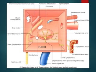


The document summarizes the anatomy of the middle ear. It describes the structures derived from the pharyngeal pouches and arches that make up the middle ear, including the ossicles, muscles, nerves and openings. It provides details on the walls, contents, blood supply and clinical relevance of the middle ear.


