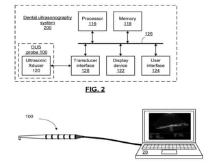
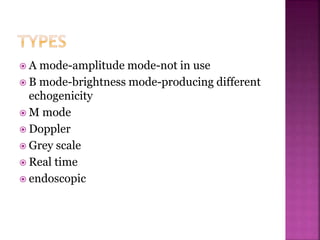
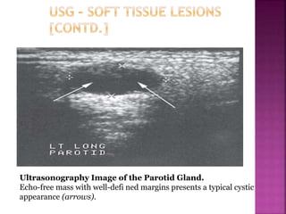
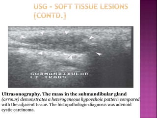
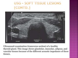
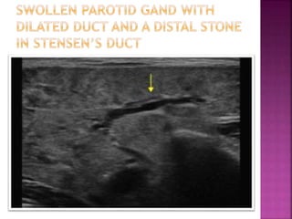
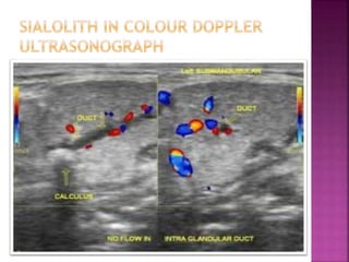
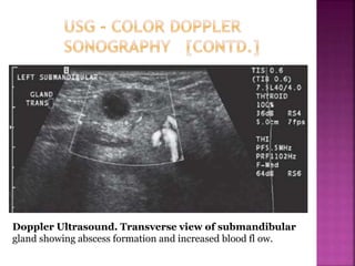
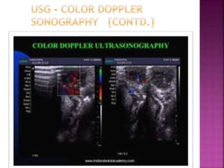
BY :
A.SELVA AROCKIAM
CRI
 Ultrasound is an oscillating sound pressure
wave with a frequency greater than upper
limit of human hearing range.thus not
separated from normal (audible) sound based
on differences in physical properties,Only the
fact humans cannot hear it
 Although this limit varies from person to
person it is approx 20kilohertz in
healthy,young adults.ultasound devices
operates from frequency from 20khz upto
several gigahertz
 Acoustic,the science of sound,starts as far back as
pythogoras in the 6th century BC who wrote on the
mathematical properties of stringed instruments
 Sir Francis Galton constructed a whistle producing
ultrasound in 1893
 The first technological application of ultrasound was
an attempt to detect icebergs by Paul Langevin in 1917
 The piezoelectric effect discovered by Jacques and
Pierre Curie in 1880 was useful in transducer to
generate and detect ultrasonic waves in air and water
 Echolocation in bats was discovered by Lazzaro
Spallanzani in 1794 ,when he demonstrated that bats
hunted and navigated by inaudible sounds and not
vision
 Scanners used for sonography generate electrical
impulses that are converted into ultra-high-
frequency sound waves by a transducer.
 Transducer is a device that can convert one form
of energy into another — in this case , electrical
energy into sonic energy.
 The most important component of the transducer
is a thin piezoelectric crystal or material made up
of a great number of dipoles arranged in a
geometric pattern.
 A dipole may be thought of as a distorted
molecule that appears to have apositive charge on
one end and a negative charge on the other.
 Currently, the most widely used piezoelectric
material is lead zirconate titanate.
 The electrical impulse generated by the
scanner causes the dipoles in the crystal to
realign themselves with the electrical fi eld
and thus suddenly change the crystal’s
thickness.
 This abrupt change begins a series of
vibrations that produce the sound waves that
are transmitted into the tissues being
examined.
 The transducer emitting ultrasound is held
against the body part being examined.
 The ultrasonic beam passes through or
interacts with tissues of different acoustic
impedance.
 Sonic waves that reflect (echo) toward the
transducer cause a change in the thickness of
the piezoelectric crystal, which in turn
produces an electrical signal that is amplified,
processed, and ultimately displayed as an
image on a monitor.
 Typically the transducer serves as both a
transmitter and a receiver.
 Current techniques permit echoes to be
processed at a sufficiently rapid rate to allow
perception of motion; this is referred to as
real-time imaging.
 The ultrasound signal transmitted into a
patient is attenuated by a combination of
absorption, refl ection, refraction, and
diffusion.
 The higher the frequency of the sound waves,
the higher the image resolution but the less
the penetration of the sound through soft
tissue.
 The fraction of the beam that is refl ected to
the transducer depends on the acoustic
impedance of the tissue, which is a product of
its density (and thus the velocity of sound
through it) and the beam ’ s angle of
incidence.
 Because of its acoustic impedance, a tissue has
a characteristic internal echo pattern.
 Consequently, not only can changes in echo
patterns distinguish between different tissues
and boundaries, but they also can be
correlated with pathologic changes within a
tissue.
 Tissues that do not produce signals, such as fl
uid-filled cysts, are said to be anechoic and
appear black.
 Tissues that produce a weak signal are
hypoechoic.
 whereas tissues that produce intense signals
such as ligaments, skin, or needles or
catheters are hyperechoic and appear bright.
 Interpretation of sonograms thus relies on
knowledge of both the physical properties of
ultrasound and the anatomy of the tissues
being scanned.
 A mode-amplitude mode-not in use
 B mode-brightness mode-producing different
echogenicity
 M mode
 Doppler
 Grey scale
 Real time
 endoscopic
 Dentistry in the modern era is emerging with
the use of advanced imaging techniques such
as computed tomography,magnetic resonance
imaging,nuclear medicine,and ultrasound of
which MRI and ultrasound are the only
imaging technique which operates without
causing radiation hazards to the patients
 It is one of the advanced imaging technique
which uses sound waves for viewing the
normal and pathological conditions involving
bone and soft tissues of the oral and
maxillofacial region
 USG has been used as an instant , non invasive
method for the observation of relatively deep
areas , recently however high frequency
echography has been developed that can provide
detail investigation of more superficial region
 USG in dentistry is used for detection of fractures
of maxillofacial region (ie) nasal bone
 Fractures ,orbital rim fractures , maxillary
fracture , mandibular fractures , zygomatic
fractures, and locating the position of mandibular
condyles . and post operative view can be instantly
seen to view the reduction and healing of
fractures.
 USG can also be used during FNAC or
FNAB,Central sampling of a solid lesion is more
reliable than peripheral sampling.So ultRa sound
guidance circumvent this problem by ensuring
that the lesion itself is sampled
 This technique offers the ability to sample non
palpable disease, gives access to different regions
of the lesion and approaches the lesion from
different angles
 The use of spring-loaded device such as biopsy or
magnum gun to discharge the needle also offers
the advantage of precise and coordinated cutting
action
 USG also used in inflammatory soft tissue
conditions of the head and neck region and
superficial tissue disorder of the maxillofacial
region
 USG can provide the content of the lesion
before any surgical procedure ,both solid and
cystic.
Ultrasonography (US) Image of Right Parotid Gland. A
well-delineated solid mass is suggested by echo returns within the
lesion (arrows). US appearance is typical of a benign salivary tumor.
Ultrasonography Image of the Parotid Gland.
Echo-free mass with well-defi ned margins presents a typical cystic
appearance (arrows).
Ultrasonography. The mass in the submandibular gland
(arrows) demonstrates a heterogeneous hypoechoic pattern compared
with the adjacent tissue. The histopathologic diagnosis was adenoid
cystic carcinoma.
Ultrasound examination (transverse section) of a healthy
thyroid gland. This image shows glandular, muscular, adipose, and
vascular tissues because of the different acoustic impedance of these
tissues.
 USG with aid of high resolution transducer
can demonstrate the internal muscle
structures more clearly than CT
 Hyper echoic bands,which corresponds to the
internal fascia are usually observed on us
image of normal muscles and are sometimes
referred to as septa
 These bands diminish or disappear with
inflammation,hence this is an important
structural index of masseteric infection
 US is also an accurate modality for measuring
the thickness of muscles,data regardinbg
thickness may provide information useful in
diagnosis and treatment especially in follow
up examination.
 US can also be used for detecting sialoliths in
parotid,submandibular and sublingual
glands,which appears as echo dense spots
with a characteristic acoustic shadow
 In USG,color doppler sonography has been
developed to identify vasculatures and to
enable evaluation of the blood flow,velocity
and vessel resistance together with
surrounding morphology
 It is also used for detecting the course of the
facial artery and for detecting hemangioma
(Doppler Effect)
 Sound reflected from a mobile structure
shows a variation in frequency which
corresponds to the speed of movement of the
structure.
 This shift in frequency, which can be
converted to an audible signal, is the principle
underlying the Doppler probe.
 The Doppler effect can also be used to image
blood flowing through heart or blood vessels.
Here the sound is reflected from the blood
cells flowing in the vessels.
 If the blood is flowing toward the transducer,
the received signal is of higher frequency.
 If the blood is flowing away the transducer,
the received signal is of low frequency.
 Colored Doppler is superimposition of
Doppler information in color on a standard
ultrasound image.
 The direction of blood flow can readily be
determined and flow toward the transducer is
by convention colored red, whereas blue
indicates flow away from the transducer.
 The turbulence signal is sometimes added as a
green component to the signal.
 The colour information gives an immediate
visual impression of the flow patterns within
the vessel and can visually identify regions of
turbulent flow within the vessel being imaged.
Doppler Ultrasound. Transverse view of submandibular
gland showing abscess formation and increased blood fl ow.
 A study was performed to evaluate the efficacy
of colour doppler USG , DIRECT DIGITAL
radiography and conventional radiograph in
diagnosis of periapical lesion
 30 patients were diagnosed with periapical
lesion in upper anterior by using CR ,DDR
,CDUSG .
 Periapical lesion diagnosed as mixed lesion by
color doppler ultrasonography were treated
non surgically while CR,DDR are treated
surgically.
 A detailed report is described below
(A ,B ) Digital
radiograph
of periapical
lesion
C ) USG of the lesion
Arrow shows
peripheral blood
supply that borders
diagnosing the lesion
as granuloma
D) Histopathological
picture of the
lesion revealing
features of
granuloma
A,B ) Pretreatment
radiograph lesion in 21
diagnosed as granuloma in
conventional radiograph
C ) USG image showing
predominantly
hypoechic area hence
diagnosed as mixed
lesion
D ) Post
treatment
radiograph
after
non surgical
treatment
 CR and DDR facilitate diagnosis of the
presence of periapical disease,but do not
provide information of its nature
 CDUSG imaging facilitate accurate
information on the pathological nature of the
lesion and hence can lead to predictable
treatment planning.
 USG is an inexpensive noninvasive and
readily available imaging technique that can
be used as an primary investigative imaging
technique so as to avoid radiation hazards
caused by x ray radiation or MRI which may
be highly economical to the patient.
 So proper application and utilisation of this
technique can be of great use in dentistry.
Ultrasonography

Ultrasonography

  • 1.
  • 2.
     Ultrasound isan oscillating sound pressure wave with a frequency greater than upper limit of human hearing range.thus not separated from normal (audible) sound based on differences in physical properties,Only the fact humans cannot hear it  Although this limit varies from person to person it is approx 20kilohertz in healthy,young adults.ultasound devices operates from frequency from 20khz upto several gigahertz
  • 3.
     Acoustic,the scienceof sound,starts as far back as pythogoras in the 6th century BC who wrote on the mathematical properties of stringed instruments  Sir Francis Galton constructed a whistle producing ultrasound in 1893  The first technological application of ultrasound was an attempt to detect icebergs by Paul Langevin in 1917  The piezoelectric effect discovered by Jacques and Pierre Curie in 1880 was useful in transducer to generate and detect ultrasonic waves in air and water  Echolocation in bats was discovered by Lazzaro Spallanzani in 1794 ,when he demonstrated that bats hunted and navigated by inaudible sounds and not vision
  • 4.
     Scanners usedfor sonography generate electrical impulses that are converted into ultra-high- frequency sound waves by a transducer.  Transducer is a device that can convert one form of energy into another — in this case , electrical energy into sonic energy.  The most important component of the transducer is a thin piezoelectric crystal or material made up of a great number of dipoles arranged in a geometric pattern.  A dipole may be thought of as a distorted molecule that appears to have apositive charge on one end and a negative charge on the other.
  • 5.
     Currently, themost widely used piezoelectric material is lead zirconate titanate.  The electrical impulse generated by the scanner causes the dipoles in the crystal to realign themselves with the electrical fi eld and thus suddenly change the crystal’s thickness.  This abrupt change begins a series of vibrations that produce the sound waves that are transmitted into the tissues being examined.
  • 6.
     The transduceremitting ultrasound is held against the body part being examined.  The ultrasonic beam passes through or interacts with tissues of different acoustic impedance.  Sonic waves that reflect (echo) toward the transducer cause a change in the thickness of the piezoelectric crystal, which in turn produces an electrical signal that is amplified, processed, and ultimately displayed as an image on a monitor.
  • 7.
     Typically thetransducer serves as both a transmitter and a receiver.  Current techniques permit echoes to be processed at a sufficiently rapid rate to allow perception of motion; this is referred to as real-time imaging.  The ultrasound signal transmitted into a patient is attenuated by a combination of absorption, refl ection, refraction, and diffusion.
  • 8.
     The higherthe frequency of the sound waves, the higher the image resolution but the less the penetration of the sound through soft tissue.  The fraction of the beam that is refl ected to the transducer depends on the acoustic impedance of the tissue, which is a product of its density (and thus the velocity of sound through it) and the beam ’ s angle of incidence.
  • 9.
     Because ofits acoustic impedance, a tissue has a characteristic internal echo pattern.  Consequently, not only can changes in echo patterns distinguish between different tissues and boundaries, but they also can be correlated with pathologic changes within a tissue.  Tissues that do not produce signals, such as fl uid-filled cysts, are said to be anechoic and appear black.
  • 10.
     Tissues thatproduce a weak signal are hypoechoic.  whereas tissues that produce intense signals such as ligaments, skin, or needles or catheters are hyperechoic and appear bright.  Interpretation of sonograms thus relies on knowledge of both the physical properties of ultrasound and the anatomy of the tissues being scanned.
  • 12.
     A mode-amplitudemode-not in use  B mode-brightness mode-producing different echogenicity  M mode  Doppler  Grey scale  Real time  endoscopic
  • 15.
     Dentistry inthe modern era is emerging with the use of advanced imaging techniques such as computed tomography,magnetic resonance imaging,nuclear medicine,and ultrasound of which MRI and ultrasound are the only imaging technique which operates without causing radiation hazards to the patients  It is one of the advanced imaging technique which uses sound waves for viewing the normal and pathological conditions involving bone and soft tissues of the oral and maxillofacial region
  • 16.
     USG hasbeen used as an instant , non invasive method for the observation of relatively deep areas , recently however high frequency echography has been developed that can provide detail investigation of more superficial region  USG in dentistry is used for detection of fractures of maxillofacial region (ie) nasal bone  Fractures ,orbital rim fractures , maxillary fracture , mandibular fractures , zygomatic fractures, and locating the position of mandibular condyles . and post operative view can be instantly seen to view the reduction and healing of fractures.
  • 17.
     USG canalso be used during FNAC or FNAB,Central sampling of a solid lesion is more reliable than peripheral sampling.So ultRa sound guidance circumvent this problem by ensuring that the lesion itself is sampled  This technique offers the ability to sample non palpable disease, gives access to different regions of the lesion and approaches the lesion from different angles  The use of spring-loaded device such as biopsy or magnum gun to discharge the needle also offers the advantage of precise and coordinated cutting action
  • 19.
     USG alsoused in inflammatory soft tissue conditions of the head and neck region and superficial tissue disorder of the maxillofacial region  USG can provide the content of the lesion before any surgical procedure ,both solid and cystic.
  • 20.
    Ultrasonography (US) Imageof Right Parotid Gland. A well-delineated solid mass is suggested by echo returns within the lesion (arrows). US appearance is typical of a benign salivary tumor.
  • 21.
    Ultrasonography Image ofthe Parotid Gland. Echo-free mass with well-defi ned margins presents a typical cystic appearance (arrows).
  • 22.
    Ultrasonography. The massin the submandibular gland (arrows) demonstrates a heterogeneous hypoechoic pattern compared with the adjacent tissue. The histopathologic diagnosis was adenoid cystic carcinoma.
  • 23.
    Ultrasound examination (transversesection) of a healthy thyroid gland. This image shows glandular, muscular, adipose, and vascular tissues because of the different acoustic impedance of these tissues.
  • 25.
     USG withaid of high resolution transducer can demonstrate the internal muscle structures more clearly than CT  Hyper echoic bands,which corresponds to the internal fascia are usually observed on us image of normal muscles and are sometimes referred to as septa  These bands diminish or disappear with inflammation,hence this is an important structural index of masseteric infection
  • 26.
     US isalso an accurate modality for measuring the thickness of muscles,data regardinbg thickness may provide information useful in diagnosis and treatment especially in follow up examination.
  • 27.
     US canalso be used for detecting sialoliths in parotid,submandibular and sublingual glands,which appears as echo dense spots with a characteristic acoustic shadow
  • 31.
     In USG,colordoppler sonography has been developed to identify vasculatures and to enable evaluation of the blood flow,velocity and vessel resistance together with surrounding morphology  It is also used for detecting the course of the facial artery and for detecting hemangioma
  • 32.
    (Doppler Effect)  Soundreflected from a mobile structure shows a variation in frequency which corresponds to the speed of movement of the structure.  This shift in frequency, which can be converted to an audible signal, is the principle underlying the Doppler probe.  The Doppler effect can also be used to image blood flowing through heart or blood vessels. Here the sound is reflected from the blood cells flowing in the vessels.
  • 33.
     If theblood is flowing toward the transducer, the received signal is of higher frequency.  If the blood is flowing away the transducer, the received signal is of low frequency.  Colored Doppler is superimposition of Doppler information in color on a standard ultrasound image.
  • 34.
     The directionof blood flow can readily be determined and flow toward the transducer is by convention colored red, whereas blue indicates flow away from the transducer.  The turbulence signal is sometimes added as a green component to the signal.  The colour information gives an immediate visual impression of the flow patterns within the vessel and can visually identify regions of turbulent flow within the vessel being imaged.
  • 35.
    Doppler Ultrasound. Transverseview of submandibular gland showing abscess formation and increased blood fl ow.
  • 38.
     A studywas performed to evaluate the efficacy of colour doppler USG , DIRECT DIGITAL radiography and conventional radiograph in diagnosis of periapical lesion  30 patients were diagnosed with periapical lesion in upper anterior by using CR ,DDR ,CDUSG .  Periapical lesion diagnosed as mixed lesion by color doppler ultrasonography were treated non surgically while CR,DDR are treated surgically.  A detailed report is described below
  • 39.
    (A ,B )Digital radiograph of periapical lesion C ) USG of the lesion Arrow shows peripheral blood supply that borders diagnosing the lesion as granuloma D) Histopathological picture of the lesion revealing features of granuloma
  • 40.
    A,B ) Pretreatment radiographlesion in 21 diagnosed as granuloma in conventional radiograph C ) USG image showing predominantly hypoechic area hence diagnosed as mixed lesion D ) Post treatment radiograph after non surgical treatment
  • 42.
     CR andDDR facilitate diagnosis of the presence of periapical disease,but do not provide information of its nature  CDUSG imaging facilitate accurate information on the pathological nature of the lesion and hence can lead to predictable treatment planning.
  • 43.
     USG isan inexpensive noninvasive and readily available imaging technique that can be used as an primary investigative imaging technique so as to avoid radiation hazards caused by x ray radiation or MRI which may be highly economical to the patient.  So proper application and utilisation of this technique can be of great use in dentistry.