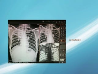
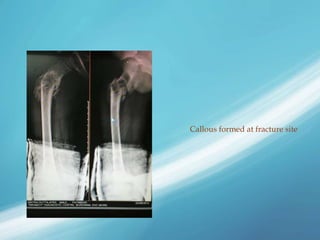
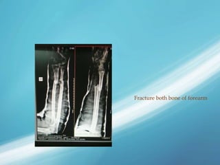
-Aurodeep Dasgupta
B.P.T. 4th Year
B.I.M.L.S.
Burdwan
 X-radiations (or x-rays) is a form of electromagnetic radiations.
Most x-rays have a wavelength of 0.01-10 nanometeres,
corresponding frequencies in the range of 30 petahertz(3X10¹⁶
Hz) to 30 exahertz(3*X10¹⁹Hz) and energies in the range of
100eV to 100 keV.
 It was invented by a German physicist Wilhelm Röntgen in
1895. He named it X-Radiations to signify an unknown type of
radiation.
 It is an important diagnostic tool for medical conditions like bone
fractures, pulmonary tuberculosis, etc.
The first x-ray image obtained was of the hands of
Wilhelm Rontgen’s wife, taken on 22nd December,1895.
 X-Ray photons carry enough energy to ionize
atoms and disrupt the molecular bonds. This
makes it a type of ionizing radiation.
 This ionizing property is used to kill the
malignant cells by radiotherapy.
 It is absorbed by solid/radio-opaque
substances like bones & metals.
 X-rays interact with matter in three main ways,
through photo-absorption, Compton scattering,
and Rayleigh scattering. The strength of these
interactions depend on the energy of the X-Rays
and the elemental composition of the material, but
not much on chemical properties since the X-Ray
photon energy is much higher than chemical
binding energies. Photo-absorption or
photoelectric absorption is the dominant
interaction mechanism in the soft X-ray regime
and for the lower hard X-ray energies. At higher
energies, Compton scattering dominates.
 Based on the energy of the radiations, x-rays
have wide range of uses like in the medical
field, industrial work places, security purposes
etc.
 Used in X-Ray scanners.
 X-Ray crystallography.
 Industrial radiography and CT scanning.
 X-Rays has an immense role in the medical
field – diagnostic as well as therapeutic.
 Diagnostic Uses-
X-ray is the most preferred tool for detecting
any bone related problem, such as fracture,
tumor, degenerations. It is also used detect
other cases like pulmonary tuberculosis, lung
abcess, etc.
 Therapeutic Uses-
It is used in cancer treatment to kill malignant
cells.
 It is a device that let us have an eye on the
amount of exposure of X-radiations to the
technician, thus enabling us to reduce the
hazardous effect of the radiation on the human
tissues.
 Helps confirm the clinical diagnosis.
 Helps study the fracture anatomy.
 Helps study the fracture displacement.
 Helps to detect crack and stress fractures.
 Helps to plan the treatment.
 Helps to detect fracture dislocation
combination, e.g. Monteggia fracture.
 Helps to ascertain post-reduction status of
fractures.
 Helps in medico legal studies.
 Before asking for an x-ray, the following points
should be kept in mind-
 Both, AP and lateral views should be taken in most
of the cases.
 Special views in some cases show the fracture better.
 X-ray requisition must specify the area of
suspension.
 X-ray of the pelvis with both hips should be asked
for in all cases of suspected pelvic injury.
 For an x-ray evaluation of the hands and feet, antero-
lateral and oblique views (not lateral) are required.
 Better no X-Ray than one view X-Ray.
 X-Ray is a shadow, it conceals and distorts. Hence
interpret X-Ray with caution.
 A joint above and joint below should be included with
the fracture under study.
 The fracture should in the middle of the film.
 Exposure should be adequate and the soft tissue
shadow be delineated properly.
 X-Ray should be read by holding the film in an
anatomical position.
 Avoid unnecessary X-Rays.
 Check X-Rays are to be taken without disturbing the
plaster cast.
 Most of the time, the body part that is to be
seen under X-Ray is to be exposed properly, i.e.
the cloth over it should be removed.
 Any accessory or jewellery on the focused part
should be removed.
 Patient needs to be calm and maintain the
posture which is instructed to him/her.
 Patient needs to follow the given instructions
strictly.
 Antero-posterior view: The X-Ray beam is
exposed from the anterior side of the body
part. The beam casts a shadow on the X-Ray
plate that is kept behind the body part.
 Lateral view: The X-Ray beam is exposed from
the lateral aspect of the body part.
 Oblique View: In this view, the X-Ray beam is
exposed from a plane which is oblique to the
body part on which the X-Ray beam is focused.
 Oblique view wrist is done to detect Scaphoid
fracture. The X-Ray beam is given in a plain
perpendicular to a plain which is slightly
slanting.
 Judet view to detect Acetabular fracture. It is a
modification of oblique view of the hip. Here
the patient’s trunk is rotated to contra-lateral
side in relation to the hip whose image is being
taken.
 Merchant view is done for patello-femoral
joint. It is taken is such a way that only the
image of the patella is obtained. The procedure
is given the pictures below.
 Skyline view for Calcaneum fracture. It is
taken by the same principle as the Merchant
view of patella. The X-Ray beam is directed
obliquely , so that it only casts a shadow of
calcaneum on the film.
 ‘Y’ view for scapula. It is taken
perpendicularly to scapular plain from the
lateral aspect. The X-Ray image looks like the
one given below.
 Dunn view is an antero-posterior view of the
hip with the patient supine and the hips and
knees flexed at 90˚, the legs abducted 15˚ -20˚,
and the femur in the neutral rotation.
 Modified Dunn view is the slight modification
of the Dunn view, where the hip is flexed to
45˚.
 Dunn view and modified Dunn view is mainly
to see the asphericity of the femoral head,
which is the cause of premature arthritis.
 Cross table lateral view is the lateral projection
radiography of a supine subject using a
horizontal X-Ray beam.
 Mainly used to detect neonatal pneumo-thorax,
hair-line fracture in the neck of the femur.
 An x-ray view box should be used in all cases. If a
fracture is obvious, one must make note of the
following points-
 Which bone is affected?
 Which part of the bone is affected? E.g., shaft etc.
 At what level is the fracture? i.e., whether the fracture
is in the upper, middle, or lower third.
 What is the pattern of the fracture?
 Is the fracture displaced?
 Is the fracture extended to nearby joints?
 Does the underlying bone appear pathological? E.g., a
cyst, abnormal texture of the whole bone, etc.
 Is it a fresh or an old fracture?
Shaft ulna fracture
Osteo-arthritis knee
Crush injury of left
foot
Scoliosis
Thoracic kyphosis
Postero-lateral subluxation of knee
Lobectomy
Callous formed at fracture site
Fracture both bone of forearm
 Diagnostic X-Rays increase the risk of
developmental problems and cancer in those
exposed cells. X-Rays are classified as a carcinogen
by both World Health Organisation’s International
Agency for Research on Cancer and the U.S.
government.
 It is estimated that 0.4% of current cancer cases in
the United States are due to diagnostic X-Rays and
CT Scans.
 That’s why when it comes to perform an X-Ray on
a pregnant woman, proper assessment of benefit
versus risk is done.
 Essential Orthopaedics
 wikipedia
 wikiRadiography
X-Ray

X-Ray

  • 1.
    -Aurodeep Dasgupta B.P.T. 4thYear B.I.M.L.S. Burdwan
  • 2.
     X-radiations (orx-rays) is a form of electromagnetic radiations. Most x-rays have a wavelength of 0.01-10 nanometeres, corresponding frequencies in the range of 30 petahertz(3X10¹⁶ Hz) to 30 exahertz(3*X10¹⁹Hz) and energies in the range of 100eV to 100 keV.  It was invented by a German physicist Wilhelm Röntgen in 1895. He named it X-Radiations to signify an unknown type of radiation.  It is an important diagnostic tool for medical conditions like bone fractures, pulmonary tuberculosis, etc.
  • 4.
    The first x-rayimage obtained was of the hands of Wilhelm Rontgen’s wife, taken on 22nd December,1895.
  • 5.
     X-Ray photonscarry enough energy to ionize atoms and disrupt the molecular bonds. This makes it a type of ionizing radiation.  This ionizing property is used to kill the malignant cells by radiotherapy.  It is absorbed by solid/radio-opaque substances like bones & metals.
  • 7.
     X-rays interactwith matter in three main ways, through photo-absorption, Compton scattering, and Rayleigh scattering. The strength of these interactions depend on the energy of the X-Rays and the elemental composition of the material, but not much on chemical properties since the X-Ray photon energy is much higher than chemical binding energies. Photo-absorption or photoelectric absorption is the dominant interaction mechanism in the soft X-ray regime and for the lower hard X-ray energies. At higher energies, Compton scattering dominates.
  • 8.
     Based onthe energy of the radiations, x-rays have wide range of uses like in the medical field, industrial work places, security purposes etc.  Used in X-Ray scanners.  X-Ray crystallography.  Industrial radiography and CT scanning.  X-Rays has an immense role in the medical field – diagnostic as well as therapeutic.
  • 9.
     Diagnostic Uses- X-rayis the most preferred tool for detecting any bone related problem, such as fracture, tumor, degenerations. It is also used detect other cases like pulmonary tuberculosis, lung abcess, etc.  Therapeutic Uses- It is used in cancer treatment to kill malignant cells.
  • 14.
     It isa device that let us have an eye on the amount of exposure of X-radiations to the technician, thus enabling us to reduce the hazardous effect of the radiation on the human tissues.
  • 15.
     Helps confirmthe clinical diagnosis.  Helps study the fracture anatomy.  Helps study the fracture displacement.  Helps to detect crack and stress fractures.  Helps to plan the treatment.  Helps to detect fracture dislocation combination, e.g. Monteggia fracture.  Helps to ascertain post-reduction status of fractures.  Helps in medico legal studies.
  • 16.
     Before askingfor an x-ray, the following points should be kept in mind-  Both, AP and lateral views should be taken in most of the cases.  Special views in some cases show the fracture better.  X-ray requisition must specify the area of suspension.  X-ray of the pelvis with both hips should be asked for in all cases of suspected pelvic injury.  For an x-ray evaluation of the hands and feet, antero- lateral and oblique views (not lateral) are required.
  • 17.
     Better noX-Ray than one view X-Ray.  X-Ray is a shadow, it conceals and distorts. Hence interpret X-Ray with caution.  A joint above and joint below should be included with the fracture under study.  The fracture should in the middle of the film.  Exposure should be adequate and the soft tissue shadow be delineated properly.  X-Ray should be read by holding the film in an anatomical position.  Avoid unnecessary X-Rays.  Check X-Rays are to be taken without disturbing the plaster cast.
  • 18.
     Most ofthe time, the body part that is to be seen under X-Ray is to be exposed properly, i.e. the cloth over it should be removed.  Any accessory or jewellery on the focused part should be removed.  Patient needs to be calm and maintain the posture which is instructed to him/her.  Patient needs to follow the given instructions strictly.
  • 19.
     Antero-posterior view:The X-Ray beam is exposed from the anterior side of the body part. The beam casts a shadow on the X-Ray plate that is kept behind the body part.
  • 20.
     Lateral view:The X-Ray beam is exposed from the lateral aspect of the body part.
  • 21.
     Oblique View:In this view, the X-Ray beam is exposed from a plane which is oblique to the body part on which the X-Ray beam is focused.
  • 23.
     Oblique viewwrist is done to detect Scaphoid fracture. The X-Ray beam is given in a plain perpendicular to a plain which is slightly slanting.
  • 24.
     Judet viewto detect Acetabular fracture. It is a modification of oblique view of the hip. Here the patient’s trunk is rotated to contra-lateral side in relation to the hip whose image is being taken.
  • 25.
     Merchant viewis done for patello-femoral joint. It is taken is such a way that only the image of the patella is obtained. The procedure is given the pictures below.
  • 26.
     Skyline viewfor Calcaneum fracture. It is taken by the same principle as the Merchant view of patella. The X-Ray beam is directed obliquely , so that it only casts a shadow of calcaneum on the film.
  • 27.
     ‘Y’ viewfor scapula. It is taken perpendicularly to scapular plain from the lateral aspect. The X-Ray image looks like the one given below.
  • 28.
     Dunn viewis an antero-posterior view of the hip with the patient supine and the hips and knees flexed at 90˚, the legs abducted 15˚ -20˚, and the femur in the neutral rotation.
  • 29.
     Modified Dunnview is the slight modification of the Dunn view, where the hip is flexed to 45˚.  Dunn view and modified Dunn view is mainly to see the asphericity of the femoral head, which is the cause of premature arthritis.
  • 30.
     Cross tablelateral view is the lateral projection radiography of a supine subject using a horizontal X-Ray beam.  Mainly used to detect neonatal pneumo-thorax, hair-line fracture in the neck of the femur.
  • 31.
     An x-rayview box should be used in all cases. If a fracture is obvious, one must make note of the following points-  Which bone is affected?  Which part of the bone is affected? E.g., shaft etc.  At what level is the fracture? i.e., whether the fracture is in the upper, middle, or lower third.  What is the pattern of the fracture?  Is the fracture displaced?  Is the fracture extended to nearby joints?  Does the underlying bone appear pathological? E.g., a cyst, abnormal texture of the whole bone, etc.  Is it a fresh or an old fracture?
  • 32.
  • 33.
  • 34.
    Crush injury ofleft foot
  • 35.
  • 36.
  • 37.
  • 38.
  • 39.
    Callous formed atfracture site
  • 40.
  • 41.
     Diagnostic X-Raysincrease the risk of developmental problems and cancer in those exposed cells. X-Rays are classified as a carcinogen by both World Health Organisation’s International Agency for Research on Cancer and the U.S. government.  It is estimated that 0.4% of current cancer cases in the United States are due to diagnostic X-Rays and CT Scans.  That’s why when it comes to perform an X-Ray on a pregnant woman, proper assessment of benefit versus risk is done.
  • 42.
     Essential Orthopaedics wikipedia  wikiRadiography