
Recommended
More Related Content
What's hot
What's hot (20)
Similar to MICROSCOPY.pptx
Similar to MICROSCOPY.pptx (20)
Recently uploaded
Recently uploaded (20)
MICROSCOPY.pptx
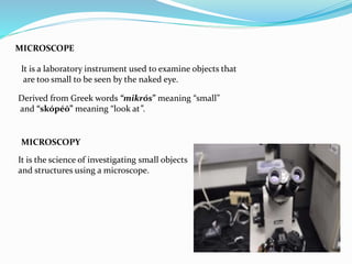
- 1. MICROSCOPE It is a laboratory instrument used to examine objects that are too small to be seen by the naked eye. MICROSCOPY It is the science of investigating small objects and structures using a microscope. Derived from Greek words “mikrós” meaning “small” and “skópéō” meaning “look at”.
- 2. MICROSCOPY TERMS MAGNIFICATION It is the process of producing an enlarged image of a specimen by using a lens system. MAGNIFYING POWER Magnifying power is how much larger a given lens can make an image appear. It can be mathematically defined as; M=1+ D/F where, M = magnifying power D = least distance of distinct vision F = Focal length of a convex lens
- 3. REFRACTIVE INDEX It is the measure of bending of a light ray when passing from one medium to another. Mathematically it can be defined as; n = c/v where, n = refractive index c = speed of light in vacuum v = velocity of light in a medium RESOLUTION It is the ability of a microscope to distinguish details on a specimen. Mathematically it is given as; r = ⋋/2NA where, r = resolution ⋋ = imaging wavelength NA = numerical aperture
- 4. The numerical aperture of a microscope objective is the measure of its ability to gather light and to resolve fine specimen detail while working at a fixed object distance. NUMERICAL APERATURE NA = n sin θ where, n=Refractive index of the medium θ is half the angular aperture Deviation of light rays by lenses or curved mirrors which causes the images to be blurred. ABERRATION
- 5. TYPES OF MICROSCOPE ELECTRON MICROSCOPE LIGHT MICROSCOPE Bright field Light Microscope Phase Contrast Light Microscope Fluorescence Light Microscope Transmission electron microscope Scanning electron microscope
- 6. Principle of a light microscope (optical microscope) When a ray of light passes through one medium into another, the ray bends at the interface causing refraction. LIGHT MICROSCOPE It uses visible light and a system of lenses to generate magnified images of Small objects. The maximum magnification power of optical microscopes is typically limited To around 1000x .
- 7. BRIGHTFIELD MICROSCOPE It is an optical microscope that uses light rays to produce a dark image against a bright background. The objectives have a magnification power of 40x-1000x depending on the type of brightfield microscope while the eyepiece lens has a standard magnification power of 10x. USES Used to visualize and study the animal cells. Used to visualize and study plant cells. Used to visualize and study the morphologies of bacterial cells. Used to identify parasitic protozoans such as Paramecium.
- 9. PHASE CONTRAST MICROSCOPE Phase-contrast microscopy is an optical microscopy technique that converts phase shifts in the light passing through a transparent specimen to brightness changes in the image To produce high-contrast images of transparent specimens, such as living cells (usually in culture) microorganisms thin tissue slices lithographic patterns fibers latex dispersions glass fragments subcellular particles (including nuclei and other organelles).
- 11. DARKFIELD MICROSCOPE USES Viewing blood cells Viewing bacteria Viewing different types of algae Viewing hairline metal fractures Viewing diamonds and other precious stones Viewing shrimp or other invertebrates Darkfield illumination is a technique in optical microscopy that eliminates scattered light from the sample image. This yields an image with a dark background around the specimen, and is essentially the complete opposite of the brightfield illumination technique
- 13. FLUORESCENCE MICROSCOPE A fluorescence microscope is an optical microscope that uses fluorescence and phosphorescence instead of, or in addition to, reflection and absorption to study the properties of organic or inorganic substances. The “fluorescence microscope” refers to any microscope that uses fluorescence to generate an image •Observing the structure of a cell •Observing DNA and RNA within a cell •Creating an image of a single molecule •Studying cell populations USES
- 15. ELECTRON MICROSCOPE It uses a beam of accelerated electron as a source of illumination. It use shaped magnetic fields to form electron optical lens system that are analogous to the glass lenses of a optical light microscope. They are used to investigate the ultrastructure of a wide range of biological and Inorganic specimens including microorganisms, cells, large molecules and crystals. Modern electron microscopes produce electron micrographs using specialized Digital cameras and frame grabbers to capture images.
- 16. TRANSMISSION ELECTRON MICROSCOPE(TEM) It uses a high voltage electron beam to illuminate the specimen and create an image.
- 17. SCANNING ELECTRON MICROSCOPE(SEM) It is a type of electron microscope that produces images of a sample by scanning The surface with a focused beam of electrons.
- 18. Bright field Phase contrast Dark field Fluorescence TEM SEM BLOOD CELLS