Download to read offline
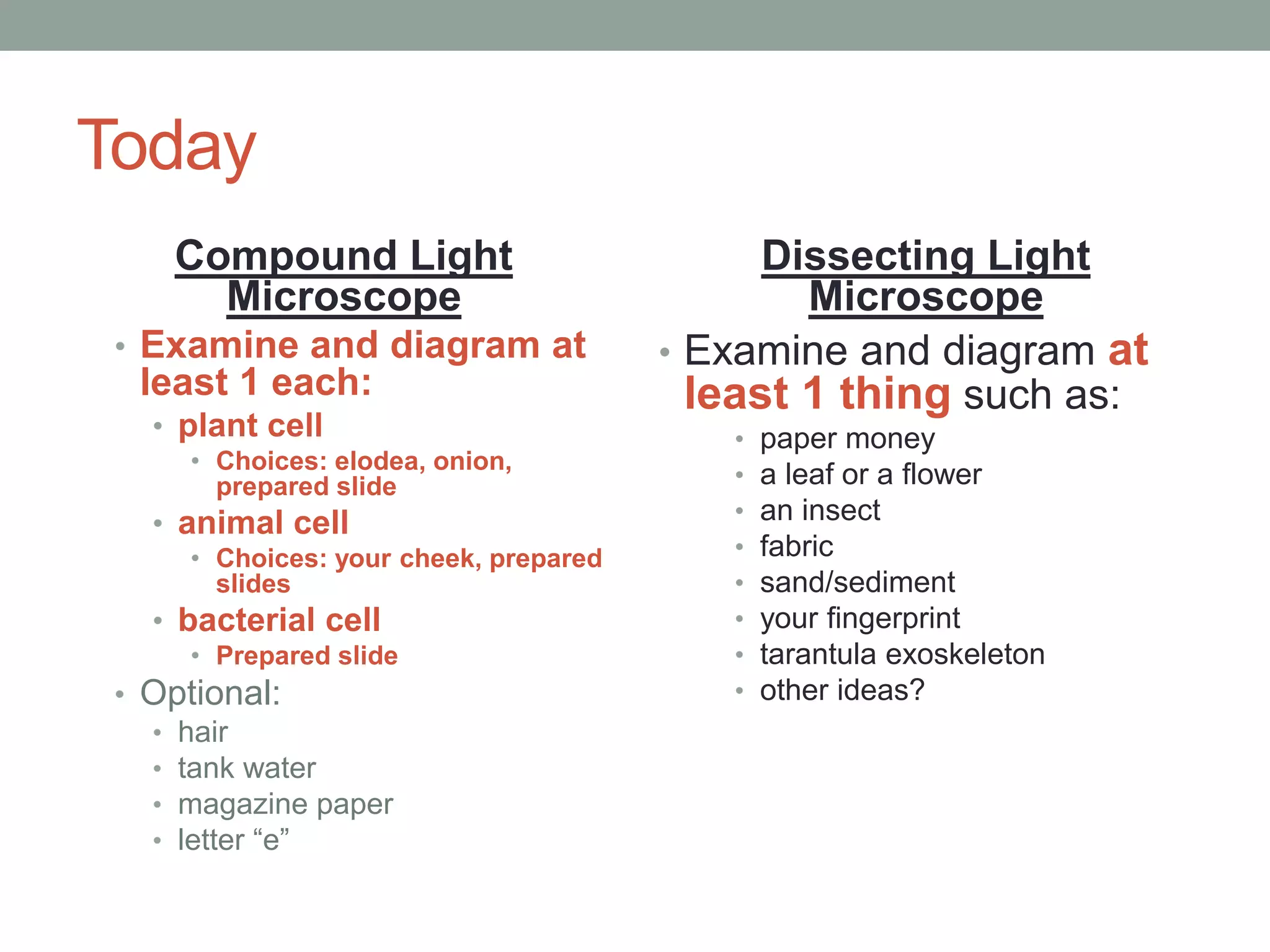
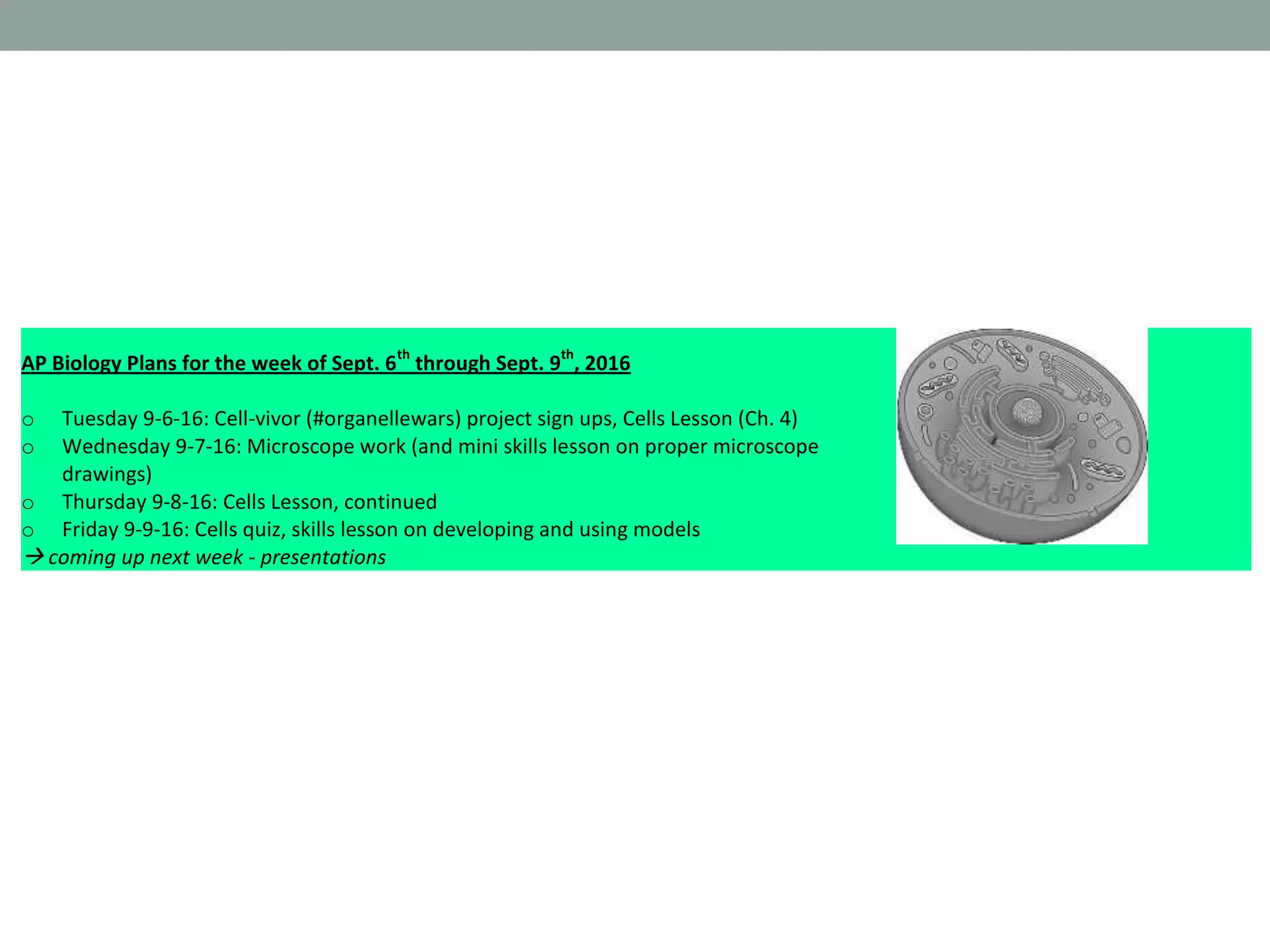
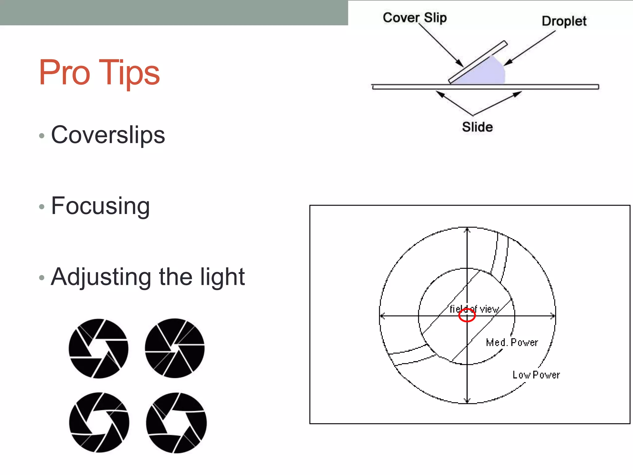
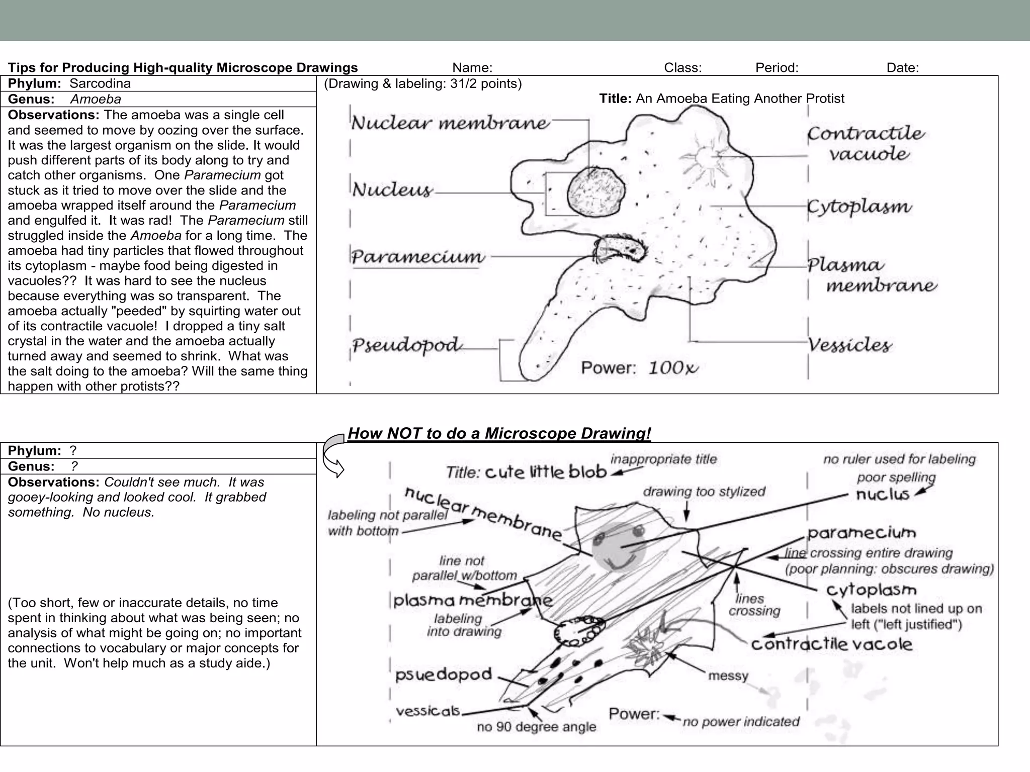


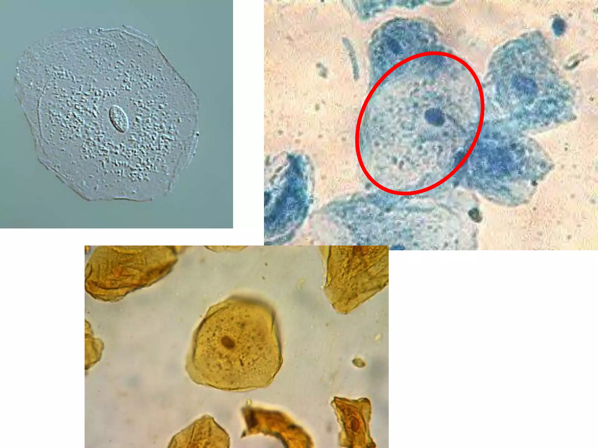
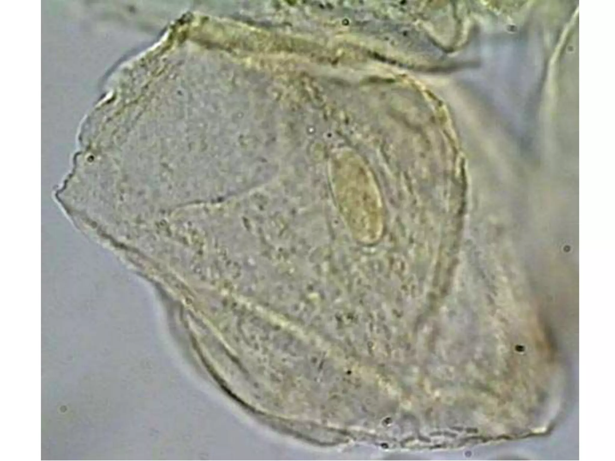

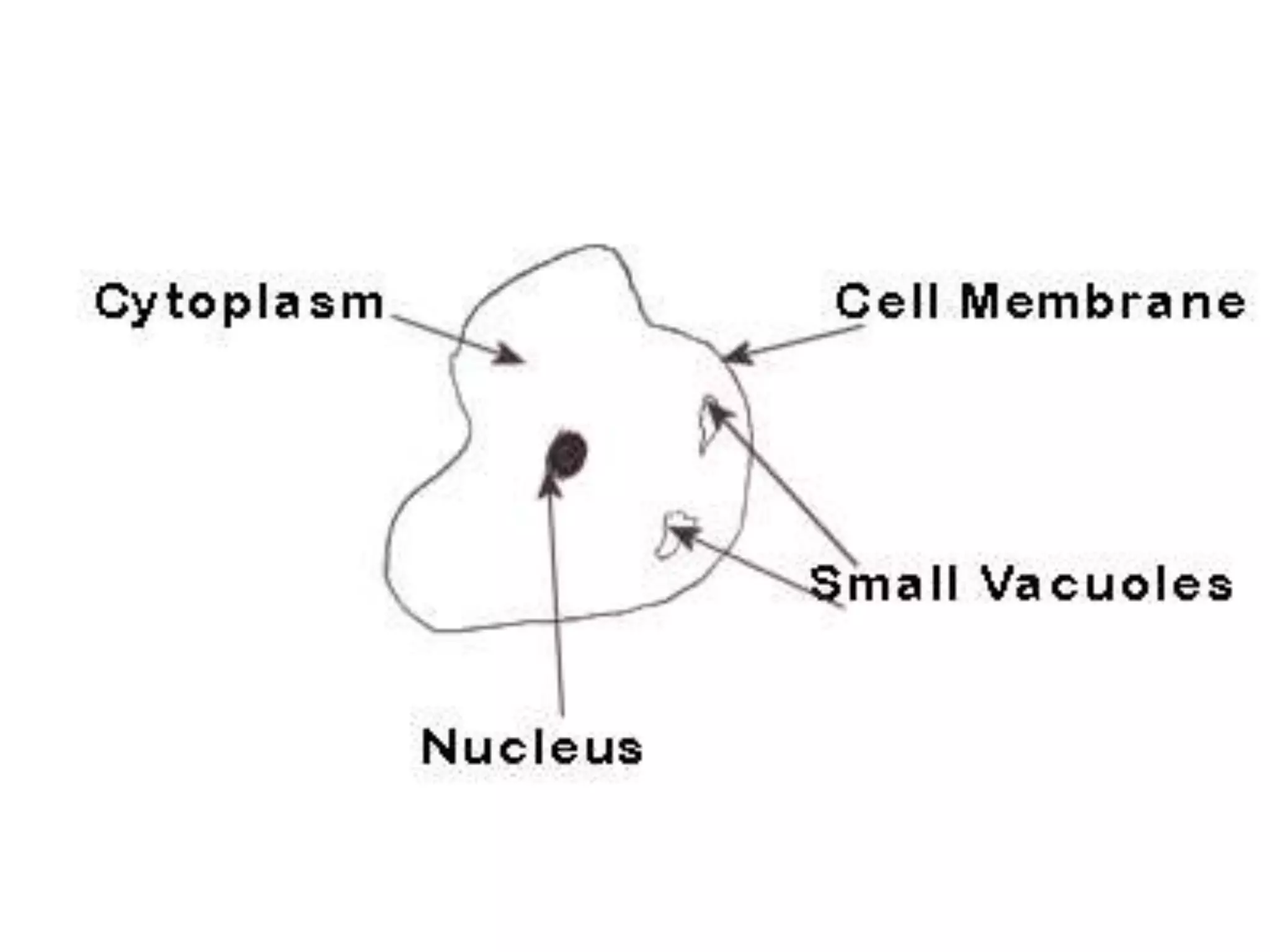
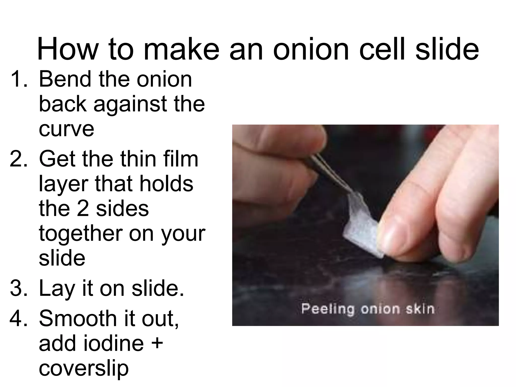


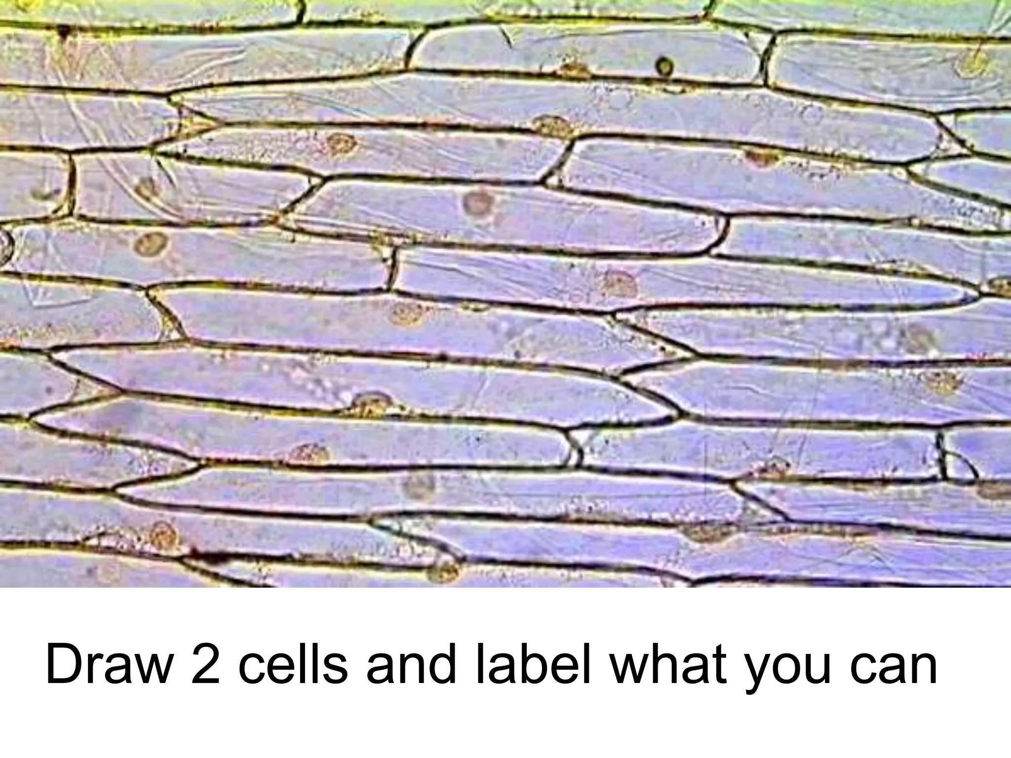
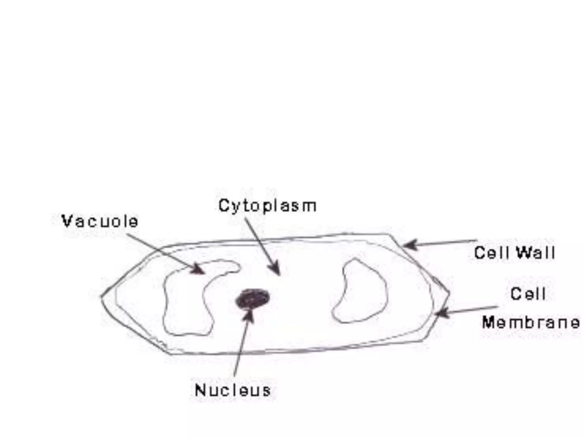

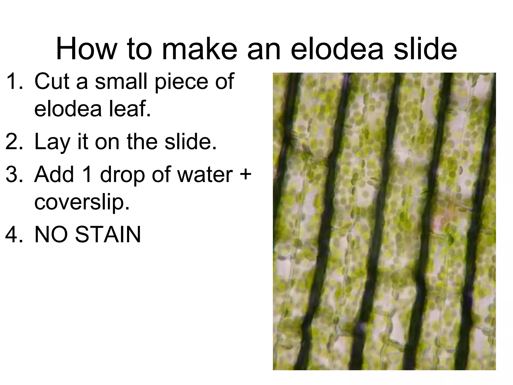


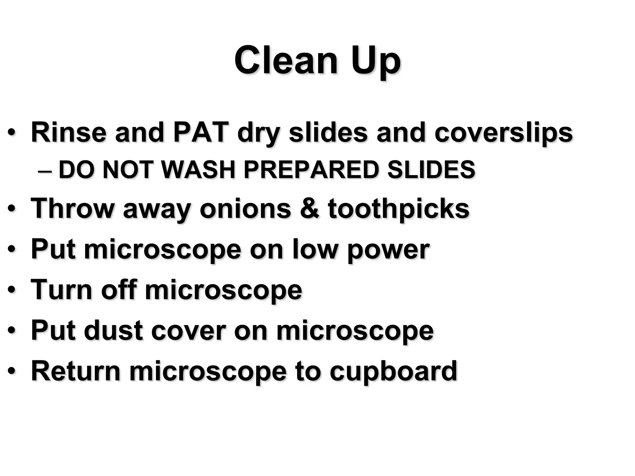


The document provides instructions for using compound light microscopes and dissecting light microscopes to examine various cell and tissue samples. It outlines activities for several days of an AP Biology class, including examining plant cells, animal cells, and bacterial cells under the compound microscope and drawing diagrams. It provides tips for producing clear microscope drawings and details how to make wet mount slides of onion cells, elodea cells, and cheek cells to view under the microscope. Basic microscope techniques like focusing and adjusting light levels are also covered.