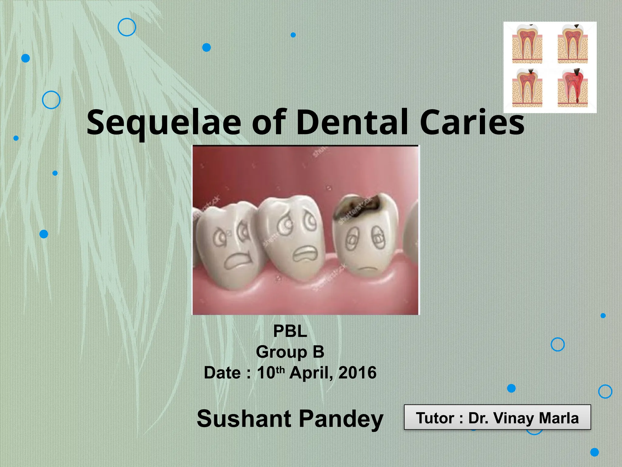
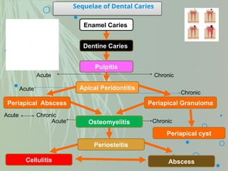
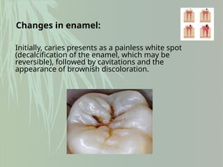
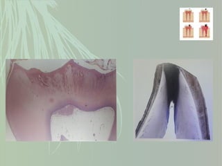
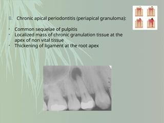
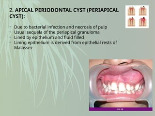
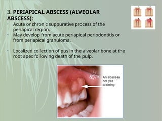
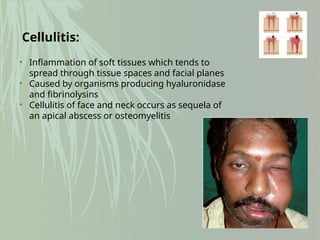
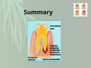
Sequelae of Dental Caries
PBL
Group B
Date : 10th
April, 2016
Sushant Pandey Tutor : Dr. Vinay Marla
Contents:
 Definition of caries
 Changes in enamel and dentin
 Pulpitis
 Periodontitis
 Summary
It is defined as “a progressive, irreversible microbial
disease of multifactorial nature affecting the
calcified tissues of the teeth, characterized by
demineralization of the inorganic portion and
destruction of the organic portion of the tooth.”
Dental Caries :
Sequelae of Dental Caries
Enamel Caries
Dentine Caries
Pulpitis
Apical Peridontitis
Periapical Abscess Periapical Granuloma
Osteomyelitis
Periosteitis
Abscess
Cellulitis
Acute Chronic
Acute
Chronic
Acute Chronic
Acute Chronic
Periapical cyst
Initially, caries presents as a painless white spot
(decalcification of the enamel, which may be
reversible), followed by cavitations and the
appearance of brownish discoloration.
Changes in enamel:
• Tubular sclerosis = Formed at halfway between pulp and DEJ.
Heavily mineralized.
• Reactionary dentine = Forms at pulp dentine interface.
• Dead tracts = Formed when odontoblasts die and their tubules
become sealed off.
Changes in dentin:
Changes in pulp:
Most common cause of dental pain.
Most cases of pulpitis are primarily a result of
dental caries in which bacteria or their
products invade the dentin and pulp tissue.
Within the rigid confines of the pulp chamber this
produces severe persistant pain and the pulp
eventually undergoes necrosis.
1. FOCAL REVERSIBLE PULPITIS:
• due to dentinal and pulpal irritation
• pulp hyperemia
C/F:
• Tooth sensitive to thermal changes
• Responds to low current in EPT
• Application of ice results in pain but disappears
on removal
2. ACUTE PULPITIS:
• extensive acute inflammation of pulp
• frequent sequel of focal reversible pulpitis
• Pain is due to pressure built up due to lack of
exudate escape
C/F:
• pain persists even after removal of thermal stimuli
• Lacinating or throbbing type pain
• Responds to low current in EPT
3. CHRONIC PULPITIS:
• Due to quiescence of a previous acute
pulpitis or may be chronic from onset
• Reduced pain and reaction to thermal
change due to degeneration of nerves
• Granulation tissue formation
• Response to high current in EPT
Effects in the periapical tissue
1. APICAL PERIODONTITIS:
• Inflammation of the periodontal ligament around the root apex
• Due to spread of infection following pulp necrosis
• May be ACUTE or CHRONIC
A. Acute apical periodontitis:
• Tenderness on mastication
• May cause reabsorption of surrounding bone
• Widening of periodontal space
B. Chronic apical periodontitis (periapical granuloma):
• Common sequelae of pulpitis
• Localized mass of chronic granulation tissue at the
apex of non vital tissue
• Thickening of ligament at the root apex
2. APICAL PERIODONTAL CYST (PERIAPICAL
CYST):
• Due to bacterial infection and necrosis of pulp
• Usual sequela of the periapical granuloma
• Lined by epithelium and fluid filled
• Lining epithelium is derived from epithelial rests of
Malassez
3. PERIAPICAL ABSCESS (ALVEOLAR
ABSCESS):
• Acute or chronic suppurative process of the
periapical region.
• May develop from acute periapical periodontitis or
from periapical granuloma.
• Localized collection of pus in the alveolar bone at the
root apex following death of the pulp.
Osteomyelitis:
• Inflammation of bone and bone marrow
• Sequela of periapical infection resulting in diffuse spread of
infection throughout the medullary space
• Necrosis of jaw bone
Cellulitis:
• Inflammation of soft tissues which tends to
spread through tissue spaces and facial planes
• Caused by organisms producing hyaluronidase
and fibrinolysins
• Cellulitis of face and neck occurs as sequela of
an apical abscess or osteomyelitis
Summary
References:
 Shafer’s textbook of oral pathology, 7th
edition
 Cawson’s essentials of oral pathology
and oral medicine, 8th
edition
final_pbl_sequeale_of_dentalcaries good ppt

final_pbl_sequeale_of_dentalcaries good ppt

  • 1.
    Sequelae of DentalCaries PBL Group B Date : 10th April, 2016 Sushant Pandey Tutor : Dr. Vinay Marla
  • 2.
    Contents:  Definition ofcaries  Changes in enamel and dentin  Pulpitis  Periodontitis  Summary
  • 3.
    It is definedas “a progressive, irreversible microbial disease of multifactorial nature affecting the calcified tissues of the teeth, characterized by demineralization of the inorganic portion and destruction of the organic portion of the tooth.” Dental Caries :
  • 4.
    Sequelae of DentalCaries Enamel Caries Dentine Caries Pulpitis Apical Peridontitis Periapical Abscess Periapical Granuloma Osteomyelitis Periosteitis Abscess Cellulitis Acute Chronic Acute Chronic Acute Chronic Acute Chronic Periapical cyst
  • 5.
    Initially, caries presentsas a painless white spot (decalcification of the enamel, which may be reversible), followed by cavitations and the appearance of brownish discoloration. Changes in enamel:
  • 6.
    • Tubular sclerosis= Formed at halfway between pulp and DEJ. Heavily mineralized. • Reactionary dentine = Forms at pulp dentine interface. • Dead tracts = Formed when odontoblasts die and their tubules become sealed off. Changes in dentin:
  • 8.
    Changes in pulp: Mostcommon cause of dental pain. Most cases of pulpitis are primarily a result of dental caries in which bacteria or their products invade the dentin and pulp tissue. Within the rigid confines of the pulp chamber this produces severe persistant pain and the pulp eventually undergoes necrosis.
  • 9.
    1. FOCAL REVERSIBLEPULPITIS: • due to dentinal and pulpal irritation • pulp hyperemia C/F: • Tooth sensitive to thermal changes • Responds to low current in EPT • Application of ice results in pain but disappears on removal
  • 10.
    2. ACUTE PULPITIS: •extensive acute inflammation of pulp • frequent sequel of focal reversible pulpitis • Pain is due to pressure built up due to lack of exudate escape C/F: • pain persists even after removal of thermal stimuli • Lacinating or throbbing type pain • Responds to low current in EPT
  • 11.
    3. CHRONIC PULPITIS: •Due to quiescence of a previous acute pulpitis or may be chronic from onset • Reduced pain and reaction to thermal change due to degeneration of nerves • Granulation tissue formation • Response to high current in EPT
  • 12.
    Effects in theperiapical tissue 1. APICAL PERIODONTITIS: • Inflammation of the periodontal ligament around the root apex • Due to spread of infection following pulp necrosis • May be ACUTE or CHRONIC A. Acute apical periodontitis: • Tenderness on mastication • May cause reabsorption of surrounding bone • Widening of periodontal space
  • 13.
    B. Chronic apicalperiodontitis (periapical granuloma): • Common sequelae of pulpitis • Localized mass of chronic granulation tissue at the apex of non vital tissue • Thickening of ligament at the root apex
  • 14.
    2. APICAL PERIODONTALCYST (PERIAPICAL CYST): • Due to bacterial infection and necrosis of pulp • Usual sequela of the periapical granuloma • Lined by epithelium and fluid filled • Lining epithelium is derived from epithelial rests of Malassez
  • 15.
    3. PERIAPICAL ABSCESS(ALVEOLAR ABSCESS): • Acute or chronic suppurative process of the periapical region. • May develop from acute periapical periodontitis or from periapical granuloma. • Localized collection of pus in the alveolar bone at the root apex following death of the pulp.
  • 16.
    Osteomyelitis: • Inflammation ofbone and bone marrow • Sequela of periapical infection resulting in diffuse spread of infection throughout the medullary space • Necrosis of jaw bone
  • 17.
    Cellulitis: • Inflammation ofsoft tissues which tends to spread through tissue spaces and facial planes • Caused by organisms producing hyaluronidase and fibrinolysins • Cellulitis of face and neck occurs as sequela of an apical abscess or osteomyelitis
  • 18.
  • 19.
    References:  Shafer’s textbookof oral pathology, 7th edition  Cawson’s essentials of oral pathology and oral medicine, 8th edition

Editor's Notes

  • #4 Once caries reaches the dentine, pain may result from thermal stimulation or from sweet or sour food or drink. Untreated, caries can progress through the dentine to the pulp, which becomes inflamed (pulpitis). Within the rigid confines of the pulp chamber this produces severe persistent pain (toothache), and the pulp eventually undergoes necrosis, when inflammation can spread around the tooth apex (periapical periodontitis), eventually forming an abscess, granuloma, or cyst.
  • #8  Within the rigid confines of the pulp chamber this produces severe persistent pain (toothache), and the pulp eventually undergoes necrosis.
  • #9 Dilation of blood vessel………edema…….thronbosis
  • #10 Edema…polymorphonuclear leukocytes…..rise in pressure due to inf exudates…..pulp destruction +abscess formation
  • #11 Mononuclear cells lymphocytes and plasma cells….fibroblastic activity and collagen sunthesis………………. CHRONIC HYPERPLASTIC PULPITIS (PULP POLYP): Excessive proliferation of chronically inflamed pulp…….Pulp protrudes from pulp chamber
  • #12 Once infection has become established in the dental pulp, spread of the process can be only in one direction through the root canals into periapical region………The involved inflammatory cells are primarily neutrophils and release prostaglandins, which activate osteoclasts to resorb the surrounding bone, leading to a detectable periapical radiolucency……………………diagram=radiolucency
  • #13 chronically or subacutely inflamed granulation tissue at the apex of a nonvital tooth
  • #14 Cyst exhibit lumen……activated t cells in the periapical granuloma produce lymphokines that cause malassez cell proliferation
  • #16 A periapical infection like abscess, granuloma or even cyst if not walled off or traumatized may undergo acute exacerbation ……..staph aureus
  • #17 Space infection infratemporal space, retropharyngeal space, parotid space common agent = streptococci………….systemic infection RHD….maxillary sinusitis Pericornitis= cellulitis due to impacted third molar