NERVE AND NERVECELLS
Presented By:
TANISHA DAS
Submitted to:
Dr. Ashish Shrestha
Dr. Vinay Marla
3.
Objectives:
Introduction about NerveAnd Nerve Cells
The Nervous System
The Nerve
The Neurons and its parts
Nerve Fibre and its Types
Clinical Considerations
Summary
4.
Introduction
o Neurons arethe functional unit in CNS and PNS.
o A nerve is a cordlike structure that contain many axons of a neuron, also
called nerve fibers.
o Conveys information in the form of electrochemical impulse carried by
indivisual neurons.
o Site of inter-communication between two neuron are called as Synapses.
o Ganglia are the group of nerve cells outside CNS.
5.
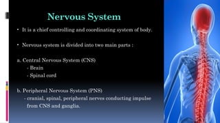
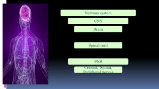
Nervous System
• Itis a chief controlling and coordinating system of body.
• Nervous system is divided into two main parts :
a. Central Nervous System (CNS)
- Brain
- Spinal cord
b. Peripheral Nervous System (PNS)
- cranial, spinal, peripheral nerves conducting impulse
from CNS and ganglia.
6.
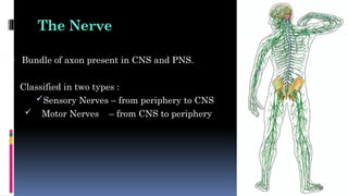
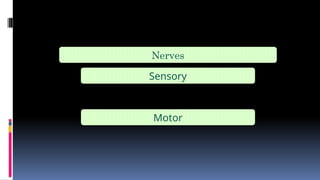
The Nerve
• Bundleof axon present in CNS and PNS.
• Classified in two types :
Sensory Nerves – from periphery to CNS
Motor Nerves – from CNS to periphery
7.
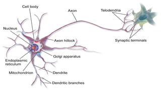
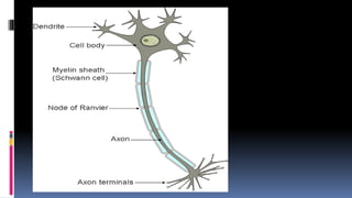
The Neurons [Nerve Cells ]
• Despite variations in shape and size in different part of nervous system
they all have same basic structures.
Cell Body [ Perikaryone ]
- large
- contain nucleus surrounding cytoplasm.
Axon
- long, slender projection from the cell body that conducts electric away
from the neuron’s cell body.
8.
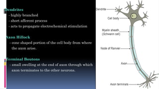
Dendrites
- highly branched
-short afferent process
- acts to propagate electrochemical stimulation
Axon Hillock
- cone shaped portion of the cell body from where
the axon arise.
Terminal Boutons
- small swelling at the end of axon through which
axon terminates to the other neurons.
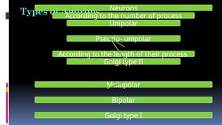
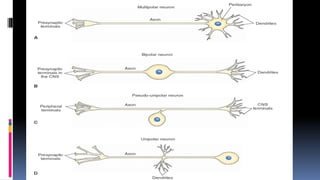
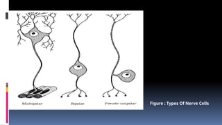
Types of NeuronsNeurons
According to the number of process
Unipolar
Pseudo- unipolar
According to the length of their process
Golgi type II
Multipolar
Bipolar
Golgi type I
12.
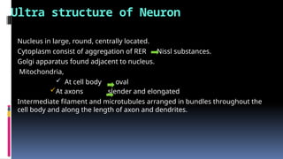
Ultra structure ofNeuron
• Nucleus in large, round, centrally located.
• Cytoplasm consist of aggregation of RER Nissl substances.
• Golgi apparatus found adjacent to nucleus.
• Mitochondria,
At cell body oval
At axons slender and elongated
• Intermediate filament and microtubules arranged in bundles throughout the
cell body and along the length of axon and dendrites.
14.
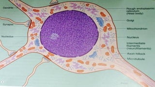
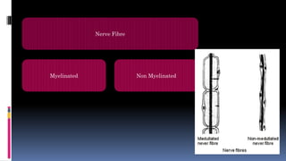
Nerve Fibers
• Nervefiber is an axon with its covering.
• Larger axons are covered with myelin sheath so called as myelinated nerve
fibers.
• Where as, smaller axons of less than 1µ diameter do not have these sheath and
are called as non myelinated nerve fibers.
Fatty nature of this myelin is responsible for glistening whiteness of the
peripheral nerve trunk and white matter of CNS.
15.
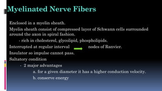
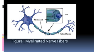
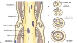
Myelinated Nerve Fibers
•Enclosed in a myelin sheath.
• Myelin sheath consist of compressed layer of Schwann cells surrounded
around the axon in spiral fashion.
- rich in cholesterol, glycolipid, phospholipids.
• Interrupted at regular interval nodes of Ranvier.
• Insulator so impulse cannot pass.
• Saltatory condition
- 2 major advantages
a. for a given diameter it has a higher conduction velocity.
b. conserve energy
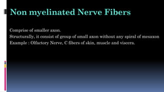
Non myelinated NerveFibers
• Comprise of smaller axon.
• Structurally, it consist of group of small axon without any spiral of mesaxon
• Example : Olfactory Nerve, C fibers of skin, muscle and viscera.
19.
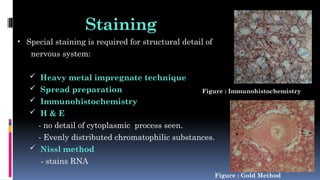
Staining
• Special stainingis required for structural detail of
nervous system:
Heavy metal impregnate technique
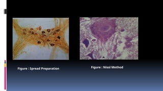
Spread preparation
Immunohistochemistry
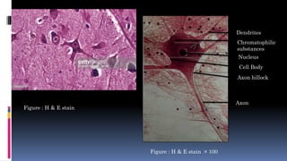
H & E
- no detail of cytoplasmic process seen.
- Evenly distributed chromatophilic substances.
Nissl method
- stains RNA
Figure : Immunohistochemistry
Figure : Gold Method
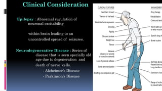
Clinical Consideration
Epilepsy :Abnormal regulation of
neuronal excitability
within brain leading to an
uncontrolled spread of seizures.
Neurodegenerative Disease : Series of
disease that is seen specially old
age due to degeneration and
death of nerve cells.
- Alzheimer’s Disease
- Parkinson’s Disease
23.
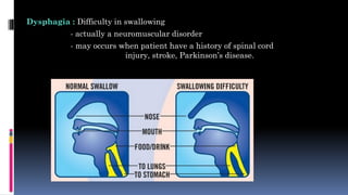
Dysphagia : Difficultyin swallowing
- actually a neuromuscular disorder
- may occurs when patient have a history of spinal cord
injury, stroke, Parkinson’s disease.
24.
Bell’s Palsy :Form of facial paralysis due
to dysfunction of Cranial
Nerve VII
- Loss of taste sensation in
anterior 2/3rd
of the tongue
in affected side.


![The Neurons [ Nerve Cells ]
• Despite variations in shape and size in different part of nervous system
they all have same basic structures.
Cell Body [ Perikaryone ]
- large
- contain nucleus surrounding cytoplasm.
Axon
- long, slender projection from the cell body that conducts electric away
from the neuron’s cell body.](https://image.slidesharecdn.com/28-250302053500-63ef10f8/85/28-Nerve-Nerve-Cells-ppt-pptx-presentation-7-320.jpg)


![REFRENCES
WWW.PHOTOBUCKET.COM
WHEATER’S FUNCTIONAL HISTOLOGY [ FIFTH
EDITION ]
JUNQUEIRA’S BASIC HISTOLOGY [ 11TH
& 12TH
EDITION ]
UNDERSTANDING MEDICAL PHYSIOLOGY BIJLANI
[ FOURTH EDITION ]](https://image.slidesharecdn.com/28-250302053500-63ef10f8/85/28-Nerve-Nerve-Cells-ppt-pptx-presentation-33-320.jpg)


![PERI-PROSTHETIC FRACTURE NAIL-PLATE CONSTRUCT [NPC].pptx](https://cdn.slidesharecdn.com/ss_thumbnails/drarunkumardrmohamedashrafperiprostheticfrasturenail-plateconstructnpc-260209164459-7e9d15a1-thumbnail.jpg?width=640&height=640&fit=bounds)


