the top three theories of root apical meristem in plants. The theories are: 1. Apical Cell Theory 2. Histogen Theory 3. Korper-Kappe Theory.The root apical meristem, or root apex, is a small region at the tip of a root in which all cells are capable of repeated division and from which all primary root tissues are derived. The root apical meristem is protected as it passes through the soil by an outer region of living parenchyma cells called the root cap.
Vascular Cambium & Seasonal activity & its Role in Stem & RootFatima Ramay
Vascular Cambium & Seasonal activity & its Role in Stem & Root:
The vascular cambium (pl. cambia or cambiums) is a lateral meristem in the vascular tissue of plants.
The vascular cambium is a cylindrical layer of cambium that runs through the stem of a plant that undergoes secondary growth.
In Dicots:
The vascular cambium is in dicot stems and roots, located between the xylem and the phloem in the stem and root of a vascular plant, and is the source of both the secondary xylem growth (inwards, towards the pith) and the secondary phloem growth (outwards).
In Monocots:
Monocot stems, such as corn, palms and bamboos, do not have a vascular cambium and do not exhibit secondary growth by the production of concentric annual rings. They cannot increase in girth by adding lateral layers of cells as in conifers and woody dicots.
Cambium of some plants remains active for the entire period of their life, i.e., cambial cells divide and resulting cells mature to form xylem and phloem elements.
This type of seasonal activity usually found in the plants present in the tropical regions, and not all plants show cambial activity.
Percentage of ringless trees in the rain forests of;India : 75%Amazon : 43%Malaysia : 15%
In regions with definite seasonal climate; seasonal activity of cambium ceased with onset of unfavorable conditions; In Autumn, it enters the dormant state and lasts for the end of summer; In Spring, cambium again becomes active.
Duration of cambial activity is also affected by day-length, e.g., In Robinia pseudoacacia, cambium is dormant under short-day condition.
The cambium cells formed in circular in cross section from the beginning onwards.
The cambial ring is partially primary (fascicular cambium) and partially secondary (interfascicular cambium).
Periderm originates from the cortical cells (extra stelar in origin).
In Dicot stem, for mechanical support xylem is with comparatively smaller vessels, greater fibers and less parenchyma.
More amount of cork is produces for protection.
Lenticels on periderm are very prominent.
The cambial ring formed is wavy in the beginning and later becomes circular.
The cambium ring is completely secondary in origin.
Periderm originates from the pericycle (intra stelar in origin).
In Dicot root, xylem is with big thin walled vessels with few fibers and more parenchyma.
Less amount of cork is produced as root is underground.
Lenticels on periderm are not very prominent.
Polyembryony is the phenomenon of two or more embryos developing from a single fertilized egg. Due to the embryos resulting from the same egg, the embryos are identical to one another, but are genetically diverse from the parents.
This PPT contains the application of Plant Anatomy in the field of Pharmacognosy & Plant systemics with number of examples to explore the beauty of this subject .
Megasporogenesis is the process of formation of megaspores from the megaspore mother cell.
In the hypodermal region of nucellus towards the micropylar end develops a primary archesporial cell.
Dr. T. Annie Sheron
Annie Sheron
Kakatiya Government College
Class 12||Chapter 2|| Sexual Reproduction in flowering plantsPrathamBiology
This chapter includes flowers, their detailed structure and developmental processess which took place durin sexual reproduction. Helpful for Board and NEET students.
Fell free for any query or suggestion
Mail us on: biologypratham@gmail.com
Website : www.prathambiology.in
The Slides contains are Female Reproductive part of Flower (Carpels/Pistils), Structure of Ovule, Types of Ovules, Microsporogenesis, Megasporogenesis, Structure of Pollen Grain, Structure of Embryo Sac
description of different types of reproductive organs, developmental stages and process of reproduction in Cycas. Various internet sources have been used.
1. INTRODUCTION
A gametophyte is a stage in the life cycle of plants and algae that undergo alternation of generations.
The gametophyte is the sexual phase in the life cycle of plants and algae.
Cell division of the zygote results in a new diploid multicellular organism, the second stage in the life cycle known as the sporophyte, the function of which is to produce haploid spores by meiosis.
2. Structure of Female Gametophyte
The female gametophyte is also called the embryo sac.
In angiosperms it mostly develops as a seven celled, eight-nucleate structure.
It consists of three antipodal cells, one central cell, two synergid cells, and one egg cell mostly.
3.Structure of Female Gametophyte
It generally contain antipodal cells at chalazal ends, an egg cell and two synergids at microplyar ends, and a center containing two polar nuclei.
Embryo sec is expect the center binucleate cell.
The process of formation of embryo sac is known as megasporogenesis.
4.The female gametophyte first arise as a tiny protuberance from the placenta, in the cavity of the overy.
As it mature,the ovule consists of a nutritive cellular mass known as nucellus, protected by two integuments, and attached to the placenta by a funiculus.
Even at an early stage, one cell becomes evident in the nucellus, which is known as the megaspore mother cell(MMC).
5.MMC is diploid (2n)
The MMC then increases in size, and divides twice (meiotically) to from 4 megaspore as ‘linear tetrad’
The first division is a reduction division
The resultant megaspores are haploid (n)
Out of the four megaspores, three degenerate, forming caps, while one remains functional.
6.This functionl megaspore is haploid (n)
The functional megaspore, then grows rapidally, by absorbing nutrition from nucellus, and forms the embryo sac.
The process of formation of embryo sac is known as megasporogenesis.
The embryo sac consists of one nucleus.
The nucleus divided into two daughter nuclei, which move towards the poles.
The pole of the embryo sac, closer to the chalaza, is the chalazal end,and the one closer to the microphyle is the micropylar end.
7.Each daughter nuclei divides again, and result in four nuclei.
Each nuclei divides again, for the last time, and result in eight nuclei.
One nuclei from each end, come to the centre, and fuse to from a diploid secondary nucleus.
The nuclei at the chalazal end, get surronded by cytoplasm, and form three distinct, haploid, antipodal cells.
Similarly, the nuclei at the micropylar end, form the egg apparatus, consisting of tow haploid synergids on either side of a large, central, haploid egg cell.
8.DEVELOPMENT OF FEMALE GAMETOPHYTE PICTURE.
Thank You.
Reproduction ensures continuity of species generation after generations as the older individuals undergo senescence and die. Flowering plants shows sexual mode of reproduction and bears complex reproductive units as male and female reproductive units along with accessary structures.
Flower is a modified stem which functions as a reproductive organ and produces ova and/or pollen. A typical angiospermic flower consists of four whorls of floral appendages attached on the receptacle: calyx, corolla, androecium (male reproductive organ consisting of stamens) and gynoecium (composed of ovary, style and stigma) .
Table of Contents:
a. Structure
b. Reproductive Structure
c. Androecium
d. Microsporogenesis
e. Gynoecium
f. Megasporogenesis
g. Pollination
h. Fertilization
i. Functions
Explore sexual reproduction in flowering plants notes to learn about the reproductive structure of the flower and the process of pollination.
Model Attribute Check Company Auto PropertyCeline George
In Odoo, the multi-company feature allows you to manage multiple companies within a single Odoo database instance. Each company can have its own configurations while still sharing common resources such as products, customers, and suppliers.
Francesca Gottschalk - How can education support child empowerment.pptxEduSkills OECD
Francesca Gottschalk from the OECD’s Centre for Educational Research and Innovation presents at the Ask an Expert Webinar: How can education support child empowerment?
2024.06.01 Introducing a competency framework for languag learning materials ...Sandy Millin
http://sandymillin.wordpress.com/iateflwebinar2024
Published classroom materials form the basis of syllabuses, drive teacher professional development, and have a potentially huge influence on learners, teachers and education systems. All teachers also create their own materials, whether a few sentences on a blackboard, a highly-structured fully-realised online course, or anything in between. Despite this, the knowledge and skills needed to create effective language learning materials are rarely part of teacher training, and are mostly learnt by trial and error.
Knowledge and skills frameworks, generally called competency frameworks, for ELT teachers, trainers and managers have existed for a few years now. However, until I created one for my MA dissertation, there wasn’t one drawing together what we need to know and do to be able to effectively produce language learning materials.
This webinar will introduce you to my framework, highlighting the key competencies I identified from my research. It will also show how anybody involved in language teaching (any language, not just English!), teacher training, managing schools or developing language learning materials can benefit from using the framework.
The French Revolution, which began in 1789, was a period of radical social and political upheaval in France. It marked the decline of absolute monarchies, the rise of secular and democratic republics, and the eventual rise of Napoleon Bonaparte. This revolutionary period is crucial in understanding the transition from feudalism to modernity in Europe.
For more information, visit-www.vavaclasses.com
Macroeconomics- Movie Location
This will be used as part of your Personal Professional Portfolio once graded.
Objective:
Prepare a presentation or a paper using research, basic comparative analysis, data organization and application of economic information. You will make an informed assessment of an economic climate outside of the United States to accomplish an entertainment industry objective.
A Strategic Approach: GenAI in EducationPeter Windle
Artificial Intelligence (AI) technologies such as Generative AI, Image Generators and Large Language Models have had a dramatic impact on teaching, learning and assessment over the past 18 months. The most immediate threat AI posed was to Academic Integrity with Higher Education Institutes (HEIs) focusing their efforts on combating the use of GenAI in assessment. Guidelines were developed for staff and students, policies put in place too. Innovative educators have forged paths in the use of Generative AI for teaching, learning and assessments leading to pockets of transformation springing up across HEIs, often with little or no top-down guidance, support or direction.
This Gasta posits a strategic approach to integrating AI into HEIs to prepare staff, students and the curriculum for an evolving world and workplace. We will highlight the advantages of working with these technologies beyond the realm of teaching, learning and assessment by considering prompt engineering skills, industry impact, curriculum changes, and the need for staff upskilling. In contrast, not engaging strategically with Generative AI poses risks, including falling behind peers, missed opportunities and failing to ensure our graduates remain employable. The rapid evolution of AI technologies necessitates a proactive and strategic approach if we are to remain relevant.
Operation “Blue Star” is the only event in the history of Independent India where the state went into war with its own people. Even after about 40 years it is not clear if it was culmination of states anger over people of the region, a political game of power or start of dictatorial chapter in the democratic setup.
The people of Punjab felt alienated from main stream due to denial of their just demands during a long democratic struggle since independence. As it happen all over the word, it led to militant struggle with great loss of lives of military, police and civilian personnel. Killing of Indira Gandhi and massacre of innocent Sikhs in Delhi and other India cities was also associated with this movement.
1. Submitted to: Dr. Sanjay Kumar Kewat
Dr.Somanjana Khatua
Dr. Rikky Rai
Submitted by: Nandani Bhartiya
M.Sc. II sem
2. What is Embryosac?
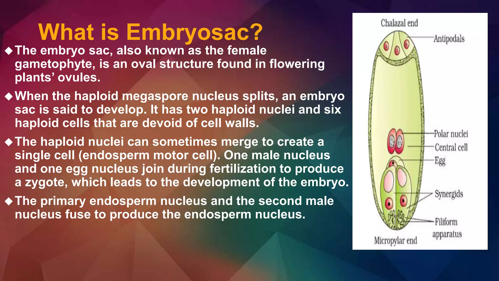
The embryo sac, also known as the female
gametophyte, is an oval structure found in flowering
plants’ ovules.
When the haploid megaspore nucleus splits, an embryo
sac is said to develop. It has two haploid nuclei and six
haploid cells that are devoid of cell walls.
The haploid nuclei can sometimes merge to create a
single cell (endosperm motor cell). One male nucleus
and one egg nucleus join during fertilization to produce
a zygote, which leads to the development of the embryo.
The primary endosperm nucleus and the second male
nucleus fuse to produce the endosperm nucleus.
3. How to develop Embryo sac?
The functional megaspore represents the first cell of the female
gametophyte.
As the functional megaspore grows, many small vacuoles appear in
its cytoplasm, which later join together to form a large vacuole. The
nucleus undergo three mitotic divisions and form eight nuclei.
After first mitotic division, two nuclei are formed and due to
enlargement of vacuole one of the nucleus is pushed towards the
micropylar end and the other towards the chalazal end.
Both these nuclei towards their respective pole undergo two mitotic
division resulting the formation of four nuclei at each pole.
4. Continue
One nucleus from each pole migrates towards the centre. These
nuclei are called as polar nuclei. The three nuclei at the chalazal
end from the antipodal cell.
Of the three nuclei at the micropylar end, one nucleus forms the
egg or female gamete and the other two as synergids.
The entire structure, the two polar nuclei, three antipodal cell, one
egg and two synergids represent the mature female gametophyte
or embryo sac.
Since this type of embryo sac develop from single megaspore and
has 8-nuclei, it is called as monosporic 8-nucleate embryo sac or
polygonum type.
It is the most common type and found in 81% of angiospermic
plant.
5.
6. Types of Embryosac
Three different type of embryo sac have been reported on the basis of
number of megaspore involve in the developing of embryo sac
a. Monosporic b. Bisporic c. Tetrasporic
A monosporic embryo sac develops from a single megaspore and as such
all the nuclei present in this type of embryo sac are genetically alike.
Monosporic embryo sacs are of the following two types.
1. Monosporic 8-nucleate or polygonum type. This type of embryo sac
develops from the chalazal megaspore. Its nucleus divides thrice to form
eight nuclei. This type is generally referred to as normal type of embryo
sac. It is also called Polygonum type as it was first time described in
Polygonum divericatum by Stransburger (1879).
2. Monosporic 4-nucleate or Oenothera type. This type of embryo sac
usually develops from the micropylar megaspore. The megaspore
nucleusdivides twice and forms only four nuclei; of these three organise
into egg apparatus and the fourth fuctions as polar nucleus. Thus
oenothera type of embryo sac does not have any antipodals. This type is
characteristic of the family Onagraceaes
7.
8. Bisporic Embryo Sac
The bisporic embryo sac develops from one of the two dyads
formed as a result of the first meiotic division (meiosis I) of
megaspore mother cell. One of the dyads degenerates. Both the
nuclei of the functional dyad take part in the formation of embryo
sac.
Each nucleus undergoes two mitotic divisions and as such the
mature embryo sac is 8-nucleate. The eight nuclei are organised
into antipodals, egg apparatus and polar nuclei as in Polygonum
type of embryo sac.
In this type of embryo sac the 4-nuclei derived from one
megaspore nucleus are genetically different from the other four
derived from the second megaspore nucleus.
On the basis of the position of functional dyad the following two
types have been recognized in bisporic embryo sacs.
1. Allium type. This type develops from the chalazal dyad.
2. Endymion type. This type develops from the micropylar dyad.
9. Tetrasporic Embryo sac
Sometimes meiotic division of the megaspore mother cell
is not accompanied by cytokinesis and hence all the four
haploid nuclei lie in a single cell called coeno-megaspore.
All the four nuclei of coeno-megaspore participate in the
formation of embryo sac. This type of embryo sac is called
tetrasporic and it is genetically more heterogeneous than
the bisporic type of embryo sac.
On the basis of (i) the position of haploid nuclei in the
coeno-megaspore, (ii) the number of times these nuclei
divide, and (iii) organisation of nuclei in the mature embryo
sac, the following types of tetrasporic embryo sacs have
been recognised.
10. PLUMBAGO TYPE- 1.Haupt(1934) in
Plimbago capensis.The 4 megaspore
nuclei are arranged in a crosswise
fashion(1+1+1+1). 1st div. of spore
nuclei –> 8 nucleate stage; arranged
in four pairs, One nucleus of the
micropylar pair is now cut off to form
the lenticular egg cell ,of the
remaining 7 nuclei, 4(one from each
pair) increase in size, approach one
another and function as secondary
nuclei ,remaining 3 nuclei
degenerates at their original
places,syneigids are completely
absent.
11. PENAEA TYPE• The four haploid nuclei of the coeno-megaspore
undergo two successive mitotic divisions forming 16 nuclei.• These
nuclei arrange themselves in four groups of four each, one at the
micropylar end, one at chalazal end and one each on the two lateral
sides.Now one nucleus from each groups migrtes to the centre, and
these four nuclei in the centre form polar nuclei.The three nuclei at the
micropylar end are cut off by membranes and form the egg
apparatus.The remaining three groups of nuclei (one chalazal and two
lateral) degenerate at maturity. Highly polyploid (5x) primary endosperm
nucleus is formed after double fertilization. Example – Family
Penaeaceae, Malpighiaceae and Euphorbiaceae.
Peperomia type -Two nuclei at the micropylar end form egg and a
synergid, eight fuse in the centre of the cell to form a polar nucleus and
the remaining six at the chalazal end formed antipodals. Example-
Peperomia and Gunnera
12.
13. DRUSA TYPE-the mature embryo sac three nuclei form egg
apparatus. Two act as polar nuclei and the remaining 11 nuclei are
cut off by membrane and form antipodal cells.Example – Drusa,
Rubia, Chrysanthemum, Ulmus etc.
FRITILLARIA TYPE- In mature embryo sac three haploid nuclei
organize into egg apparatus, three triploid into antipodal and
remaining one haploid and one triploid nuclei move to the centre
where they fuse to form a tetraploid polar nucleus.Example –
Fritillaria, Lilium, Piper and Gaillardia
PLUMBAGELLA TYPE- One triploid nuclei from chalazal end and one
haploid nucleus from the micropylar end fuse at the centre and form
tetraploid polar nucleus.One haploid nucleus at the micropylar end
forms the egg and one triploid nucleus at the chalazal end the single
antipodal. There is no synergids.