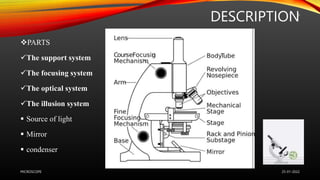
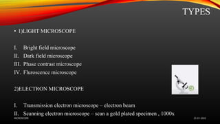
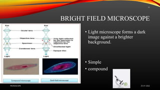
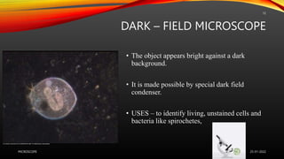
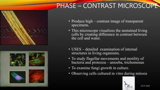
MICROSCOPE
Dr Komal M Jadhav
2nd Year PG Scholar
Dept Of Agada Tantra
CONTENTS
• Introduction
• Objectives
• History
• Types
• Description of Instrument
• Method of use & Applied aspects
• Limitations
• Precautionary measures
• Summary
• Conclusion
25-01-2022
MICROSCOPE
2
OBJECTIVE
To understand the operative procedure of Microscope & its applications in
brief.
25-01-2022
MICROSCOPE
3
HISTORY
1590- Hans Janssen and his son Zacharias Janssen,
developed first microscope .
1609- Galileo Galilei – occhiolino or compound
microscope.
1620 – Christian Huygens, another Dutchman,
developed a simple 2 lens ocular system that was
chromatically corrected.
25-01-2022
MICROSCOPE
4
INTRODUCTION
Microscope – is an instrument for viewing objects that are too small to be seen by
the naked eye.
Most commonly used instrument in medical and life science colleges and clinical
laboratories.
It is a system of suitably arranged lenses.
It magnifies the objects that were invisible to the naked eye.
25-01-2022
MICROSCOPE
5
MICRO – Extremely small
SCOPE – Assess/
Investigating
DESCRIPTION
PARTS
The support system
The focusing system
The optical system
The illusion system
 Source of light
 Mirror
 condenser
25-01-2022
MICROSCOPE
6
PRINCIPLE
• A focused beam of light passes through the specimen.
The rays passing through the specimen is gathered by the object & a
magnified image is formed.
This image further magnified by the ocular lens to produce the final
magnified virtual image
25-01-2022
MICROSCOPE
7
PROCEDURE FOR THE USE
Focusing under low power (100x)
Focusing under high power (450x)
Focusing under oil immersion(100x)
Racking the microscope ( locking)
25-01-2022
MICROSCOPE
8
TYPES
• 1)LIGHT MICROSCOPE
I. Bright field microscope
II. Dark field microscope
III. Phase contrast microscope
IV. Fluroscence microscope
2)ELECTRON MICROSCOPE
I. Transmission electron microscope – electron beam
II. Scanning electron microscope – scan a gold plated specimen , 1000x
25-01-2022
MICROSCOPE
9
MICROSCOPE
Binocular
Dissection
Dark - field
Phase -
contrast
fluroscence
polarizing
25-01-2022
MICROSCOPE
10
BRIGHT FIELD MICROSCOPE
• Light microscope forms a dark
image against a brighter
background.
• Simple
• compound
25-01-2022
MICROSCOPE
11
DARK – FIELD MICROSCOPE
• The object appears bright against a dark
background.
• It is made possible by special dark field
condenser.
• USES – to identify living, unstained cells and
bacteria like spirochetes,
25-01-2022
MICROSCOPE
12
BINOCULAR MICROSCOPE
• There are two eyepieces for visualizing
the image.
• Compare any 2 images.
25-01-2022
MICROSCOPE
13
PHASE – CONTRAST MICROSCOPE
• Produce high – contrast image of transparent
specimens.
• This microscope visualizes the unstained living
cells by creating difference in contrast between
the cell and water.
• USES – detailed examination of internal
structures in living organisms.
• To study flagellar movements and motility of
bacteria and protozoa – amoeba, trichomonas
• To examine fungi growth in culture.
• Observing cells cultured in vitro during mitosis
25-01-2022
MICROSCOPE
14
FLUROSCENCE MICROSCOPE
• This microscope uses fluroscence property to
generate an image.
• UV rays convert invisible, short wavelength rays
into longer wavelengths.
• Specimen is stained with florescent dyes.
• USES – critical tool for academic and
pharmaceutical research, pathology, clinical
medicine.
• some microbes directly fluoresce when placed
under UV lamp e.g. Cyclospora
• Acridine orange R – gives orange red fluroscence
with RNA and yellow green fluroscence with DNA
25-01-2022
MICROSCOPE
15
POLARIZING MICROSCOPE
• Used for mineral identification
25-01-2022
MICROSCOPE
16
APPLICATION
• To visualize the anatomical structure of any object like blood, cells, hair,
fiber, sperm…
• At cellular level – nucleus, mitochondria etc.
• For WBC, RBC counts.
• Striations in bullets ( to match bullet shot from a particular gun).
• To identify the number and diversity of organisms in a particular region
over time.
• Study of proteins.
25-01-2022
MICROSCOPE
17
LIMITATIONS
• Specimens must be very thin.
• Specimens small enough to fit inside the chamber.
• Certain specimens such as viruses, atoms can’t be viewed with it.
• Black and white images.
• Can’t operate in darkness.
25-01-2022
MICROSCOPE
18
PRECAUTIONARY MEASURES
Select a stool or table with appropriate height for comfortable working
for long periods.
Keep all lenses clean.
Do not touch with fingers, nor blow on them to remove dust.
Check position of object, condenser and diaphragm.
At the end clean the microscope.
25-01-2022
MICROSCOPE
19
25-01-2022
MICROSCOPE
20
25-01-2022
MICROSCOPE
21
25-01-2022
MICROSCOPE
22
25-01-2022
MICROSCOPE
23
REFERENCES
• A textbook of practical physiology by CL GHAI
25-01-2022
MICROSCOPE
24
25-01-2022
MICROSCOPE
25

Microscope.pptx

  • 1.
    MICROSCOPE Dr Komal MJadhav 2nd Year PG Scholar Dept Of Agada Tantra
  • 2.
    CONTENTS • Introduction • Objectives •History • Types • Description of Instrument • Method of use & Applied aspects • Limitations • Precautionary measures • Summary • Conclusion 25-01-2022 MICROSCOPE 2
  • 3.
    OBJECTIVE To understand theoperative procedure of Microscope & its applications in brief. 25-01-2022 MICROSCOPE 3
  • 4.
    HISTORY 1590- Hans Janssenand his son Zacharias Janssen, developed first microscope . 1609- Galileo Galilei – occhiolino or compound microscope. 1620 – Christian Huygens, another Dutchman, developed a simple 2 lens ocular system that was chromatically corrected. 25-01-2022 MICROSCOPE 4
  • 5.
    INTRODUCTION Microscope – isan instrument for viewing objects that are too small to be seen by the naked eye. Most commonly used instrument in medical and life science colleges and clinical laboratories. It is a system of suitably arranged lenses. It magnifies the objects that were invisible to the naked eye. 25-01-2022 MICROSCOPE 5 MICRO – Extremely small SCOPE – Assess/ Investigating
  • 6.
    DESCRIPTION PARTS The support system Thefocusing system The optical system The illusion system  Source of light  Mirror  condenser 25-01-2022 MICROSCOPE 6
  • 7.
    PRINCIPLE • A focusedbeam of light passes through the specimen. The rays passing through the specimen is gathered by the object & a magnified image is formed. This image further magnified by the ocular lens to produce the final magnified virtual image 25-01-2022 MICROSCOPE 7
  • 8.
    PROCEDURE FOR THEUSE Focusing under low power (100x) Focusing under high power (450x) Focusing under oil immersion(100x) Racking the microscope ( locking) 25-01-2022 MICROSCOPE 8
  • 9.
    TYPES • 1)LIGHT MICROSCOPE I.Bright field microscope II. Dark field microscope III. Phase contrast microscope IV. Fluroscence microscope 2)ELECTRON MICROSCOPE I. Transmission electron microscope – electron beam II. Scanning electron microscope – scan a gold plated specimen , 1000x 25-01-2022 MICROSCOPE 9
  • 10.
    MICROSCOPE Binocular Dissection Dark - field Phase- contrast fluroscence polarizing 25-01-2022 MICROSCOPE 10
  • 11.
    BRIGHT FIELD MICROSCOPE •Light microscope forms a dark image against a brighter background. • Simple • compound 25-01-2022 MICROSCOPE 11
  • 12.
    DARK – FIELDMICROSCOPE • The object appears bright against a dark background. • It is made possible by special dark field condenser. • USES – to identify living, unstained cells and bacteria like spirochetes, 25-01-2022 MICROSCOPE 12
  • 13.
    BINOCULAR MICROSCOPE • Thereare two eyepieces for visualizing the image. • Compare any 2 images. 25-01-2022 MICROSCOPE 13
  • 14.
    PHASE – CONTRASTMICROSCOPE • Produce high – contrast image of transparent specimens. • This microscope visualizes the unstained living cells by creating difference in contrast between the cell and water. • USES – detailed examination of internal structures in living organisms. • To study flagellar movements and motility of bacteria and protozoa – amoeba, trichomonas • To examine fungi growth in culture. • Observing cells cultured in vitro during mitosis 25-01-2022 MICROSCOPE 14
  • 15.
    FLUROSCENCE MICROSCOPE • Thismicroscope uses fluroscence property to generate an image. • UV rays convert invisible, short wavelength rays into longer wavelengths. • Specimen is stained with florescent dyes. • USES – critical tool for academic and pharmaceutical research, pathology, clinical medicine. • some microbes directly fluoresce when placed under UV lamp e.g. Cyclospora • Acridine orange R – gives orange red fluroscence with RNA and yellow green fluroscence with DNA 25-01-2022 MICROSCOPE 15
  • 16.
    POLARIZING MICROSCOPE • Usedfor mineral identification 25-01-2022 MICROSCOPE 16
  • 17.
    APPLICATION • To visualizethe anatomical structure of any object like blood, cells, hair, fiber, sperm… • At cellular level – nucleus, mitochondria etc. • For WBC, RBC counts. • Striations in bullets ( to match bullet shot from a particular gun). • To identify the number and diversity of organisms in a particular region over time. • Study of proteins. 25-01-2022 MICROSCOPE 17
  • 18.
    LIMITATIONS • Specimens mustbe very thin. • Specimens small enough to fit inside the chamber. • Certain specimens such as viruses, atoms can’t be viewed with it. • Black and white images. • Can’t operate in darkness. 25-01-2022 MICROSCOPE 18
  • 19.
    PRECAUTIONARY MEASURES Select astool or table with appropriate height for comfortable working for long periods. Keep all lenses clean. Do not touch with fingers, nor blow on them to remove dust. Check position of object, condenser and diaphragm. At the end clean the microscope. 25-01-2022 MICROSCOPE 19
  • 20.
  • 21.
  • 22.
  • 23.
  • 24.
    REFERENCES • A textbookof practical physiology by CL GHAI 25-01-2022 MICROSCOPE 24
  • 25.