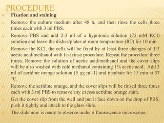
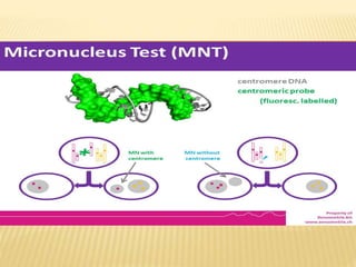
NOIDA INSTITUTE OF ENGINEERING AND
TECHNOLOGY (PHARMACY INSTITUTE)
Genotoxicity studies (in vitro and in vivo
micronucleus)
Presented by:
Shobhini Chandel
M.Pharm
2nd semester
Submitted to:
Dr. Saumya Das
Associate Professor
NIET(Pharmacy
Institute)
TABLE OF CONTENT
 Introduction
 Characteristics
 Materials and Reagents
 Equipments
 Procedure
 References
INTRODUCTION
 Genotoxicity studies can be defined as various in-vitro and in-vivo tests
designed to identify any substance or compounds which may induce
damage to genetic material either directly or indirectly by various
mechanisms. These tests should enable the identification of hazard with
respect to DNA damage and fixation.
 A micronucleus test is a test used in toxicological screening for
potential genotoxic compounds. The assay is now recognized as one of
the most successful and reliable assays for genotoxic carcinogens, i.e.,
carcinogens that act by causing genetic damage and is recommended by
the OECD guideline for the testing of chemicals. There are two major
versions of this test, one in vivo and the other in vitro
CHARACTERISTICS
 The characteristics of the micronucleus test compared to the metaphase
analysis-
1. any dividing cell population can be used regardless of its karyotype
2. accurate data can be obtained because its endpoint is simple and easy to
identify
3. response can be detected for longer duration
4. spindle poisons can also be detected
5. the background frequencies of micronucleated cells are usually stable
6. an additional treatment of chemicals other than test articles, e.g.,
colcemid or BrdU, is not required
7. types of chromosomal aberration cannot be classified by micronuclei
8. possibility of appearance of pseudo-micronucleus under some
circumstances.
MATERIAL AND REAGENTS
 Latex gloves
 Pipette tips
 Petri dishes
 Mammalian cell culture
containers for adherent cells
 Microscope slides
 Cover glass
 6-well tissue culture treated
plate
 24-well tissue culture plate
 Mammalian non cancerous
adherent cells
Required complete culture media
a) DMEM
b) Fetal bovine serum (FBS)
c) Penicillin/streptomycin
d) Trypsin
e) Phosphate buffer saline
f) Potassium chloride (KCl)
g) Acetic acid
h) Methanol
i) Acridine orange
j) Metallodrugs
k) FBS
EQUIPMENTS
 Laminar flow hood
 Hemocytometer
 Pipettes
 Incubator
 Fluorescent microscope with highly sensitive camera controlled by
Nikon NIS-Elements software
PROCEDURE
 Cell preparation
a) Culture the cells according to their requirement
b) Remove medium, and then twice rinse cells with 10 ml PBS, removing
the PBS after each washing. The PBS should be autoclaved before use.
c) Add 1 ml 0.25% trypsin to the cells and incubate at 37 °C for 1-5 min
until the cells appear round.
d) Add 10 ml culture medium with 10% FBS, and detach the cells by
pipetting.
e) Count the cells using a hemocytometer. Note: A relatively accurate
number of cells each time is crucial.
f) Prepare desired seeding concentration, and then seed cell into dishes or
6-well plates.
PROCEDURE
 Growing cells on cover glasses
a) Harvest cells and plate an appropriate number of cells per dish or per well
in a 6-well plate, at least in duplicate in a final volume of culture medium 1
ml.
b) The number of cells for seeding should be determined by the
aggressiveness of the treatment.
c) Incubate cells for 24 h in a CO2 incubator at 37 °C and allow them to
attach to the plate/dish.
d) Before the plating, place the sterile cover glasses slip flat into each well of
a 6-well plate.
e) Pipette up and down or shake the dish to make sure cells are not
concentrating in the center of the dish well. Make sure there are no air
bubbles between the cover glasses and the tissue culture dish.
f) Treat the cells as necessary with the metallodrugs (at their IC50 values,
against immortalized non-cancer cells).
g) Incubate the cells in a CO2 incubator at 37 °C for 48 h.
PROCEDURE
 Fixation and staining
a) Remove the culture medium after 48 h, and then rinse the cells three
times each with 3 ml PBS.
b) Remove PBS and add 2-3 ml of a hypotonic solution (75 mM KCl)
solution and leave the dishes/plates at room temperature (RT) for 10 min.
c) Remove the KCl, the cells will be fixed by at least three changes of 1/3
acetic acid/methanol with fast rinse procedure. Repeat the procedure three
times. Remove the solution of acetic acid/methanol and the cover slips
will be also washed with cold methanol containing 1% acetic acid. Add 3
ml of acridine orange solution (5 μg ml-1) and incubate for 15 min at 37
°C.
d) Remove the acridine orange, and the cover slips will be rinsed three times
each with 3 ml PBS to remove any excess acridine orange stain.
e) Get the cover slip from the well and put it face down on the drop of PBS,
push it tightly and attach to the glass slide.
f) The slide now is ready to observe under a fluorescence microscope.
REFERENCES
 academic.oup.com/mutage/article/15/3/245/1066769?login=false
 bio-protocol.org/pdf/Bio-protocol3311.pdf
 genesenvironment.biomedcentral.com/articles/10.1186/s41021-016-
0044-x

micronucleus assay.pptx

  • 1.
    NOIDA INSTITUTE OFENGINEERING AND TECHNOLOGY (PHARMACY INSTITUTE) Genotoxicity studies (in vitro and in vivo micronucleus) Presented by: Shobhini Chandel M.Pharm 2nd semester Submitted to: Dr. Saumya Das Associate Professor NIET(Pharmacy Institute)
  • 2.
    TABLE OF CONTENT Introduction  Characteristics  Materials and Reagents  Equipments  Procedure  References
  • 3.
    INTRODUCTION  Genotoxicity studiescan be defined as various in-vitro and in-vivo tests designed to identify any substance or compounds which may induce damage to genetic material either directly or indirectly by various mechanisms. These tests should enable the identification of hazard with respect to DNA damage and fixation.  A micronucleus test is a test used in toxicological screening for potential genotoxic compounds. The assay is now recognized as one of the most successful and reliable assays for genotoxic carcinogens, i.e., carcinogens that act by causing genetic damage and is recommended by the OECD guideline for the testing of chemicals. There are two major versions of this test, one in vivo and the other in vitro
  • 4.
    CHARACTERISTICS  The characteristicsof the micronucleus test compared to the metaphase analysis- 1. any dividing cell population can be used regardless of its karyotype 2. accurate data can be obtained because its endpoint is simple and easy to identify 3. response can be detected for longer duration 4. spindle poisons can also be detected 5. the background frequencies of micronucleated cells are usually stable 6. an additional treatment of chemicals other than test articles, e.g., colcemid or BrdU, is not required 7. types of chromosomal aberration cannot be classified by micronuclei 8. possibility of appearance of pseudo-micronucleus under some circumstances.
  • 5.
    MATERIAL AND REAGENTS Latex gloves  Pipette tips  Petri dishes  Mammalian cell culture containers for adherent cells  Microscope slides  Cover glass  6-well tissue culture treated plate  24-well tissue culture plate  Mammalian non cancerous adherent cells Required complete culture media a) DMEM b) Fetal bovine serum (FBS) c) Penicillin/streptomycin d) Trypsin e) Phosphate buffer saline f) Potassium chloride (KCl) g) Acetic acid h) Methanol i) Acridine orange j) Metallodrugs k) FBS
  • 6.
    EQUIPMENTS  Laminar flowhood  Hemocytometer  Pipettes  Incubator  Fluorescent microscope with highly sensitive camera controlled by Nikon NIS-Elements software
  • 7.
    PROCEDURE  Cell preparation a)Culture the cells according to their requirement b) Remove medium, and then twice rinse cells with 10 ml PBS, removing the PBS after each washing. The PBS should be autoclaved before use. c) Add 1 ml 0.25% trypsin to the cells and incubate at 37 °C for 1-5 min until the cells appear round. d) Add 10 ml culture medium with 10% FBS, and detach the cells by pipetting. e) Count the cells using a hemocytometer. Note: A relatively accurate number of cells each time is crucial. f) Prepare desired seeding concentration, and then seed cell into dishes or 6-well plates.
  • 8.
    PROCEDURE  Growing cellson cover glasses a) Harvest cells and plate an appropriate number of cells per dish or per well in a 6-well plate, at least in duplicate in a final volume of culture medium 1 ml. b) The number of cells for seeding should be determined by the aggressiveness of the treatment. c) Incubate cells for 24 h in a CO2 incubator at 37 °C and allow them to attach to the plate/dish. d) Before the plating, place the sterile cover glasses slip flat into each well of a 6-well plate. e) Pipette up and down or shake the dish to make sure cells are not concentrating in the center of the dish well. Make sure there are no air bubbles between the cover glasses and the tissue culture dish. f) Treat the cells as necessary with the metallodrugs (at their IC50 values, against immortalized non-cancer cells). g) Incubate the cells in a CO2 incubator at 37 °C for 48 h.
  • 9.
    PROCEDURE  Fixation andstaining a) Remove the culture medium after 48 h, and then rinse the cells three times each with 3 ml PBS. b) Remove PBS and add 2-3 ml of a hypotonic solution (75 mM KCl) solution and leave the dishes/plates at room temperature (RT) for 10 min. c) Remove the KCl, the cells will be fixed by at least three changes of 1/3 acetic acid/methanol with fast rinse procedure. Repeat the procedure three times. Remove the solution of acetic acid/methanol and the cover slips will be also washed with cold methanol containing 1% acetic acid. Add 3 ml of acridine orange solution (5 μg ml-1) and incubate for 15 min at 37 °C. d) Remove the acridine orange, and the cover slips will be rinsed three times each with 3 ml PBS to remove any excess acridine orange stain. e) Get the cover slip from the well and put it face down on the drop of PBS, push it tightly and attach to the glass slide. f) The slide now is ready to observe under a fluorescence microscope.
  • 11.