NOIDAINSTITUTEOFENGINEERINGANDTECHNOLOGY
(PHARMACY INSTITUTE)
Presented by:
Shobhini Chandel
M.Pharm
1stsemester
Submitted to:
Dr. Saumya Das
Associate Professor
NIET(Pharmacy Institute)
Hepatoprotective Screening Methods
TABLE OF CONTENT
Introduction
Liver toxicity
Drugs causing DILI
Hepatoprotective drug classification
Invitro models
Invivo models
References
Introduction:
 The liver is the body's largest internal organ that sits on the
right-hand side of the belly.
 The liver is an essential organ that has many functions in the
body, including making proteins and blood clotting factors, , it
breaks down and detoxifies substances in the body, and it also
acts as a storage unit, manufacturing
triglyceride and cholesterol, glycogen synthesis, and bile
production.
 Many different disease processes can occur in the liver,
including infections such
as hepatitis, cirrhosis (scarring), cancers, and damage by
medications or toxins.
Introduction:
 Hepatoprotective agents are those compounds, which
mitigate the liver injury caused by hepatotoxic agents thus
can prevent damage to the liver.
Examples:
Ursodeoxycholic acid (UDCA), silibinin, and N-acetyl
cysteine (NAC).
Liver toxicity:
Main causes of liver or hepatic toxicity are as follows:
 Alcohol consumption (ethanol)
 Chemicals (carbon tetrachloride, phosphorus, alfatoxin,
chlorinated hydrocarbon, etc)
 Drug-Induced Liver Injury
 Autoimmune disorder
 Infection (hepatitis A, B, C)
 Cancer and other growths (Liver cancer, Bile duct cancer, Liver
adenoma)
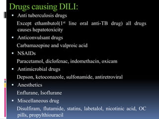
Drugs causing DILI:
 Anti tuberculosis drugs
Except ethambutol(1st line oral anti-TB drug) all drugs
causes hepatotoxicity
 Anticonvulsant drugs
Carbamazepine and valproic acid
 NSAIDs
Paracetamol, diclofenac, indomethacin, oxicam
 Antimicrobial drugs
Depson, ketoconazole, sulfonamide, antiretroviral
 Anesthetics
Enflurane, Isoflurane
 Miscellaneous drug
Disulfiram, flutamide, statins, labetalol, nicotinic acid, OC
pills, propylthiouracil
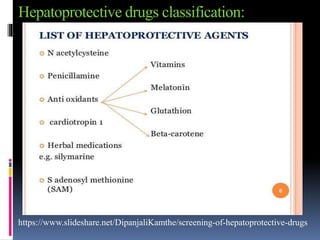
Hepatoprotective drugs classification:
https://www.slideshare.net/DipanjaliKamthe/screening-of-hepatoprotective-drugs
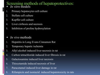
Screening methods of hepatoprotectives:
 In vitro models
1. Primary hepatocytes cell culture
2. Stellate cell culture
3. Kupffer cell culture
4. Liver cirrhosis and necrosis
5. Inhibition of proline hydroxylation
 In vivo methods
1. Hepatitis in Long Evans Cinnamon Rat
2. Temporary hepatic ischemia
3. Allyl alcohol induced liver necrosis in rat
4. Carbon tetrachloride induced liver fibrosis in rat
5. Galactosamine induced liver necrosis
6. Thiocetamide induced necrosis of liver
7. Paracetamol induced liver damage in rat
8. Rifampicin and isoniazid induced hepatotoxicity in rats
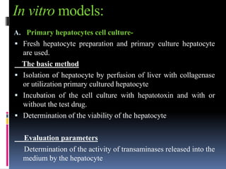
In vitro models:
A. Primary hepatocytes cell culture-
 Fresh hepatocyte preparation and primary culture hepatocyte
are used.
The basic method
 Isolation of hepatocyte by perfusion of liver with collagenase
or utilization primary cultured hepatocyte
 Incubation of the cell culture with hepatotoxin and with or
without the test drug.
 Determination of the viability of the hepatocyte
Evaluation parameters
Determination of the activity of transaminases released into the
medium by the hepatocyte
B. Stellate cell culture
 In chronic injury there is stellate cell activation
 There is secretion of matrix by activated stellate cell results in
liver fibrosis and ultimately cirrhosis
 Stellate cell are isolated from rat liver by collagenase or
pronase digestion
 Test drugs are incubated with activated stellate cell culture by
hepatotoxin
Evaluation parameters
Morphology of cell, Alpha SMA expression, cell count with
thymidine incorporation and inhibition in synthesis of collagen
type 1 and 3
C. Inhibition of proline hydroxylation
 The thermal stability of the triple helix of collagen is depend
on intramolecular hydrogen bond synthesized by the enzyme
propyl-4-hydroxylase
Procedure
 Reaction volume of 1 ml contain tris buffer, glutarate ,ferrous
oxide, ascorbate, catalase, BSA, dithiotreitol inhibitor
 After incubation at 37 degrees celsius for 30 minutes the
generated CO2 is trapped and determined
In vivo methods
A. Hepatitis and Long Evans Cinnamon Rat
Purpose and rationale
 The Long Evans Cinnamon Rat is useful model to study genetically
transmitted hepatitis and chronic liver disease
 Due to excessive copper accumulation in the liver of Long Evans Cinnamon
Rat making this animal a model for Wilson’s disease in human(excess
copper accumulation in body)
Procedure
 Long Evans Cinnamon Rat of age of 5 weeks are housed in temperature and
humidity controlled room
 Group of 6 to 10 rats are given different diets on a 15% purified protein diet
and supplemented with vitamin or drugs
 Drugs are supplied via mini pumps intraperitoneal implanted under
anesthesia
 The occurrence of jaundice is easily observable as the time when the ear and
tail turn yellow and the urine become bright ending in death of animal
within about a week.
B. Temporary hepatic ischemia
 Hepatocellular function is altered by temporary hepatic ischemia as occurring
during surgical management of acute hepatic trauma and being essential during
hepatic transplantation.
Purpose and rationale
 Total hepatic ischemia in rats is produced by placing a ligature around the
hepatic artery portal vein and common bile duct
Procedure
 Male albino rat 300 to 350 g is fasted for 16 hours prior to the experiment but
allow water ad libitum
 The rats are anesthetized lightly with ether and abdomen cavity is opened
through a midline incision
 Portal vein as a hepatic artery and the bile duct are occluded by placing a
tourniquets around the vessels
 Blood pressure is measured by a catheter is inserted into the right
femoral artery. During the ischemic period, 0.7ml of saline is given
intravenous at 20 minute interval for volume replacement
 At the end of 60 minute ischemic period the, tourniquet around the
portal vein, hepatic artery and the bile duct is removed in order to re
stabilize blood flow to the liver
 The abdominal incision is then closed and the animal receive either
saline or the drug
C. Allyl alcohol induced liver necrosis in rat
Purpose and rationale
 Administration of allyl alcohol induced liver necrosis in rat which
can be partially prevented by treatment with several drugs such as
antibiotic .
Procedure
 Female Wistar rat weighing 120-150 g are fasted overnight with
water ad libitum
 On following morning test drug is given orally or intraperitoneally to
10 rats. After 1 hour 0.4 ml per kg of 1.25% allyl alcohol solution is
given orally
 Test drug given again on second day and sacrifice on 3rd day liver is
removed
Evaluation parameter
 The parietal sides of the liver are checked using a stereo
microscope with 25 times magnification
 Focal necrosis is observed as white green or yellowish
hemorrhagic area clearly separated from an infected tissue
 The diameter of the necrotic area is determined using ocular
micro meter
 The value added for each animal to obtain an index of necrosis
 Mean of necrosis index is calculated and compared with student t
test
 The protective effect is expressed as percentage decrease of the
necrosis index versus control
D. Carbon tetrachloride induced liver fibrosis in rat
Purpose and rationale
 chronic administration of tetrachloride to rat induced severe
disturbance of hepatic function together with histologically observable
liver fibrosis.
Procedure
 Group of 20 female Wistar rat is used weighing 100- 150g
 The animal are treated orally twice a week with 1 mg per kg carbon
tetrachloride dissolved in olive oil 1:1 over a period of 8 week
 Animal are fed on chow diet with water ad libitum. Control group
receive only olive oil. Test and standard drug given twice daily except
on Sunday when only one dose is given
 Animal weighed every week and at the end of 8 week animal sacrifice
using and anesthetic ether
Evaluation parameter
 Total bilirubin
 Total bile acid
 7S fragment of type 4 collagen
 Procollagen 3 N- peptide
E. Galactosamine induced liver necrosis
 Single dose or few repeated dose of D galactosamine cause acute
hepatic necrosis in rat
 Prolonged administration lead to cirrhosis
Procedure
 Divided doses of 100 to 400 mg/ kg D galactosamine are injected
to rat IP or IV during day one
 for induction of liver cirrhosis male Wistar rat weighing 110-180
gm are injected IP three time weekly with 500 mg/ kg D
galactosamine over a period of 1 to 3 months
 Potential protective substance are administered orally with the
food every day
 The rat sacrifice at various time interval and the liver obtained by
autopsy
Evaluation
 The liver are evaluated by light microscope and
immunohistology using antibodies against macrophages,
lymphocytes and the extracellular matrix component
 The extent of liver cell necrosis and Immuno reactivity for
macrophages, lymphocytes and extracellular matrix component
is graded semi-quantitatively on 0-4 scale
 0= absent
 1 –trace
 2- weak
 3- moderate
 4- strong
References -
 https://www.ncbi.nlm.nih.gov/pmc/articles/PMC4265599/
 https://www.spandidos-publications.com/ijmm/42/4/2020
 https://www.sciencedirect.com/science/article/abs/pii/S03788741
20336552?via%3Dihub
 https://pubmed.ncbi.nlm.nih.gov/?term=hepatoprotective+agents
 https://www.sciencedirect.com/topics/medicine-and-
dentistry/liver-toxicity
 https://www.ncbi.nlm.nih.gov/pmc/articles/PMC4202426/
 https://www.slideshare.net/DipanjaliKamthe/screening-of-
hepatoprotective-drugs
Hepatoprotective screening methods