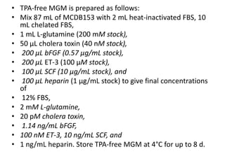
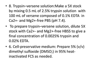
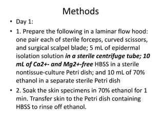
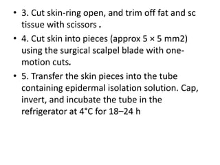
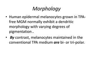
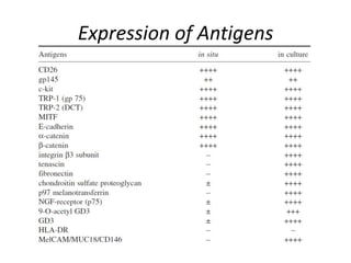
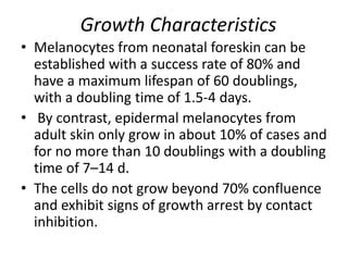
Tutorial presentation

Melanocyte culture-Technique
presented by :Dr. Mahesh prajapat
Introduction
• First attempt -1957
• Since 1982 have pure normal human
melanocyte cultures been reproducibly
established to yield cells in sufficient quantity
for biological, biochemical, and molecular
analyses .
• Selective growth of melanocytes, which
comprise only 3–7% of epidermal cells in
normal human skin, was initially achieved by
suppressing the growth of keratinocytes and
fibroblasts in epidermal cell suspensions with
• the tumor promoter 12-O-tetradecanoyl
phorbol-13-acetate (TPA) and
• the intracellular cyclic adenosine 3′, 5′
monophosphate (cAMP) enhancer cholera
toxin, respectively, which both also act as
melanocyte growth promoters.
• However, phorbol ester has prolonged effects
on multiple cellular responses.
• Recent progress in basic cell-culture
technology, has led to an effective and
standardized isolation method, and special
TPA-free culture media for selective growth
and long-term maintenance of human
melanocytes.
• Altered cell adhesion may be expressed as increased
attachment by cells that otherwise grow in suspension
or as decreased attachment in cells already growing
attached to substrate.
• Evidence of two classes of membrane associated
phorbol esters receptors in fibroblast culture.
• Can inhibit or delay terminal differentiation in both
normal embryo cultures and several tumor cell lines
• Higher doses irreversibly prevent pigmentation of
some cells.
• shown to reduce the numbers of melanosomes in
human melanocytes in culture and to delay the onset
of melanization .
•

•
M. EISINGER AND 0. MARKO
Memorial Sloan-Kettering Cancer Center, New York, New York 10021
Procedure
• 1. Tissue source and collection:
• human neonatal foreskins obtained from routine
circumcision and
• normal adult skin acquired from reduction
mammoplasty.
• the skin is then placed into a sterile container with 20
mL of normal skin-transporting medium supplied in
advance and kept near the surgical area at 4°C.
• Specimens are delivered immediately to the tissueculture laboratory or stored at 4°C.
• Neonatal foreskins can be kept for up to 48 h, and
normal adult skin, for up to 24 h.
composed of Hanks balanced salt solution
(HBSS) without Ca2+ and Mg2+ (HBSS;)
supplemented with penicillin (100 U/mL),
streptomycin (100 μg/mL), gentamicin (100
μg/mL) and fungizone (0.25 μg/mL).
• After sterilization through a 0.2-μm filter, the
skin transporting medium is transferred into
sterile containers in 20-mL aliquots and stored
at 4°C for up to 1 mo.
• 2. Sterilization of skin specimens:
• Reduce contamination by a short treatment (1
min) of intact skin with 70% ethanol in a laminar
flow hood. After sterilization, rinse samples
thoroughly with Ca2+- and Mg2+-free HBSS.
• 3. Preliminary tissue preparation: Place tissue on
a 100-mm nontissue-culture Petri dish, and
remove most of the sc fat and membranous
material with curved scissors.
• 4. Adjustment of tissue size for enzymatic digest:
To improve reagent penetration,cut the skin
samples into small pieces (approx 5 × 5 mm2)
rinsed in Ca2+- and Mg2+-free HBSS.
• 5. Dispase treatment: Because melanocytes
are located just above the basement
membrane in the epidermis, successful
isolation requires effective separation of
epidermis from dermis.
• Pieces of skin are incubated in epidermal
isolation solution for up to 24 h at 4°C to allow
detachment of epidermis from dermis.
• dispase splits epidermis from dermis along
the basement membrane.
• Each piece of skin is secured with two pairs of
forceps; one holds the epidermis and the
other the dermis.
Dissolve 0.48 g of dispase (grade II, 0.5 U/mg;)
in 100 mL of phosphate buffered saline (PBS)
without Ca2+ and Mg2+. containing 0.1%
bovine serum albumin (BSA) (fraction V;) to
yield a final dispase activity of 2.4 U/mL.
Sterilize the enzyme solution through a 0.2μm filter, aliquot into 5-mL tube, and store at
–20°C for up to 3 mo.
• The epidermal sheet is then peeled apart from
the dermis, transferred to a Petri dish, and
minced with a scalpel blade to smaller
fragments to expedite the subsequent cell
dispersal.
• To prevent the epidermal sheets from drying,
a drop of Ca2+- and Mg2+-free HBSS can be
added to the Petri dish.
• To avoid potential sources of fibroblast
contamination, dermal pieces should be
discarded immediately once they are
separated from the epidermis, and the forceps
used to hold the dermis should never come in
contact with the epidermal sheets and vise
versa.
• Contaminated dermal fragments are easily
recognized by their white opaque color in
contrast to the yellowish-brown
semitransluscent epidermis.
• 6. Cell dispersal techniques: A single-cell
suspension is generated from clumps of
epidermal tissue by enzymatic treatment with
cell-dispersal solution containing trypsin at
37°C for 5 min followed by mechanical
dissociation.
• After washing the cells once with Ca2+- and
Mg2+-free HBSS to remove the enzyme, cells
are then pelleted by centrifugation,
resuspended and seeded in a T25 culture
vessel.
contains 0.25% trypsin and 0.1% ethylene
diamine tetraacetic acid (EDTA) and Store at
4°C for up to 1 mo.
• 7. TPA-free melanocyte growth medium (TPAfree MGM):
• a. MCDB153 : Dissolve MCDB153 powder in
~approx 700 mL ddH2O, add 1.18 g sodium
bicarbonate, adjust pH to 7.4 ± 0.02, bring the
total volume to 1 L with ddH2O, sterilize
through a 0.2-μm filter, and store lightprotected at 4°C for up to 3 wk. Use 87 mL per
100 mL complete MGM.
• TPA-free MGM is prepared as follows:
• Mix 87 mL of MCDB153 with 2 mL heat-inactivated FBS, 10
mL chelated FBS,
• 1 mL L-glutamine (200 mM stock),
• 50 μL cholera toxin (40 nM stock),
• 200 μL bFGF (0.57 μg/mL stock),
• 200 μL ET-3 (100 μM stock),
• 100 μL SCF (10 μg/mL stock), and
• 100 μL heparin (1 μg/mL stock) to give final concentrations
of
• 12% FBS,
• 2 mM L-glutamine,
• 20 pM cholera toxin,
• 1.14 ng/mL bFGF,
• 100 nM ET-3, 10 ng/mL SCF, and
• 1 ng/mL heparin. Store TPA-free MGM at 4°C for up to 8 d.
• 8. Trypsin–versene solution:Make a 5X stock
by mixing 0.5 mL of 2.5% trypsin solution with
100 mL of versene composed of 0.1% EDTA in
Ca2+- and Mg2+-free PBS (pH 7.4).
• To prepare trypsin–versene solution, dilute 5X
stock with Ca2+- and Mg2+-free HBSS to give a
final concentration of 0.0025% trypsin and
0.02% EDTA.
• 6. Cell-preservative medium: Prepare 5% (v/v)
dimethyl sulfoxide (DMSO;) in 95% heatinactivated FCS as needed.
Methods
• Day 1:
• 1. Prepare the following in a laminar flow hood:
one pair each of sterile forceps, curved scissors,
and surgical scalpel blade; 5 mL of epidermal
isolation solution in a sterile centrifuge tube; 10
mL of Ca2+- and Mg2+-free HBSS in a sterile
nontissue-culture Petri dish; and 10 mL of 70%
ethanol in a separate sterile Petri dish
• 2. Soak the skin specimens in 70% ethanol for 1
min. Transfer skin to the Petri dish containing
HBSS to rinse off ethanol.
• 3. Cut skin-ring open, and trim off fat and sc
tissue with scissors .
• 4. Cut skin into pieces (approx 5 × 5 mm2)
using the surgical scalpel blade with onemotion cuts.
• 5. Transfer the skin pieces into the tube
containing epidermal isolation solution. Cap,
invert, and incubate the tube in the
refrigerator at 4°C for 18–24 h
Day 2
• 1. Remove the tube containing the sample
from the refrigerator and incubate at 37°C for
5 min.
• 2. Prepare the following in a laminar flow
hood: two pairs of sterile forceps and a
surgical scalpel blade; two empty sterile
nontissue-culture Petri dishes; 5 mL of celldispersal solution; and 10 mL of Ca2+- and
Mg2+-free HBSS in a 15-mL centrifuge tube.
• 3. Pour tissue in epidermal isolation solution into
one of the empty Petri dishes.
• Separate the epidermis (thin, brownish,
translucent layer) from the dermis (thick,white,
opaque layer) using the forceps.
• Hold the dermal part of the skin piece with one
pair of forceps, and the epidermal side another.
Gently tease them apart.
• Discard the dermis immediately . Transfer the
harvested epidermal sheets to an empty Petri
dish, add a drop of Ca2+- and Mg2+-free HBSS to
prevent tissue from drying. Repeat the above
described procedure for each piece of tissue and
then mince them into smaller pieces (approx 2 ×
2 mm2) with a surgical scalpel blade
• 4. Transfer the collected epidermal sheets from
the Petri dish to the centrifuge tube containing 5
mL of cell-dispersal solution. Incubate the tube at
37°C for 5 min.
• Vortex the tube vigorously or use repetitive pipet
motions to release single cells from epidermal
sheets.
• Wash the resulting single-cell suspension once
with 10 mL of Ca2+- and Mg2+-free HBSS.
• Centrifuge for 5 min at 800g at room
temperature.
• Carefully aspirate the supernatant, which may
contain remaining stratum corneum. Resuspend
the pellet with 5 mL TPA-free MGM.
• 5. Plate the resulting epidermal cell
suspension in a T25 cell-culture vessel.
Incubate at 37°C in 5% CO2/5% air for 48–72 h
without disturbance.
Subsequent Maintenance,
Subcultivation, Cryopreservation,
and Thawing
• 1. Wash culture with MGM on d 4 to remove
nonadherent cells, which may include but are
not limited to keratinocytes and fragments of
stratum corneum.
• Medium change should be performed twice a
week thereafter. Seventy percent confluent
primary melanocyte cultures can be obtained
in approx 1 wk.
• 2. Subcultivation: Primary cultures established
from foreskins usually reach 70% confluence
within 7–9 d after plating.
• At this point, cultures are treated with
trypsin–versene solution at room
temperature for 2–3 min, harvested with
Leibovittz’s L-15 containing 10% heatinactivated FBS, centrifuged at 2000 rpm for 3
min, resuspended in TPA-free MGM,
reinoculated at approx 104 cells/cm2, and
serially passaged.
• Medium is changed twice each week.
• 3. Cryopreservation:Melanocyte suspensions harvested
by trypsin–versene and Leibovitz’s L-15 containing 10%
heat-inactivated FBS are centrifuged at 800g for 5 min
and resuspended in cell-preservative medium
containing 5% DMSO as a cryopreservative.
• Cells are normally suspended at a density of 106/mL
and transferred to cryotubes.
• The tubes are then placed in a plastic sandwich box,
which is immediately transferred to a –70°C freezer.
• The insulation of the freezing container ensures
gradual cooling of the cryotubes and results in more
than 80% viability of cells upon thawing.
• After overnight storage in the –70°C freezer, the
cryotubes are placed in permanent storage in liquid
nitrogen.
• 4. Thawing : The melanocyte suspension is
thawed by incubating the cryotube in a 37°C
water bath. When the cell-preservative medium
is almost, but not totally, defrosted, the outside
of the tube is wiped with 70% alcohol.
• The cell suspension is then withdrawn, quickly
diluted in TPA-free MGM at room temperature,
centrifuged, and resuspended in fresh TPA-free
MGM. Cell viability is determined by Trypan blue
exclusion.
• The resulting melanocytes are then seeded at a
density of 104 cells/cm2.
Morphology
• Human epidermal melanocytes grown in TPAfree MGM normally exhibit a dendritic
morphology with varying degrees of
pigmentation .
• By contrast, melanocytes maintained in the
conventional TPA medium are bi- or tri-polar.
Expression of Antigens
Growth Characteristics
• Melanocytes from neonatal foreskin can be
established with a success rate of 80% and
have a maximum lifespan of 60 doublings,
with a doubling time of 1.5-4 days.
• By contrast, epidermal melanocytes from
adult skin only grow in about 10% of cases and
for no more than 10 doublings with a doubling
time of 7–14 d.
• The cells do not grow beyond 70% confluence
and exhibit signs of growth arrest by contact
inhibition.
• thanx

Melanocyte culture technique

  • 1.
  • 2.
    Introduction • First attempt-1957 • Since 1982 have pure normal human melanocyte cultures been reproducibly established to yield cells in sufficient quantity for biological, biochemical, and molecular analyses .
  • 3.
    • Selective growthof melanocytes, which comprise only 3–7% of epidermal cells in normal human skin, was initially achieved by suppressing the growth of keratinocytes and fibroblasts in epidermal cell suspensions with • the tumor promoter 12-O-tetradecanoyl phorbol-13-acetate (TPA) and • the intracellular cyclic adenosine 3′, 5′ monophosphate (cAMP) enhancer cholera toxin, respectively, which both also act as melanocyte growth promoters.
  • 4.
    • However, phorbolester has prolonged effects on multiple cellular responses. • Recent progress in basic cell-culture technology, has led to an effective and standardized isolation method, and special TPA-free culture media for selective growth and long-term maintenance of human melanocytes.
  • 5.
    • Altered celladhesion may be expressed as increased attachment by cells that otherwise grow in suspension or as decreased attachment in cells already growing attached to substrate. • Evidence of two classes of membrane associated phorbol esters receptors in fibroblast culture. • Can inhibit or delay terminal differentiation in both normal embryo cultures and several tumor cell lines • Higher doses irreversibly prevent pigmentation of some cells. • shown to reduce the numbers of melanosomes in human melanocytes in culture and to delay the onset of melanization . • • M. EISINGER AND 0. MARKO Memorial Sloan-Kettering Cancer Center, New York, New York 10021
  • 6.
    Procedure • 1. Tissuesource and collection: • human neonatal foreskins obtained from routine circumcision and • normal adult skin acquired from reduction mammoplasty. • the skin is then placed into a sterile container with 20 mL of normal skin-transporting medium supplied in advance and kept near the surgical area at 4°C. • Specimens are delivered immediately to the tissueculture laboratory or stored at 4°C. • Neonatal foreskins can be kept for up to 48 h, and normal adult skin, for up to 24 h.
  • 7.
    composed of Hanksbalanced salt solution (HBSS) without Ca2+ and Mg2+ (HBSS;) supplemented with penicillin (100 U/mL), streptomycin (100 μg/mL), gentamicin (100 μg/mL) and fungizone (0.25 μg/mL). • After sterilization through a 0.2-μm filter, the skin transporting medium is transferred into sterile containers in 20-mL aliquots and stored at 4°C for up to 1 mo.
  • 8.
    • 2. Sterilizationof skin specimens: • Reduce contamination by a short treatment (1 min) of intact skin with 70% ethanol in a laminar flow hood. After sterilization, rinse samples thoroughly with Ca2+- and Mg2+-free HBSS. • 3. Preliminary tissue preparation: Place tissue on a 100-mm nontissue-culture Petri dish, and remove most of the sc fat and membranous material with curved scissors. • 4. Adjustment of tissue size for enzymatic digest: To improve reagent penetration,cut the skin samples into small pieces (approx 5 × 5 mm2) rinsed in Ca2+- and Mg2+-free HBSS.
  • 9.
    • 5. Dispasetreatment: Because melanocytes are located just above the basement membrane in the epidermis, successful isolation requires effective separation of epidermis from dermis. • Pieces of skin are incubated in epidermal isolation solution for up to 24 h at 4°C to allow detachment of epidermis from dermis. • dispase splits epidermis from dermis along the basement membrane. • Each piece of skin is secured with two pairs of forceps; one holds the epidermis and the other the dermis.
  • 10.
    Dissolve 0.48 gof dispase (grade II, 0.5 U/mg;) in 100 mL of phosphate buffered saline (PBS) without Ca2+ and Mg2+. containing 0.1% bovine serum albumin (BSA) (fraction V;) to yield a final dispase activity of 2.4 U/mL. Sterilize the enzyme solution through a 0.2μm filter, aliquot into 5-mL tube, and store at –20°C for up to 3 mo.
  • 11.
    • The epidermalsheet is then peeled apart from the dermis, transferred to a Petri dish, and minced with a scalpel blade to smaller fragments to expedite the subsequent cell dispersal. • To prevent the epidermal sheets from drying, a drop of Ca2+- and Mg2+-free HBSS can be added to the Petri dish.
  • 12.
    • To avoidpotential sources of fibroblast contamination, dermal pieces should be discarded immediately once they are separated from the epidermis, and the forceps used to hold the dermis should never come in contact with the epidermal sheets and vise versa. • Contaminated dermal fragments are easily recognized by their white opaque color in contrast to the yellowish-brown semitransluscent epidermis.
  • 13.
    • 6. Celldispersal techniques: A single-cell suspension is generated from clumps of epidermal tissue by enzymatic treatment with cell-dispersal solution containing trypsin at 37°C for 5 min followed by mechanical dissociation. • After washing the cells once with Ca2+- and Mg2+-free HBSS to remove the enzyme, cells are then pelleted by centrifugation, resuspended and seeded in a T25 culture vessel.
  • 14.
    contains 0.25% trypsinand 0.1% ethylene diamine tetraacetic acid (EDTA) and Store at 4°C for up to 1 mo.
  • 15.
    • 7. TPA-freemelanocyte growth medium (TPAfree MGM): • a. MCDB153 : Dissolve MCDB153 powder in ~approx 700 mL ddH2O, add 1.18 g sodium bicarbonate, adjust pH to 7.4 ± 0.02, bring the total volume to 1 L with ddH2O, sterilize through a 0.2-μm filter, and store lightprotected at 4°C for up to 3 wk. Use 87 mL per 100 mL complete MGM.
  • 16.
    • TPA-free MGMis prepared as follows: • Mix 87 mL of MCDB153 with 2 mL heat-inactivated FBS, 10 mL chelated FBS, • 1 mL L-glutamine (200 mM stock), • 50 μL cholera toxin (40 nM stock), • 200 μL bFGF (0.57 μg/mL stock), • 200 μL ET-3 (100 μM stock), • 100 μL SCF (10 μg/mL stock), and • 100 μL heparin (1 μg/mL stock) to give final concentrations of • 12% FBS, • 2 mM L-glutamine, • 20 pM cholera toxin, • 1.14 ng/mL bFGF, • 100 nM ET-3, 10 ng/mL SCF, and • 1 ng/mL heparin. Store TPA-free MGM at 4°C for up to 8 d.
  • 17.
    • 8. Trypsin–versenesolution:Make a 5X stock by mixing 0.5 mL of 2.5% trypsin solution with 100 mL of versene composed of 0.1% EDTA in Ca2+- and Mg2+-free PBS (pH 7.4). • To prepare trypsin–versene solution, dilute 5X stock with Ca2+- and Mg2+-free HBSS to give a final concentration of 0.0025% trypsin and 0.02% EDTA. • 6. Cell-preservative medium: Prepare 5% (v/v) dimethyl sulfoxide (DMSO;) in 95% heatinactivated FCS as needed.
  • 18.
    Methods • Day 1: •1. Prepare the following in a laminar flow hood: one pair each of sterile forceps, curved scissors, and surgical scalpel blade; 5 mL of epidermal isolation solution in a sterile centrifuge tube; 10 mL of Ca2+- and Mg2+-free HBSS in a sterile nontissue-culture Petri dish; and 10 mL of 70% ethanol in a separate sterile Petri dish • 2. Soak the skin specimens in 70% ethanol for 1 min. Transfer skin to the Petri dish containing HBSS to rinse off ethanol.
  • 19.
    • 3. Cutskin-ring open, and trim off fat and sc tissue with scissors . • 4. Cut skin into pieces (approx 5 × 5 mm2) using the surgical scalpel blade with onemotion cuts. • 5. Transfer the skin pieces into the tube containing epidermal isolation solution. Cap, invert, and incubate the tube in the refrigerator at 4°C for 18–24 h
  • 20.
    Day 2 • 1.Remove the tube containing the sample from the refrigerator and incubate at 37°C for 5 min. • 2. Prepare the following in a laminar flow hood: two pairs of sterile forceps and a surgical scalpel blade; two empty sterile nontissue-culture Petri dishes; 5 mL of celldispersal solution; and 10 mL of Ca2+- and Mg2+-free HBSS in a 15-mL centrifuge tube.
  • 21.
    • 3. Pourtissue in epidermal isolation solution into one of the empty Petri dishes. • Separate the epidermis (thin, brownish, translucent layer) from the dermis (thick,white, opaque layer) using the forceps. • Hold the dermal part of the skin piece with one pair of forceps, and the epidermal side another. Gently tease them apart. • Discard the dermis immediately . Transfer the harvested epidermal sheets to an empty Petri dish, add a drop of Ca2+- and Mg2+-free HBSS to prevent tissue from drying. Repeat the above described procedure for each piece of tissue and then mince them into smaller pieces (approx 2 × 2 mm2) with a surgical scalpel blade
  • 22.
    • 4. Transferthe collected epidermal sheets from the Petri dish to the centrifuge tube containing 5 mL of cell-dispersal solution. Incubate the tube at 37°C for 5 min. • Vortex the tube vigorously or use repetitive pipet motions to release single cells from epidermal sheets. • Wash the resulting single-cell suspension once with 10 mL of Ca2+- and Mg2+-free HBSS. • Centrifuge for 5 min at 800g at room temperature. • Carefully aspirate the supernatant, which may contain remaining stratum corneum. Resuspend the pellet with 5 mL TPA-free MGM.
  • 23.
    • 5. Platethe resulting epidermal cell suspension in a T25 cell-culture vessel. Incubate at 37°C in 5% CO2/5% air for 48–72 h without disturbance.
  • 24.
    Subsequent Maintenance, Subcultivation, Cryopreservation, andThawing • 1. Wash culture with MGM on d 4 to remove nonadherent cells, which may include but are not limited to keratinocytes and fragments of stratum corneum. • Medium change should be performed twice a week thereafter. Seventy percent confluent primary melanocyte cultures can be obtained in approx 1 wk.
  • 25.
    • 2. Subcultivation:Primary cultures established from foreskins usually reach 70% confluence within 7–9 d after plating. • At this point, cultures are treated with trypsin–versene solution at room temperature for 2–3 min, harvested with Leibovittz’s L-15 containing 10% heatinactivated FBS, centrifuged at 2000 rpm for 3 min, resuspended in TPA-free MGM, reinoculated at approx 104 cells/cm2, and serially passaged. • Medium is changed twice each week.
  • 26.
    • 3. Cryopreservation:Melanocytesuspensions harvested by trypsin–versene and Leibovitz’s L-15 containing 10% heat-inactivated FBS are centrifuged at 800g for 5 min and resuspended in cell-preservative medium containing 5% DMSO as a cryopreservative. • Cells are normally suspended at a density of 106/mL and transferred to cryotubes. • The tubes are then placed in a plastic sandwich box, which is immediately transferred to a –70°C freezer. • The insulation of the freezing container ensures gradual cooling of the cryotubes and results in more than 80% viability of cells upon thawing. • After overnight storage in the –70°C freezer, the cryotubes are placed in permanent storage in liquid nitrogen.
  • 27.
    • 4. Thawing: The melanocyte suspension is thawed by incubating the cryotube in a 37°C water bath. When the cell-preservative medium is almost, but not totally, defrosted, the outside of the tube is wiped with 70% alcohol. • The cell suspension is then withdrawn, quickly diluted in TPA-free MGM at room temperature, centrifuged, and resuspended in fresh TPA-free MGM. Cell viability is determined by Trypan blue exclusion. • The resulting melanocytes are then seeded at a density of 104 cells/cm2.
  • 28.
    Morphology • Human epidermalmelanocytes grown in TPAfree MGM normally exhibit a dendritic morphology with varying degrees of pigmentation . • By contrast, melanocytes maintained in the conventional TPA medium are bi- or tri-polar.
  • 29.
  • 30.
    Growth Characteristics • Melanocytesfrom neonatal foreskin can be established with a success rate of 80% and have a maximum lifespan of 60 doublings, with a doubling time of 1.5-4 days. • By contrast, epidermal melanocytes from adult skin only grow in about 10% of cases and for no more than 10 doublings with a doubling time of 7–14 d. • The cells do not grow beyond 70% confluence and exhibit signs of growth arrest by contact inhibition.
  • 31.