Download to read offline
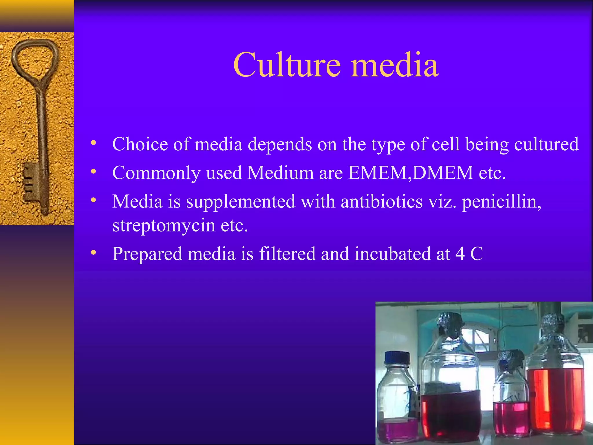
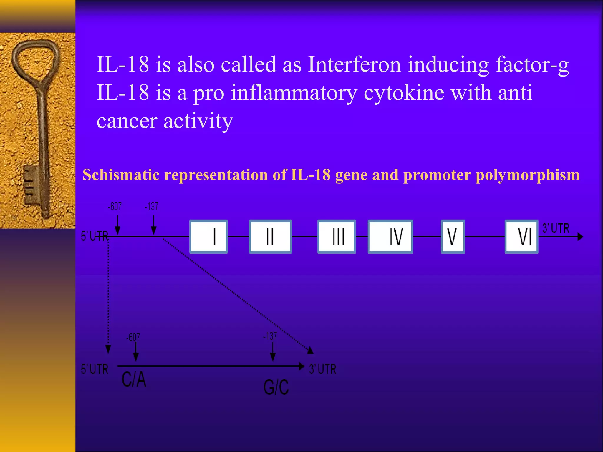
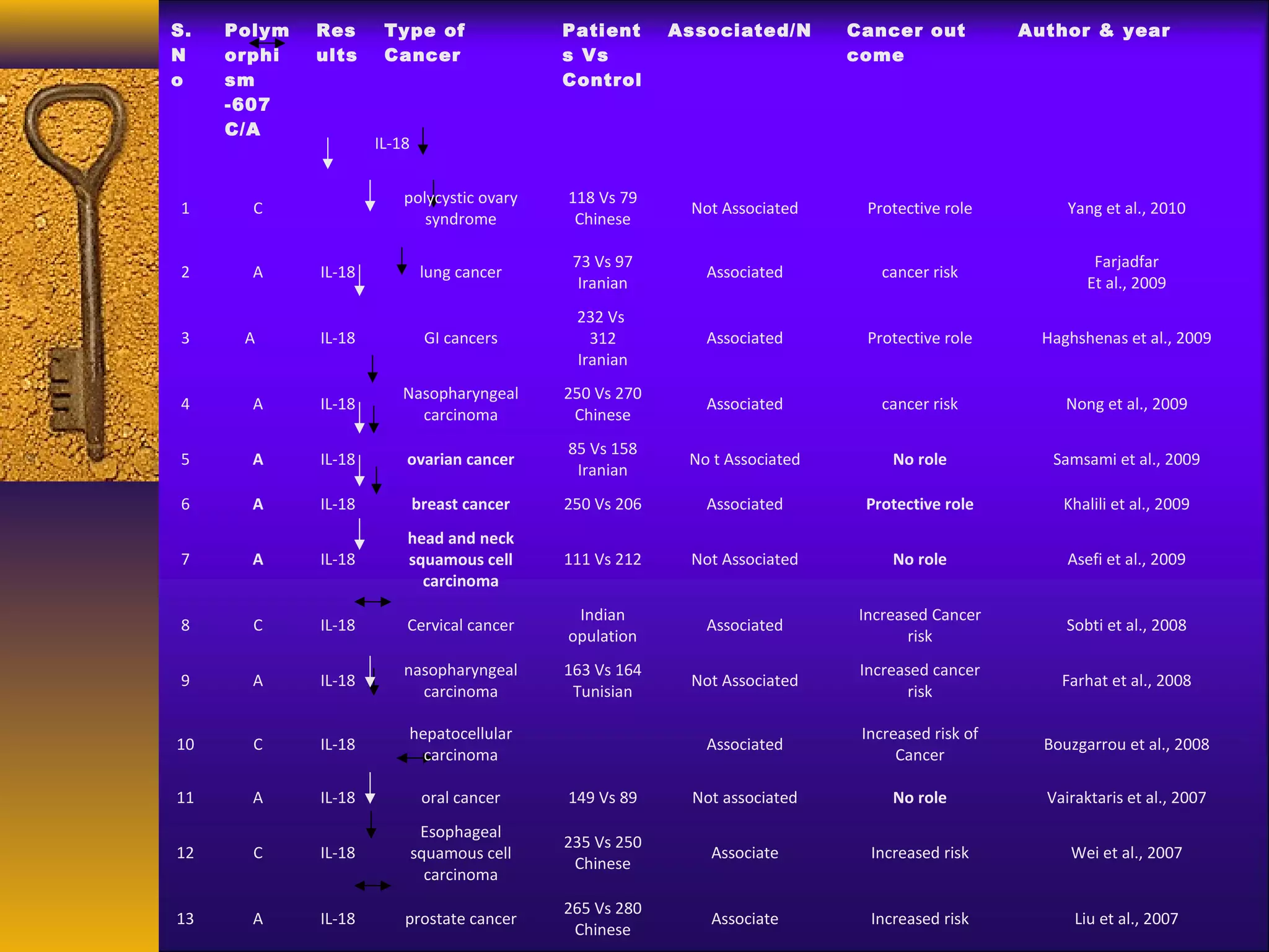
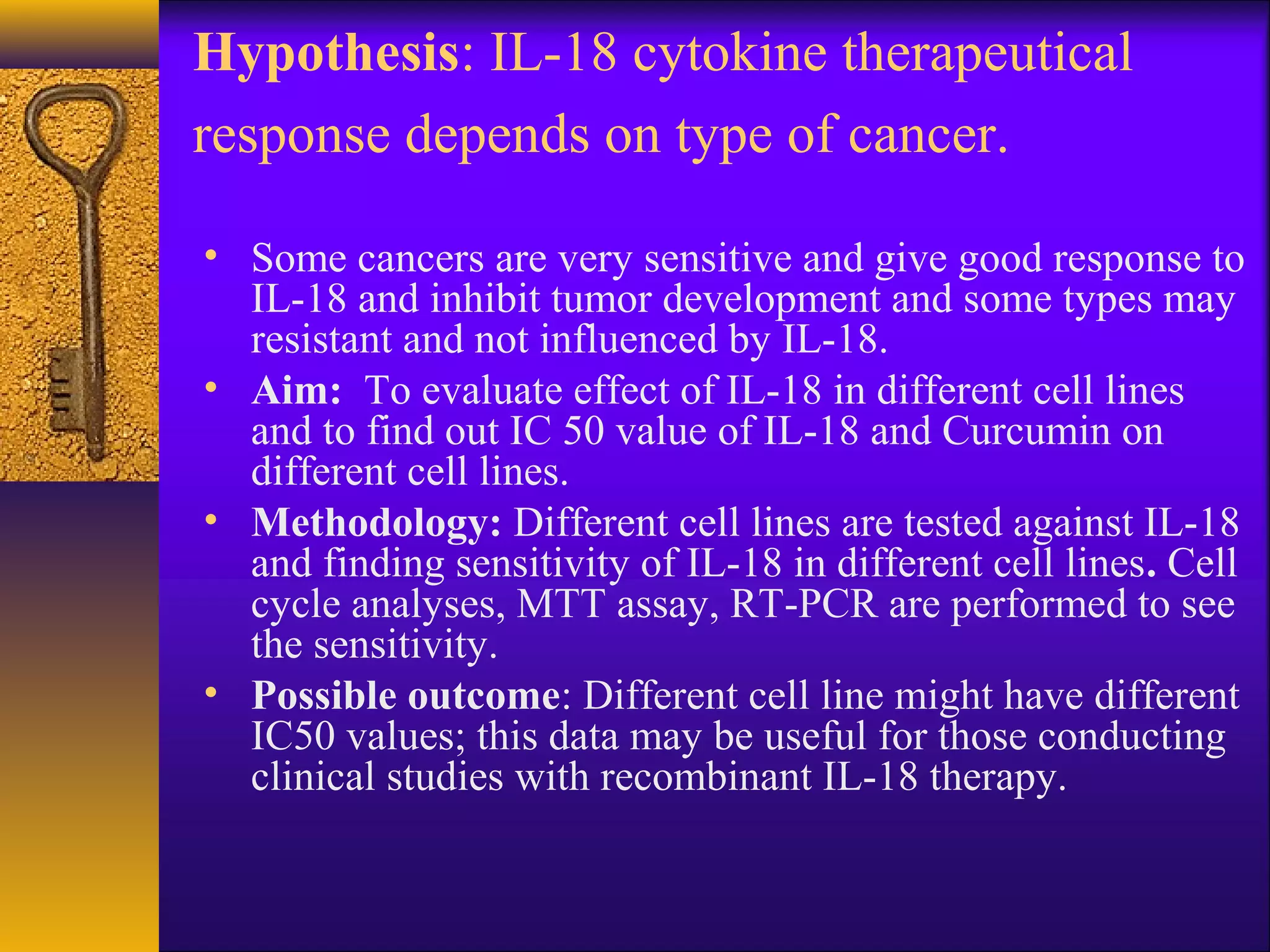
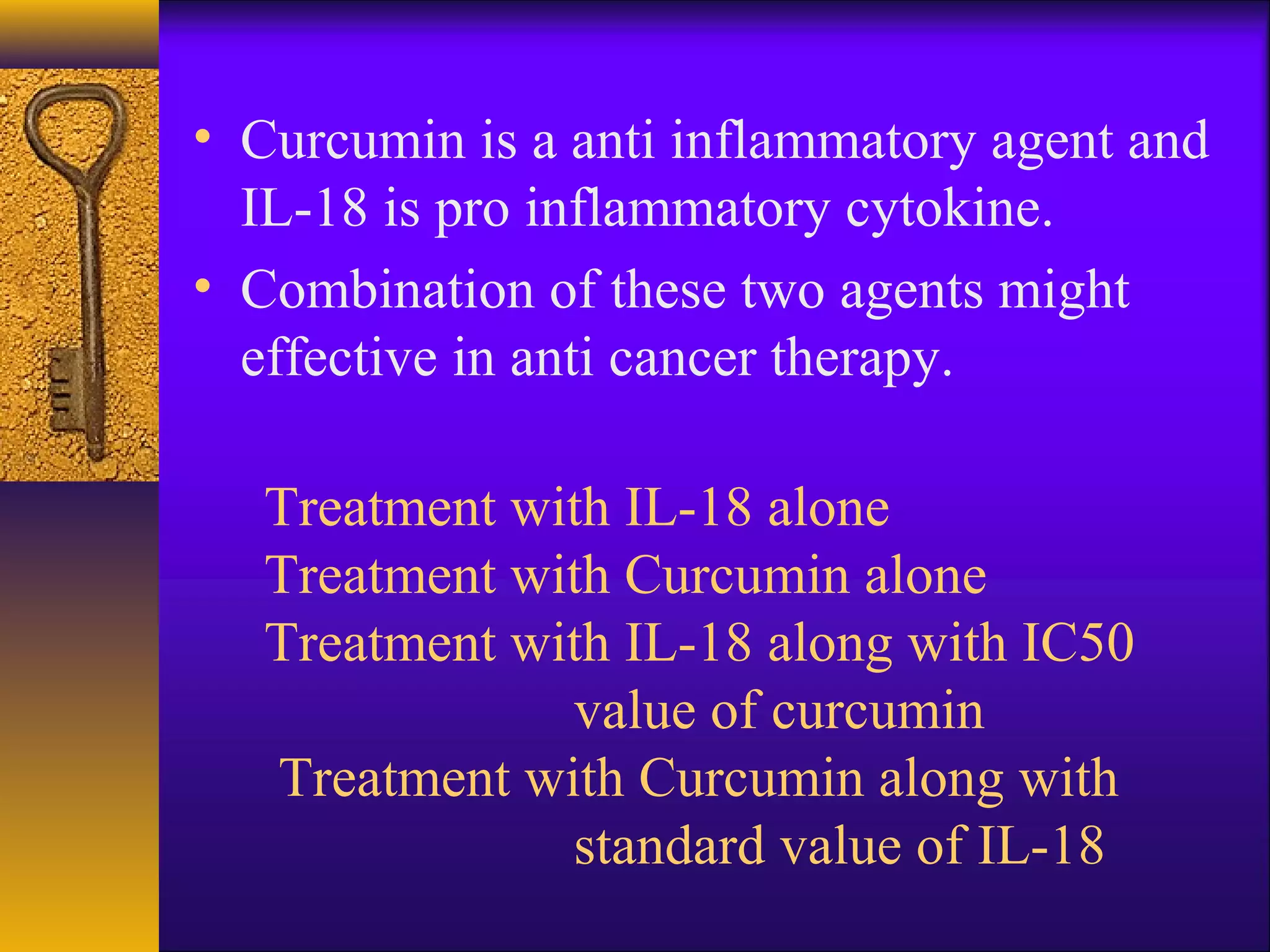
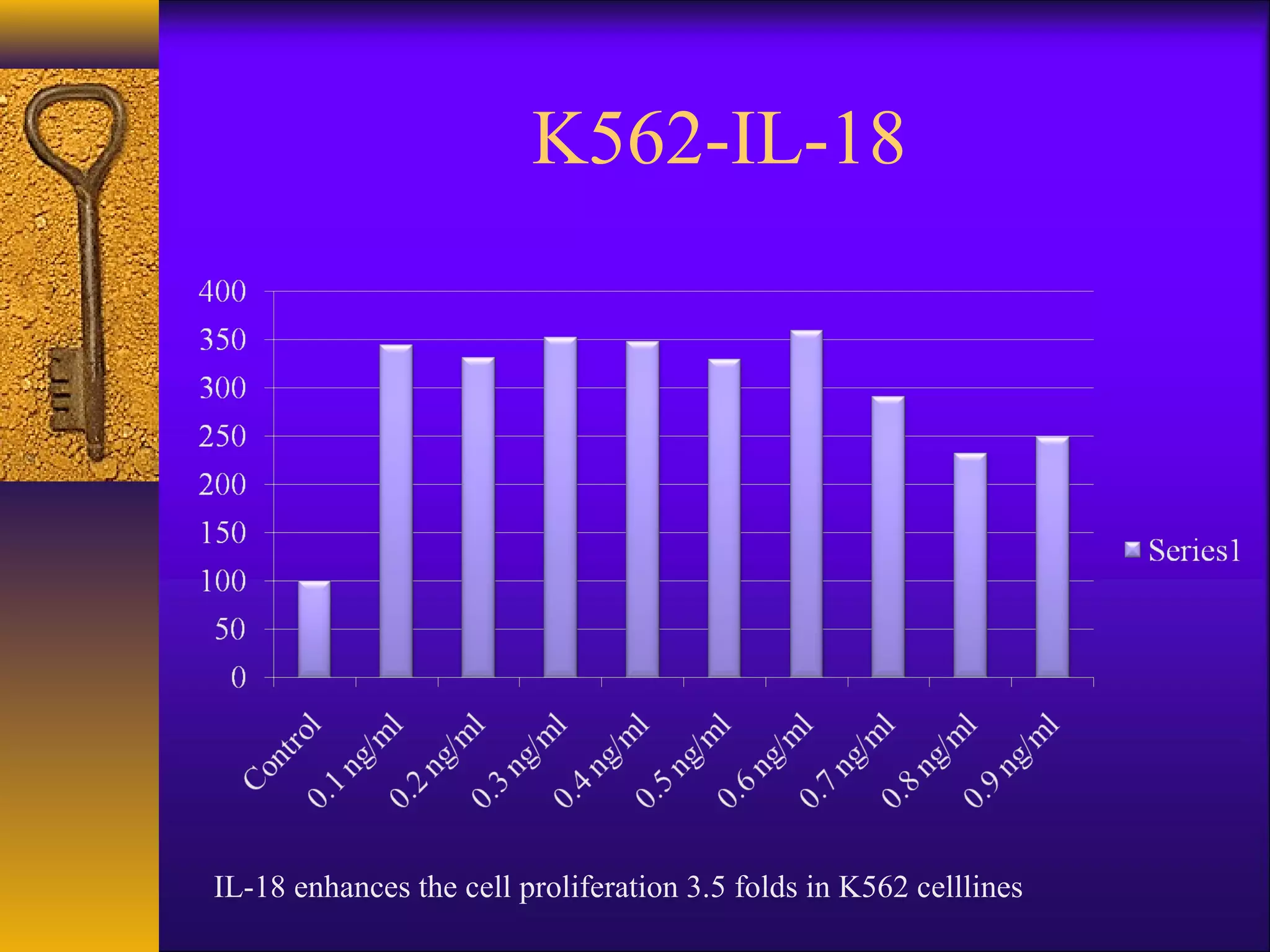
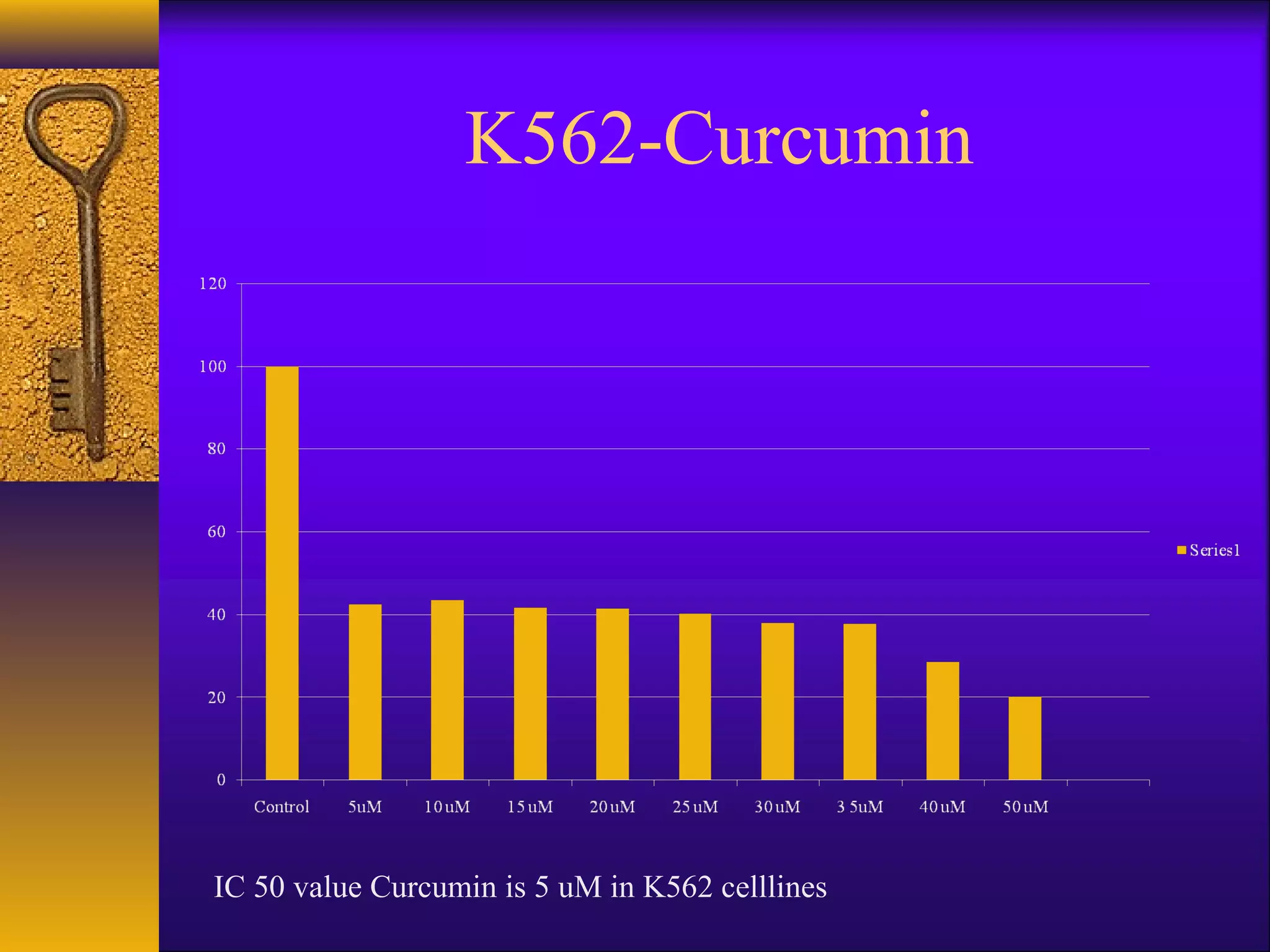
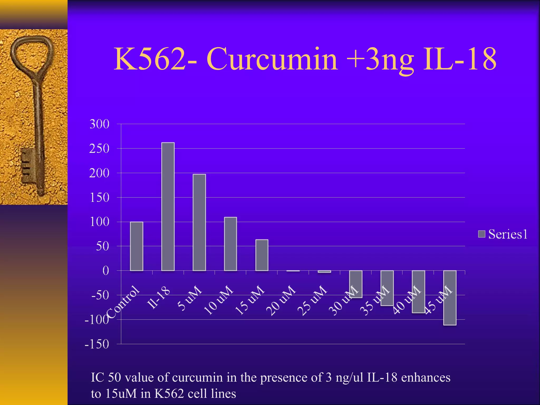
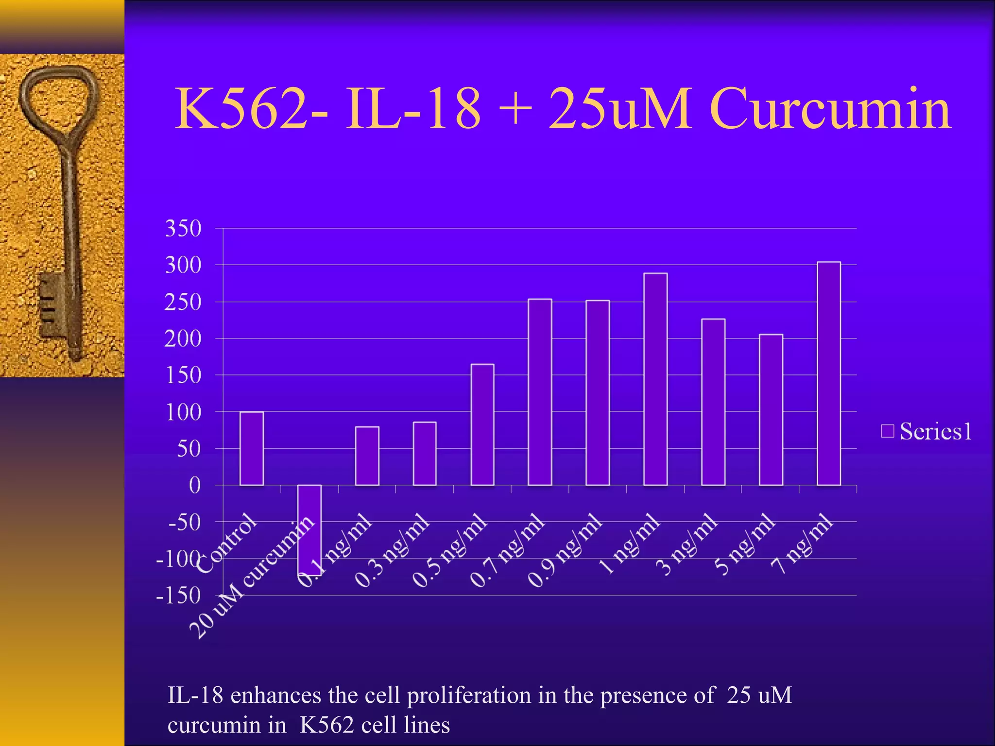
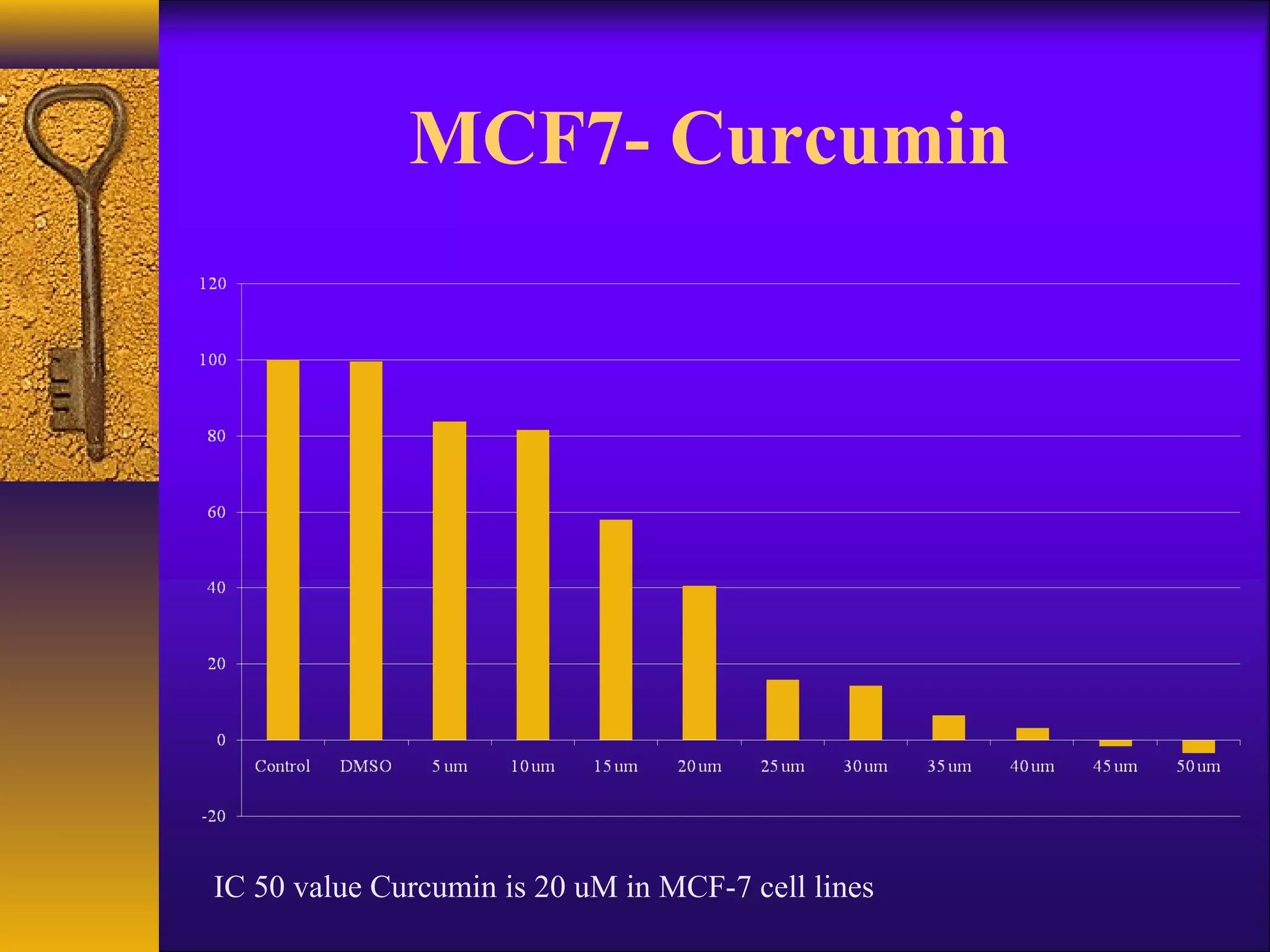
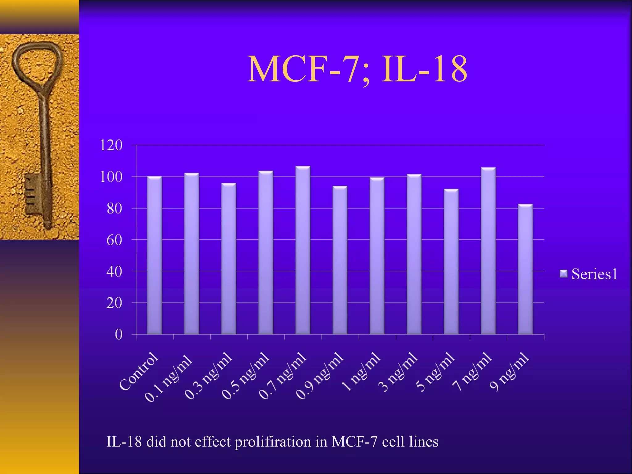
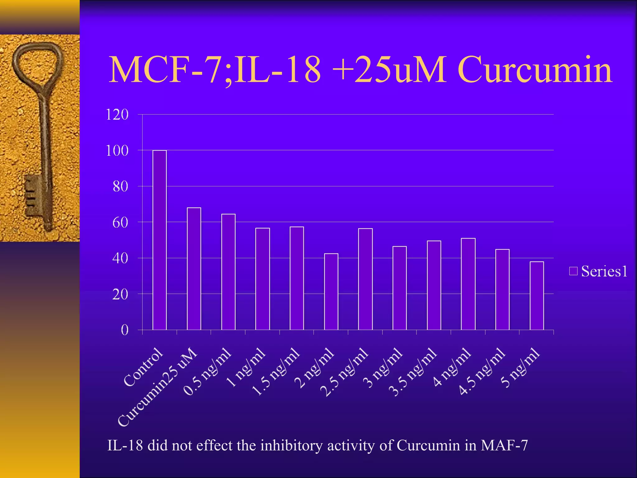
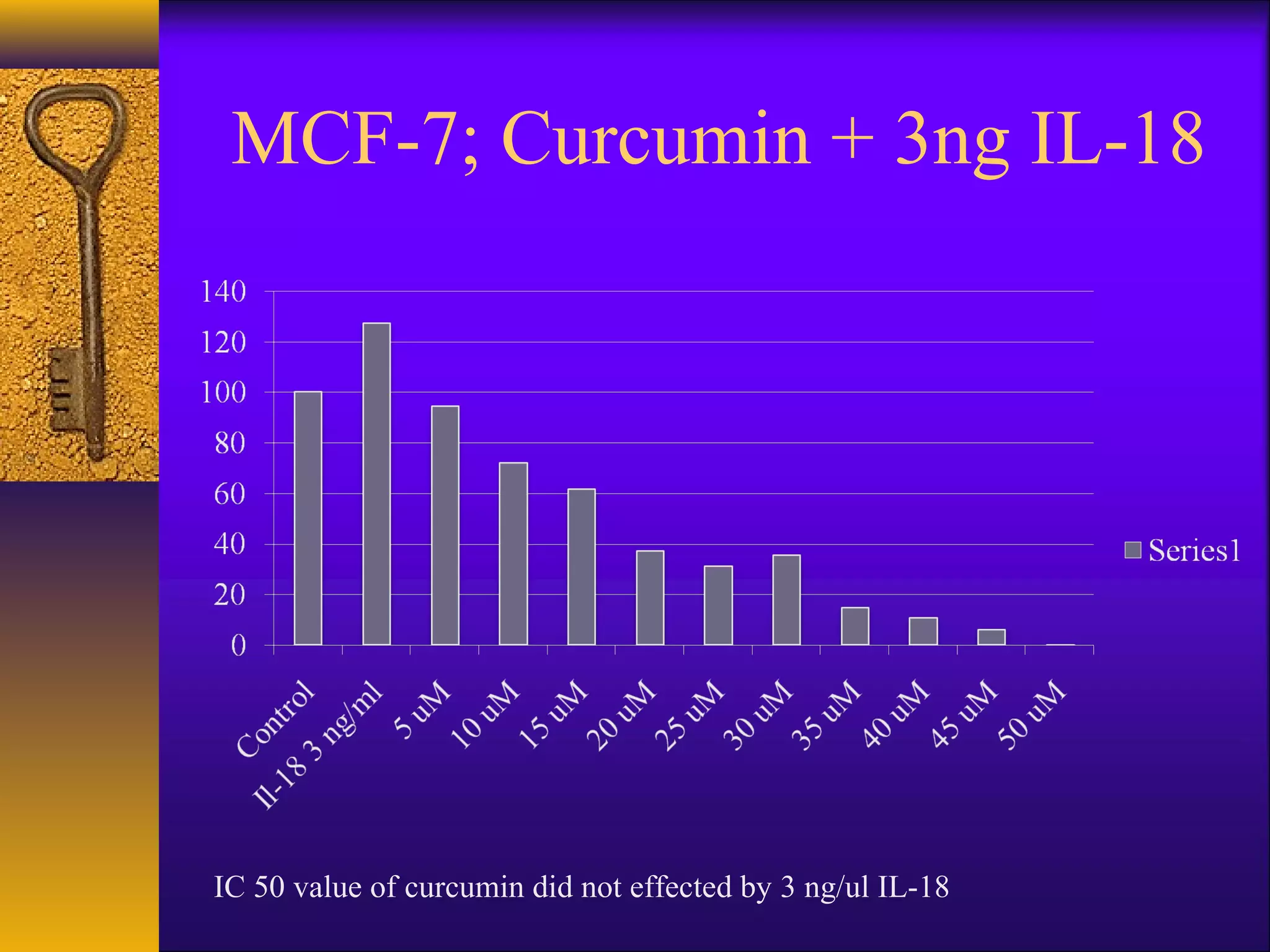
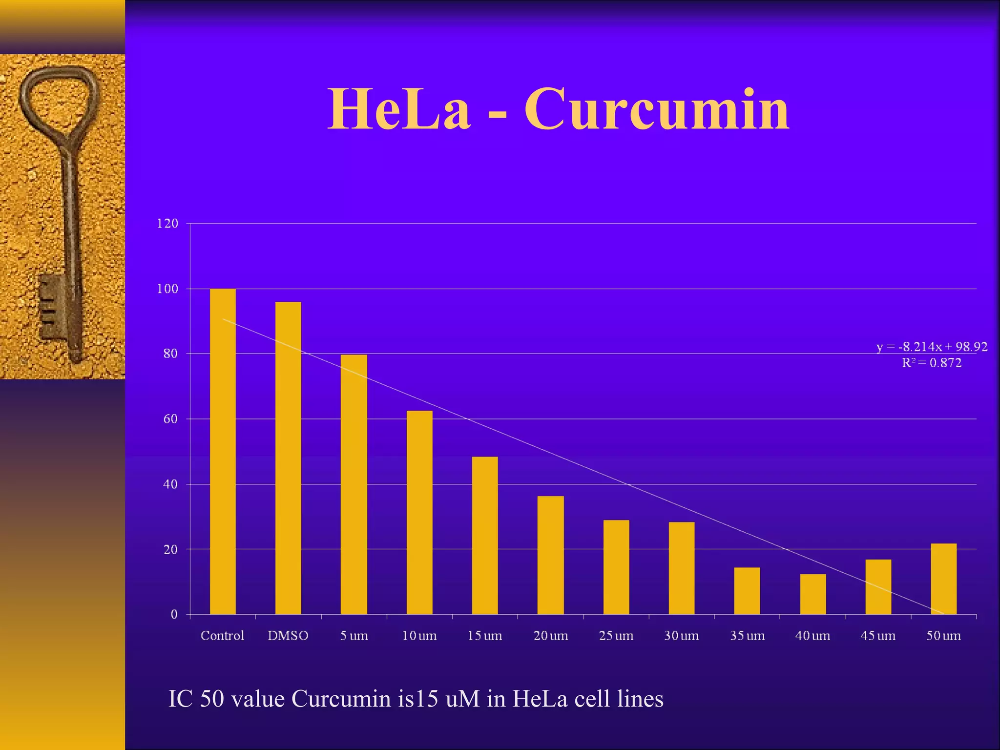
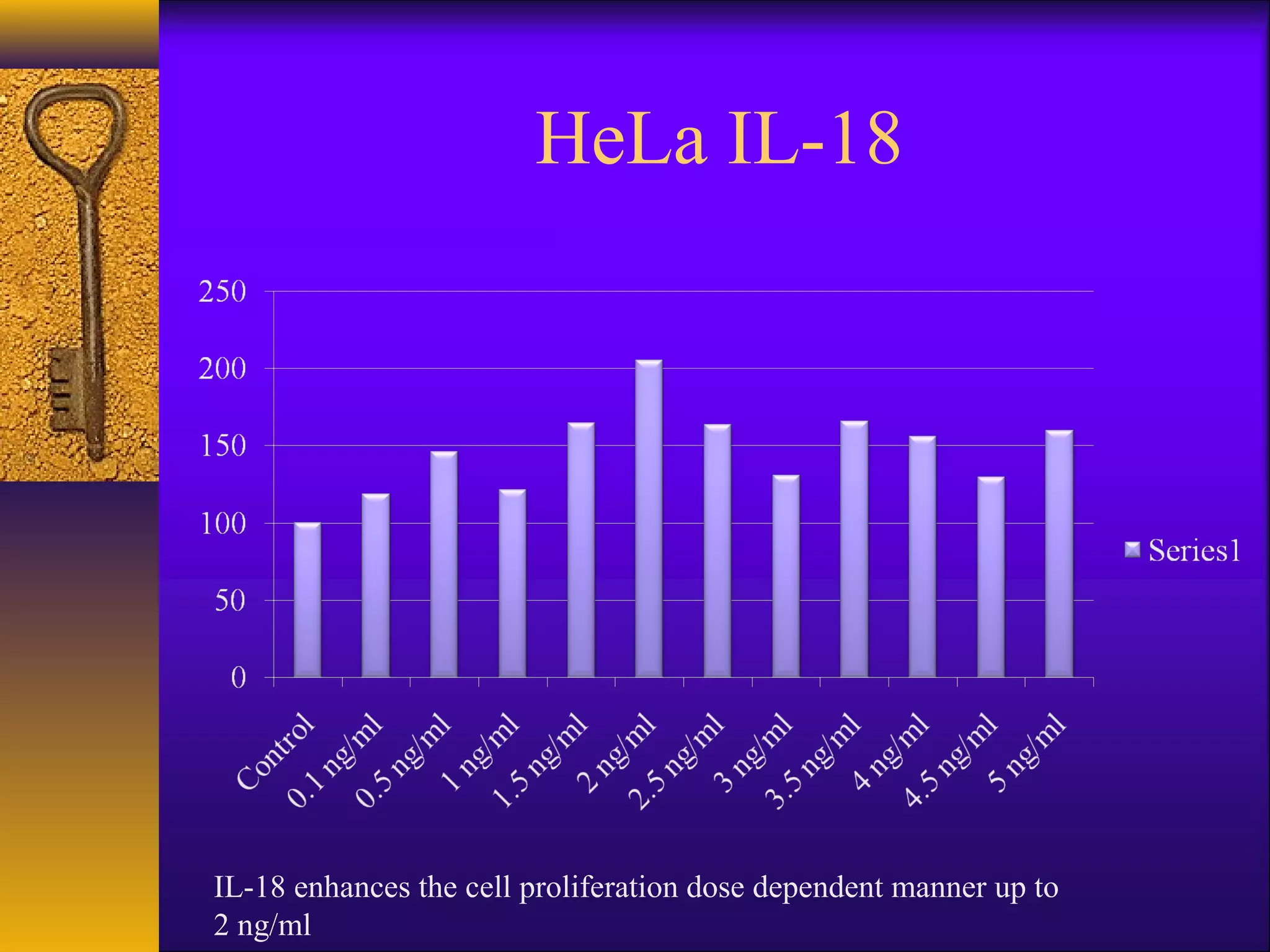
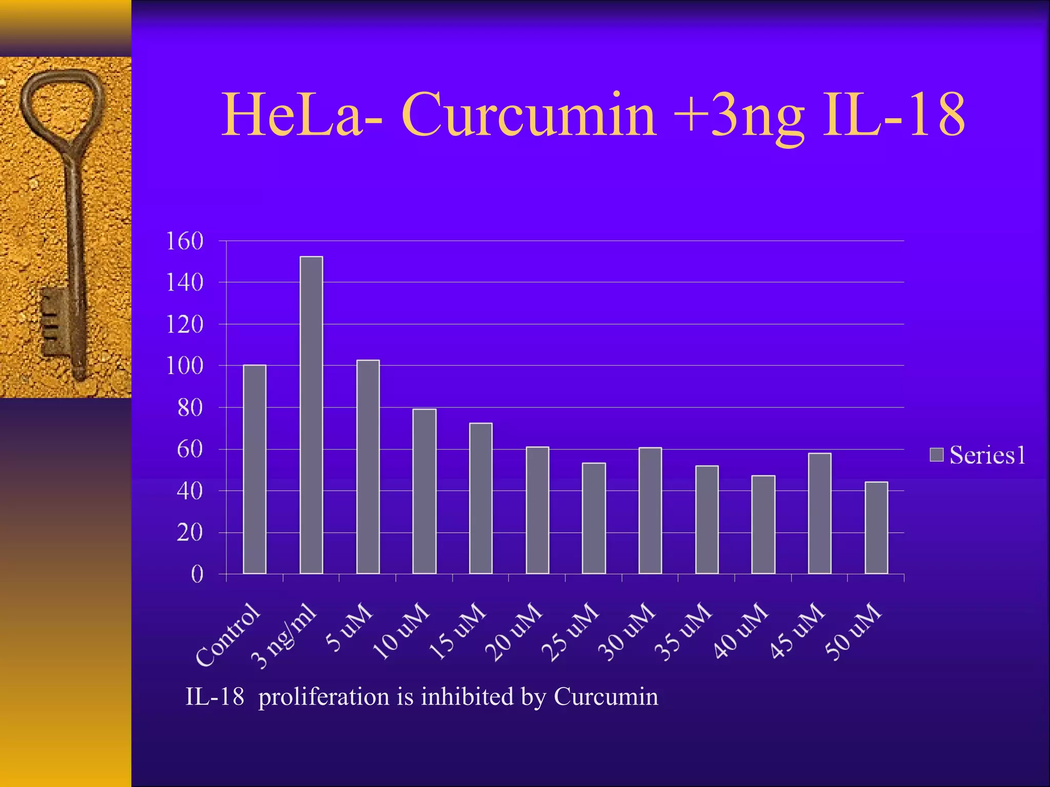
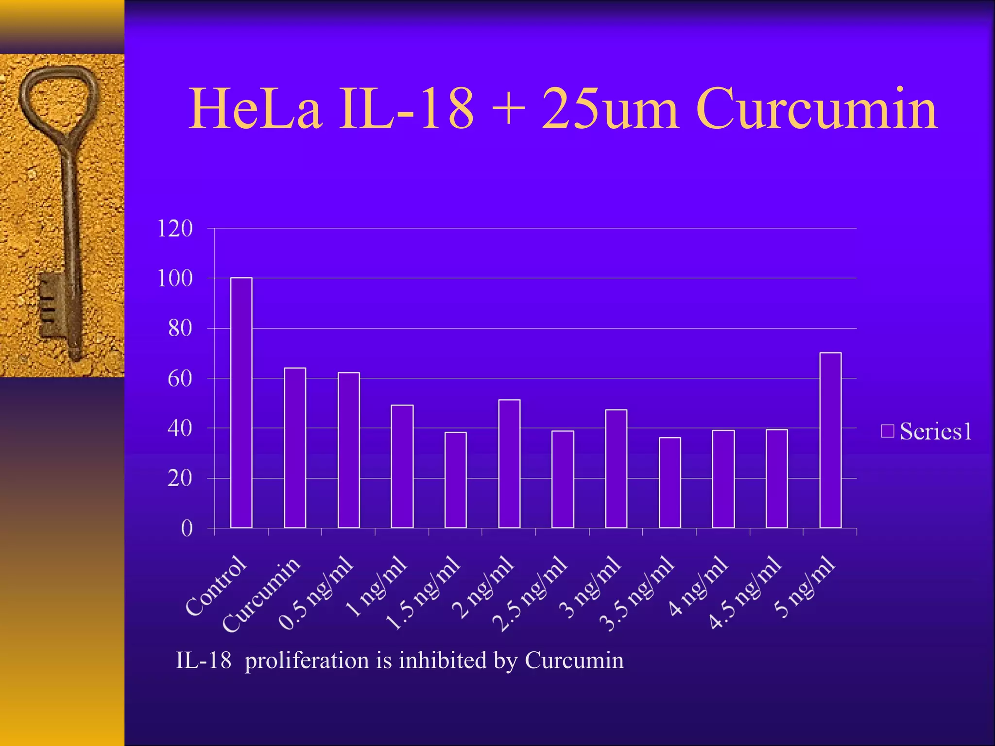
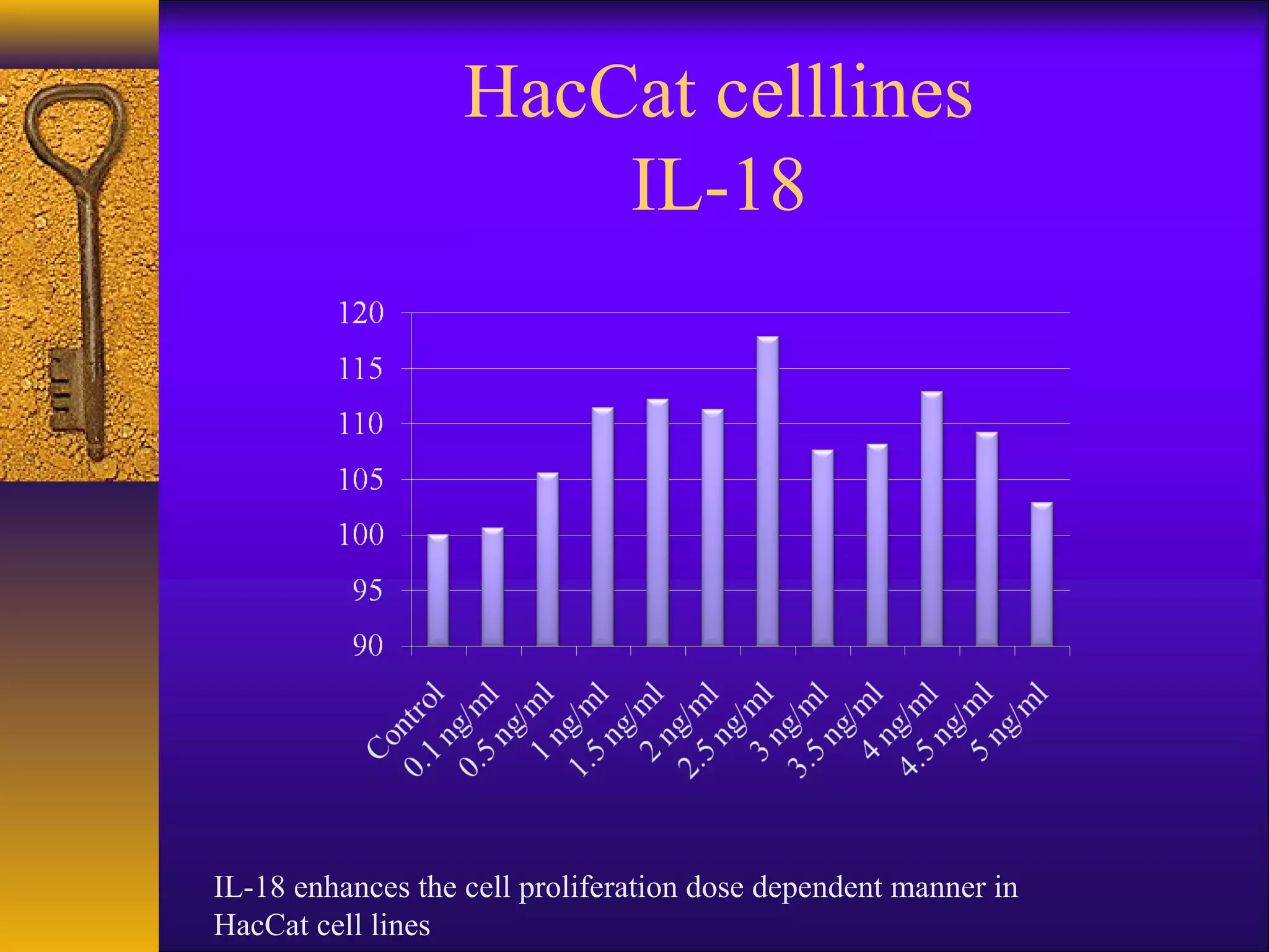
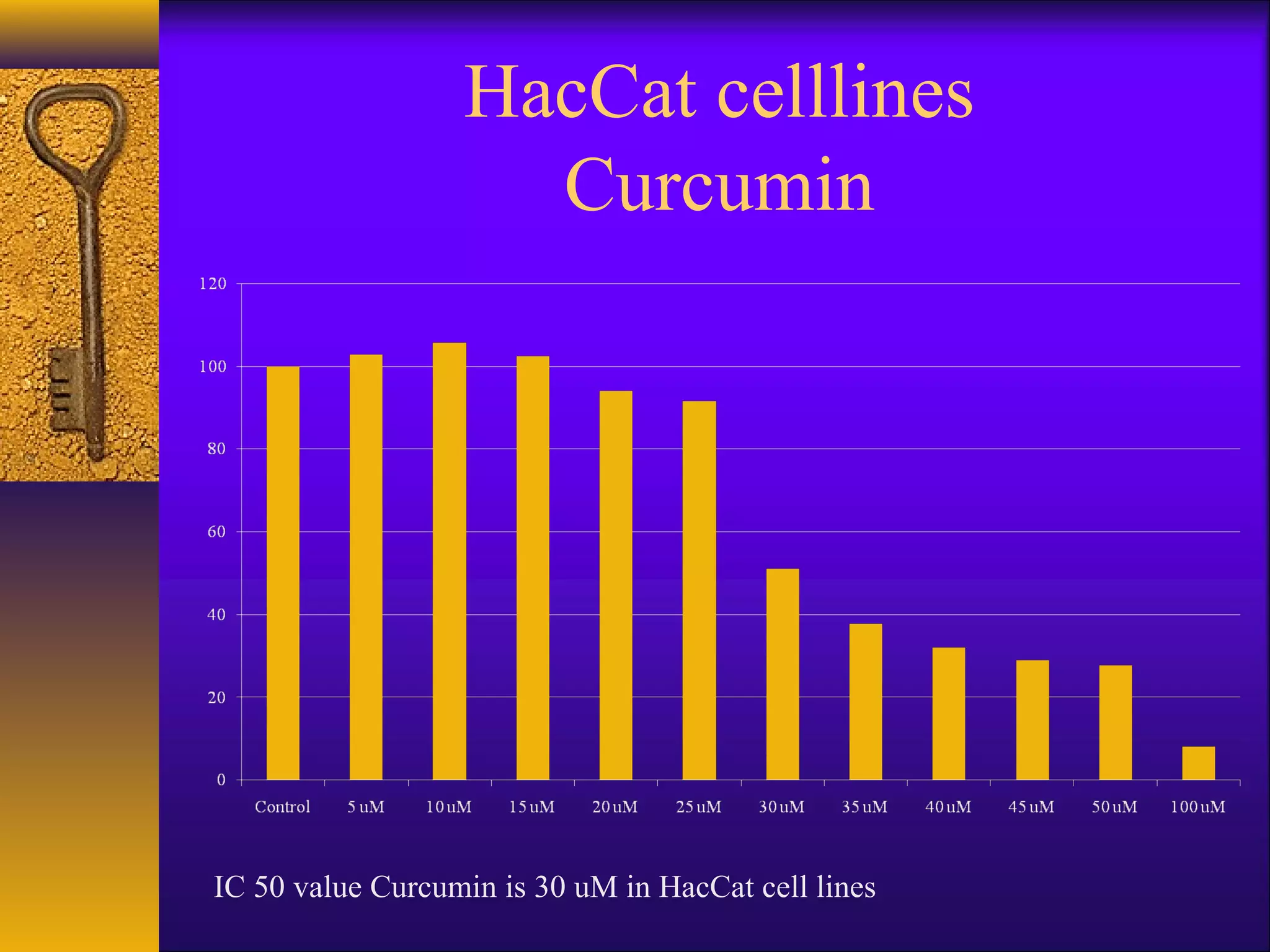
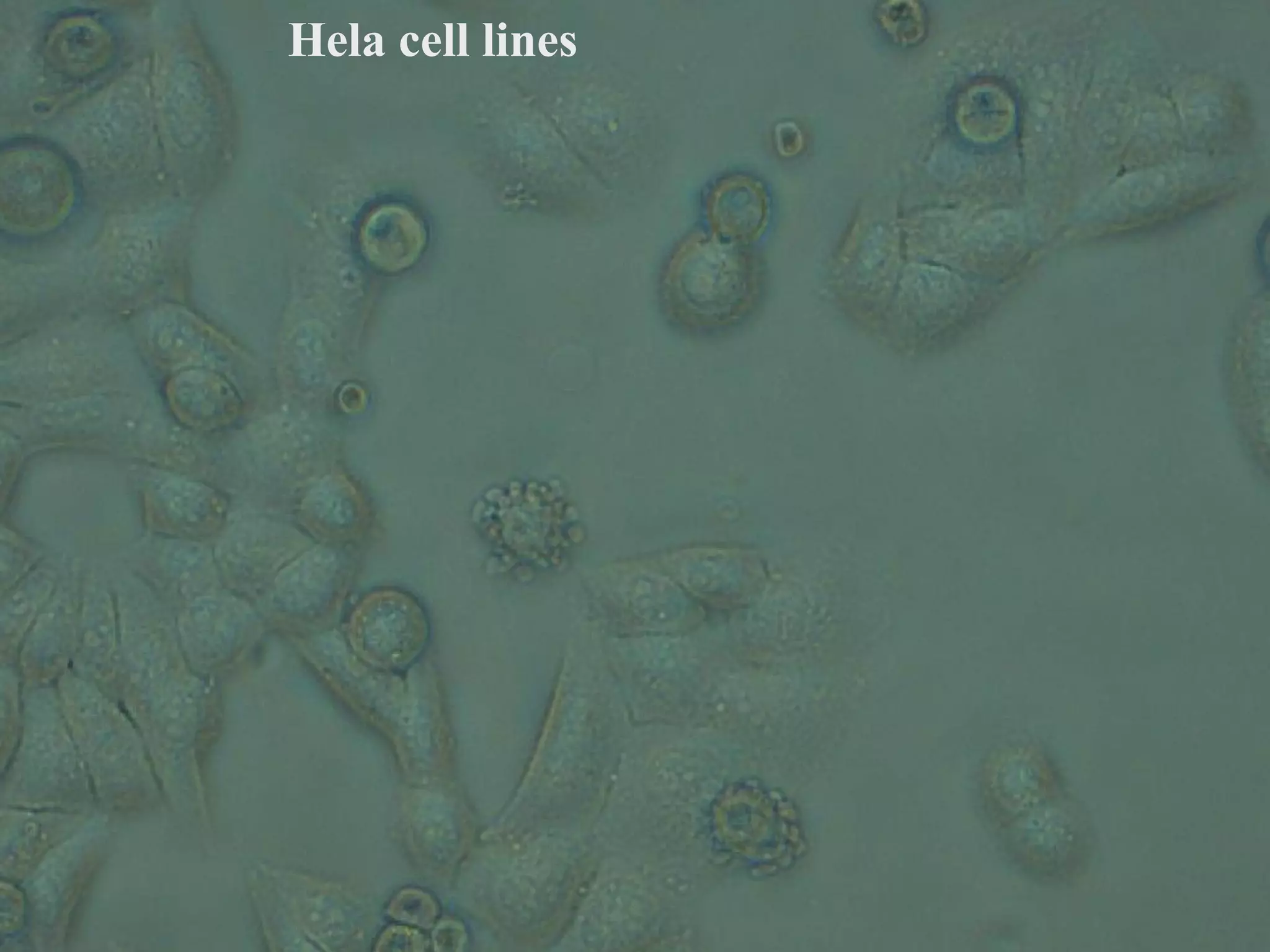
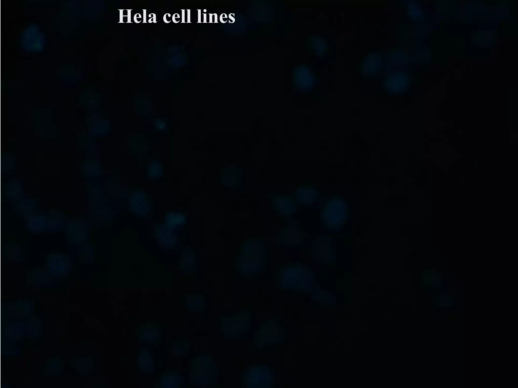
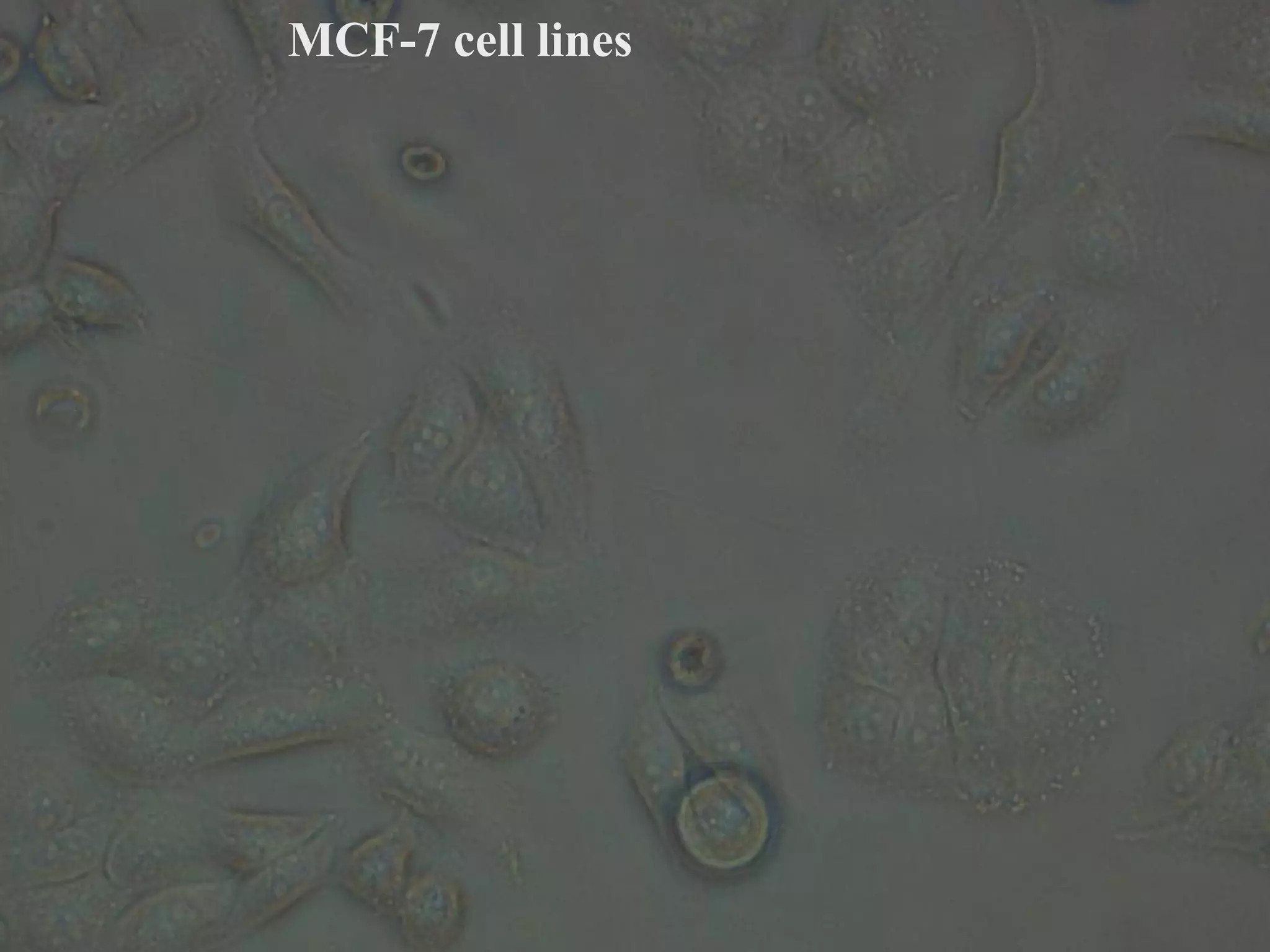
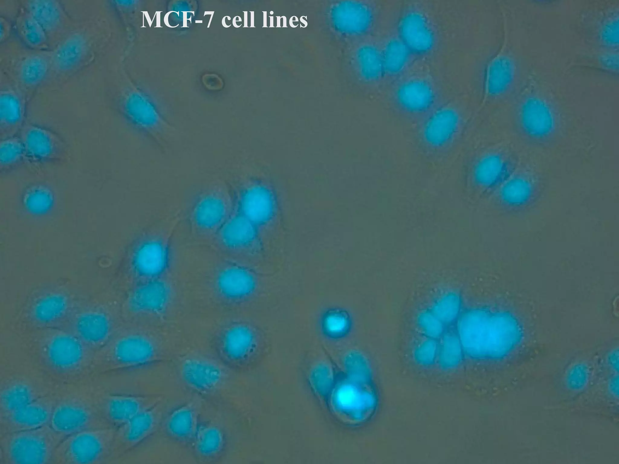
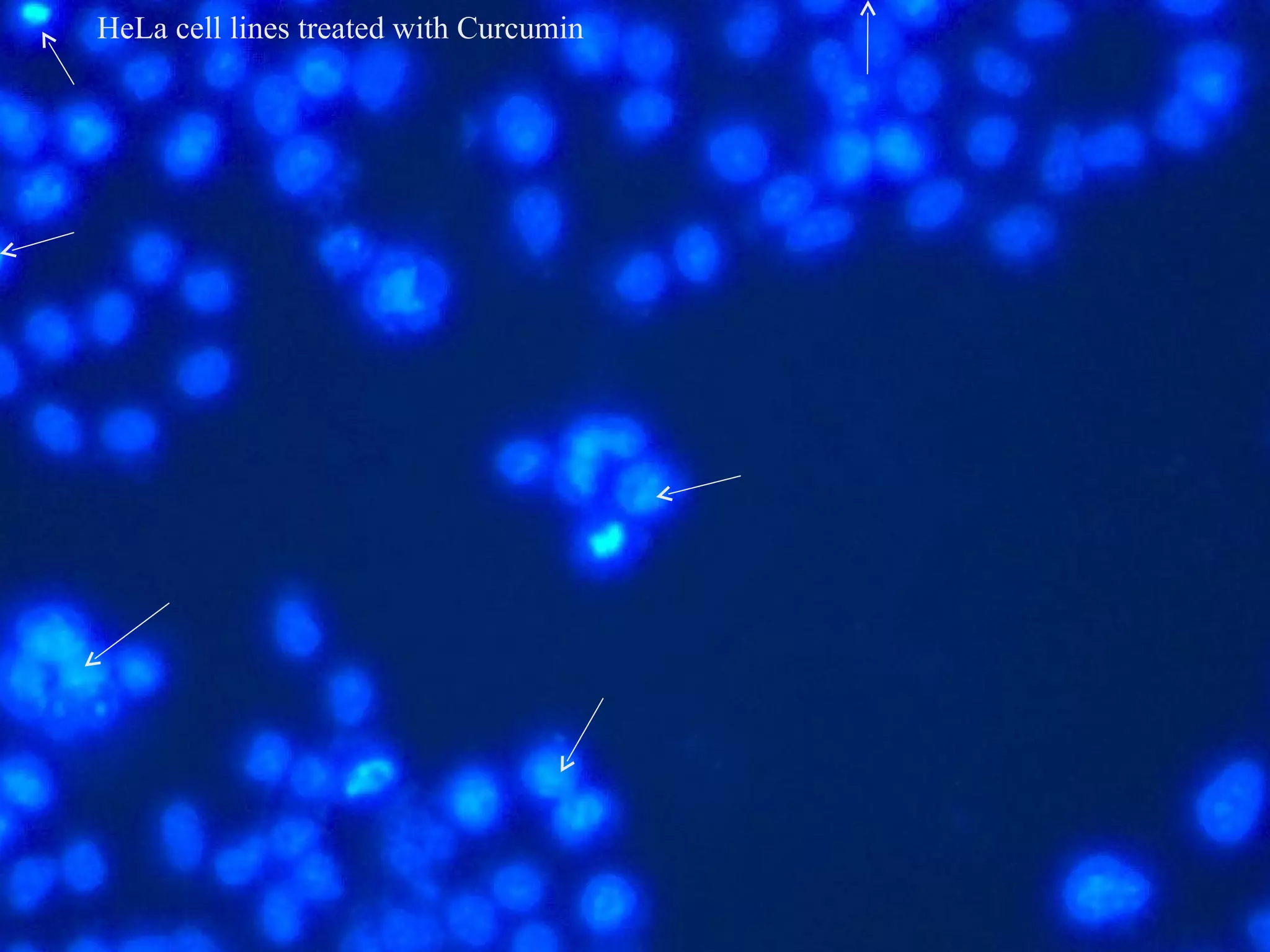
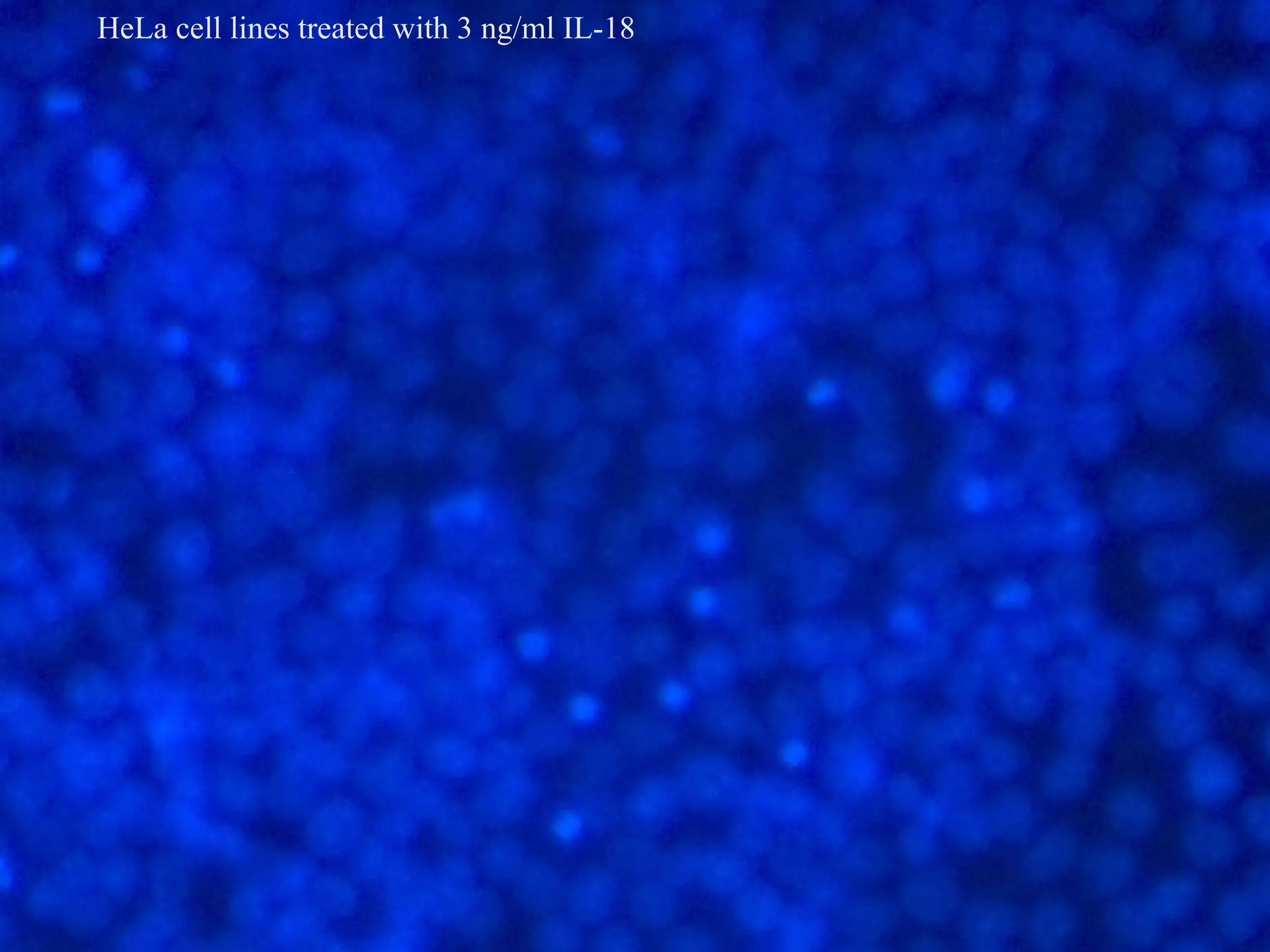
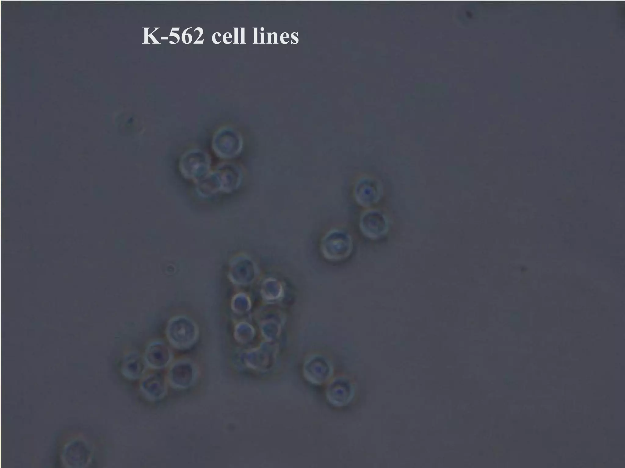

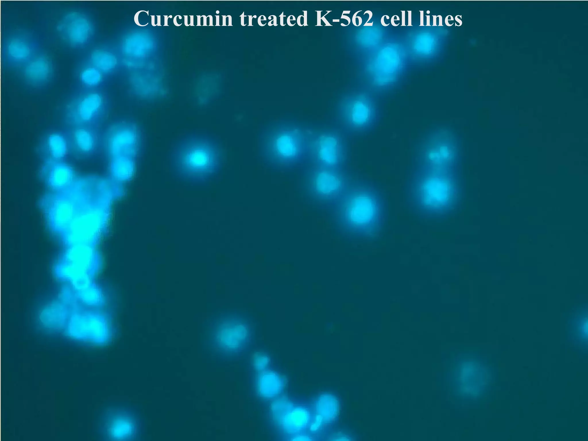


The document outlines the principles and practices of cell culture, including types of cell lines, maintenance, media preparation, and various assays for evaluating cell viability and drug sensitivity. It details methodologies like trypsinization, cryopreservation, and contamination detection, along with applications in cancer research, genetic engineering, and virology. Additionally, it discusses the effects of the IL-18 cytokine on cancer cell lines and the potential for therapeutic responses based on cell type sensitivity.


![Apporach to lung biopsy [Auto-saved].pptx latest](https://cdn.slidesharecdn.com/ss_thumbnails/apporachtolungbiopsyauto-saved-251211225655-93258539-thumbnail.jpg?width=640&height=640&fit=bounds)


