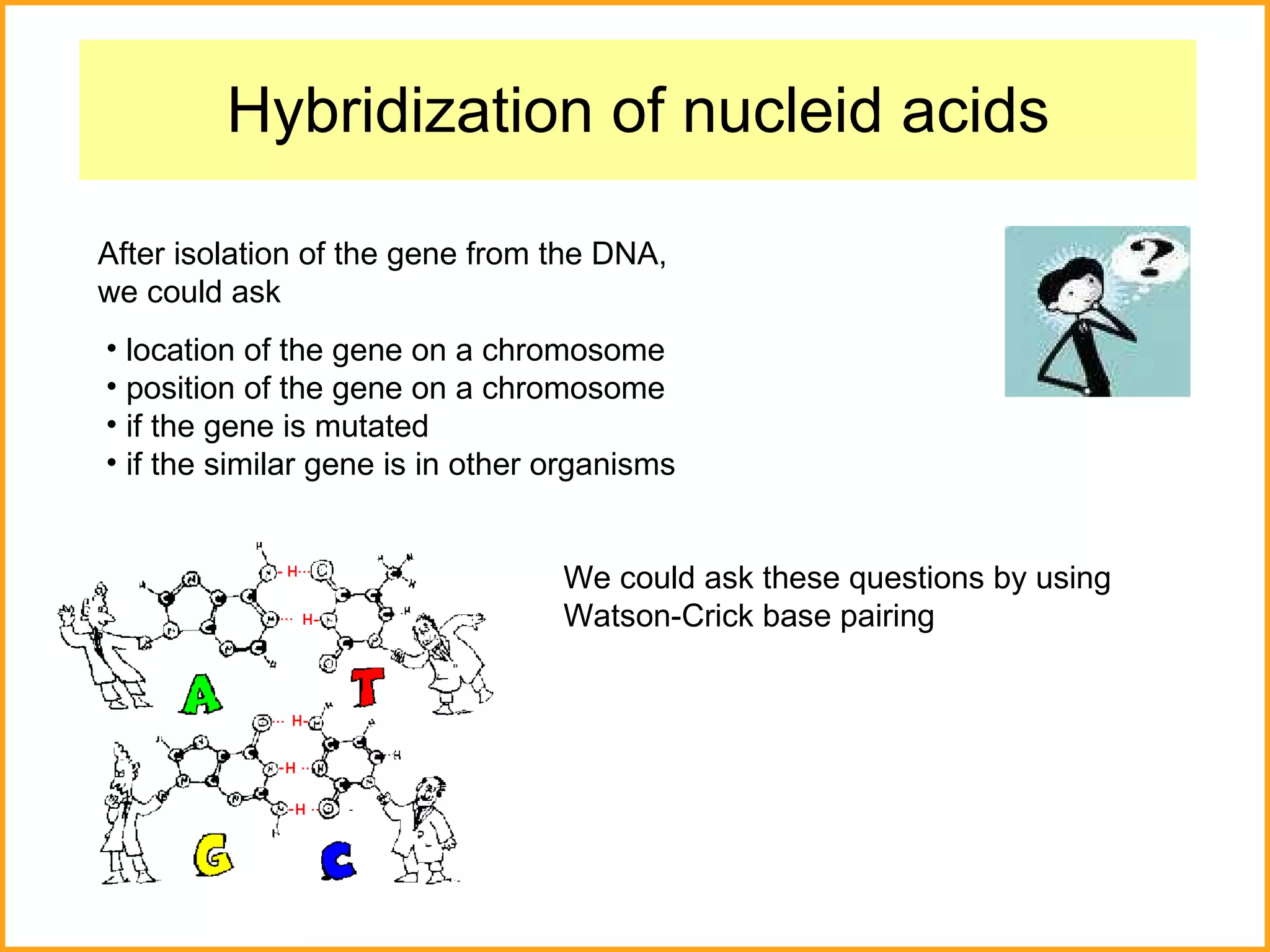
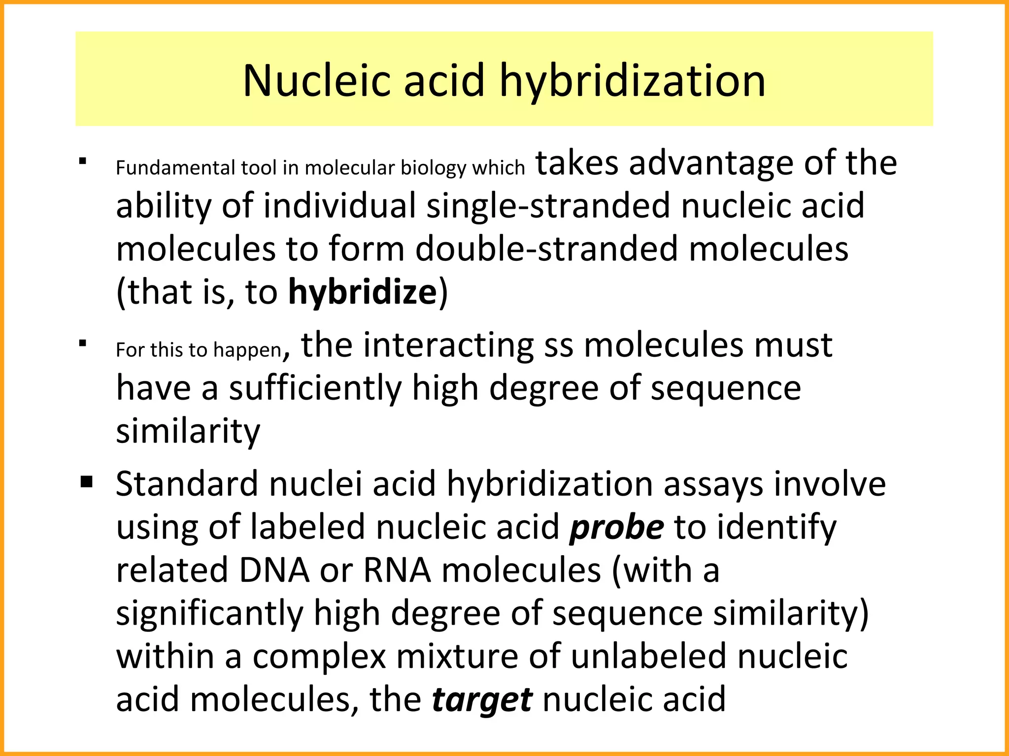
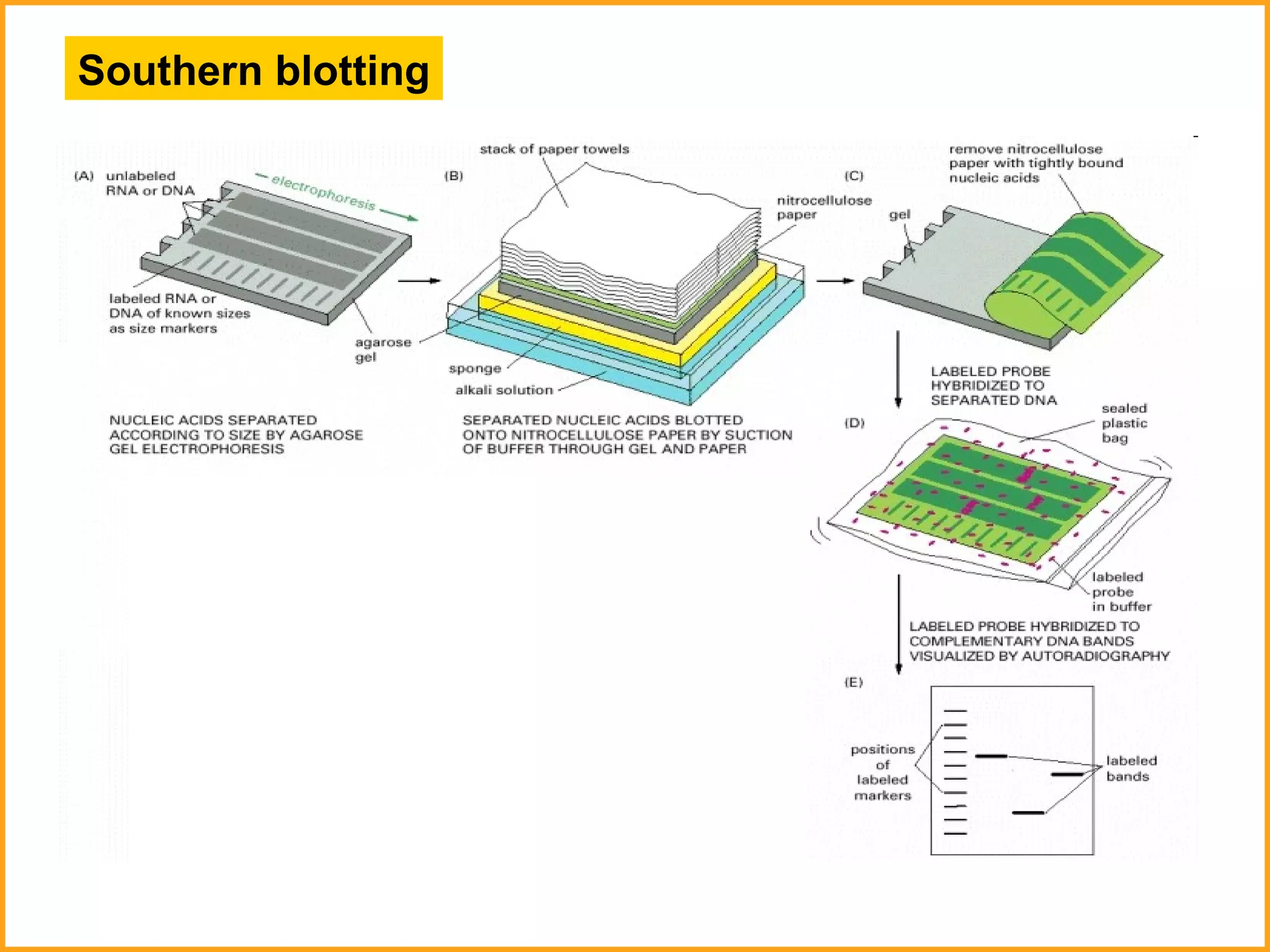
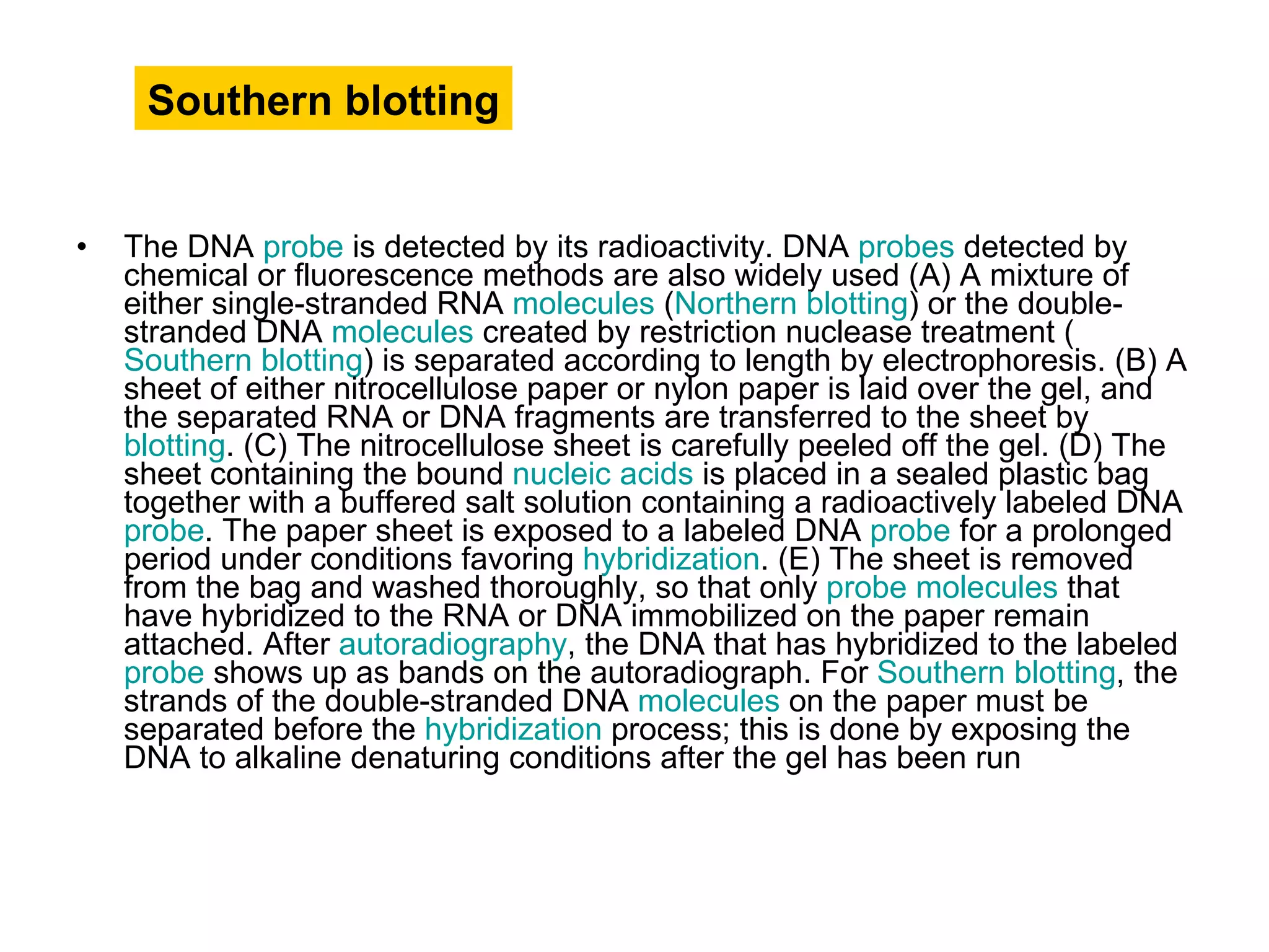
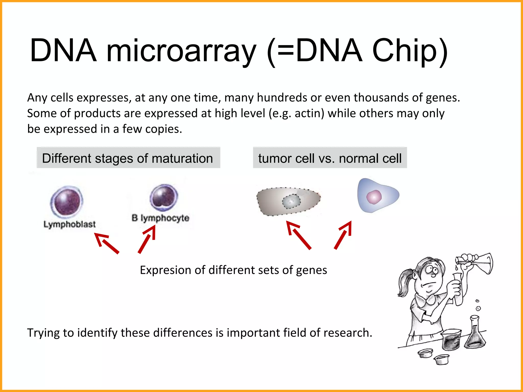
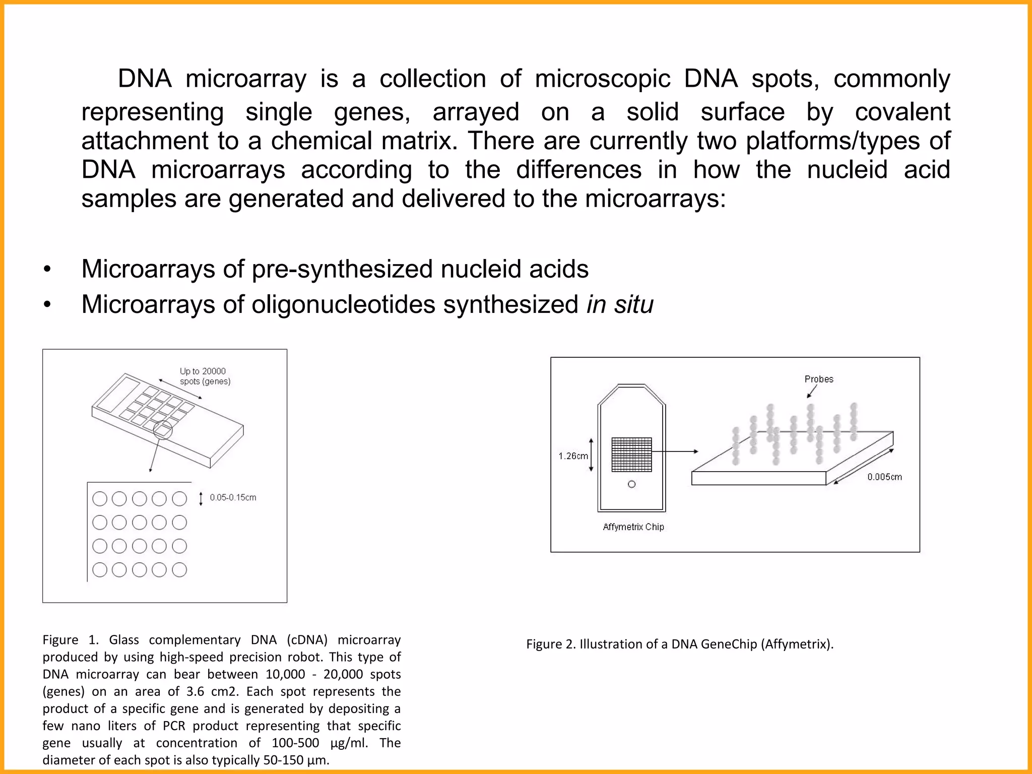
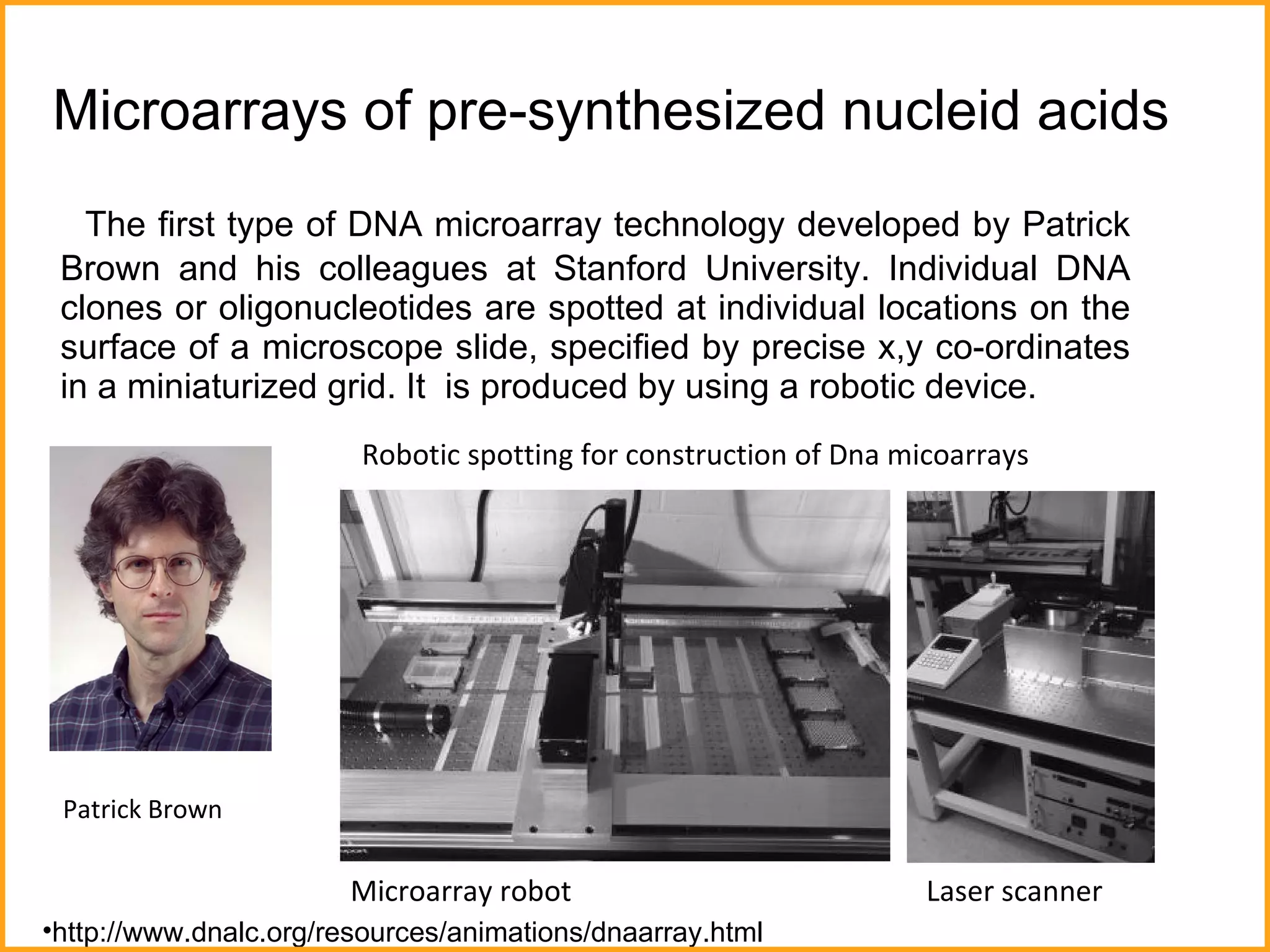
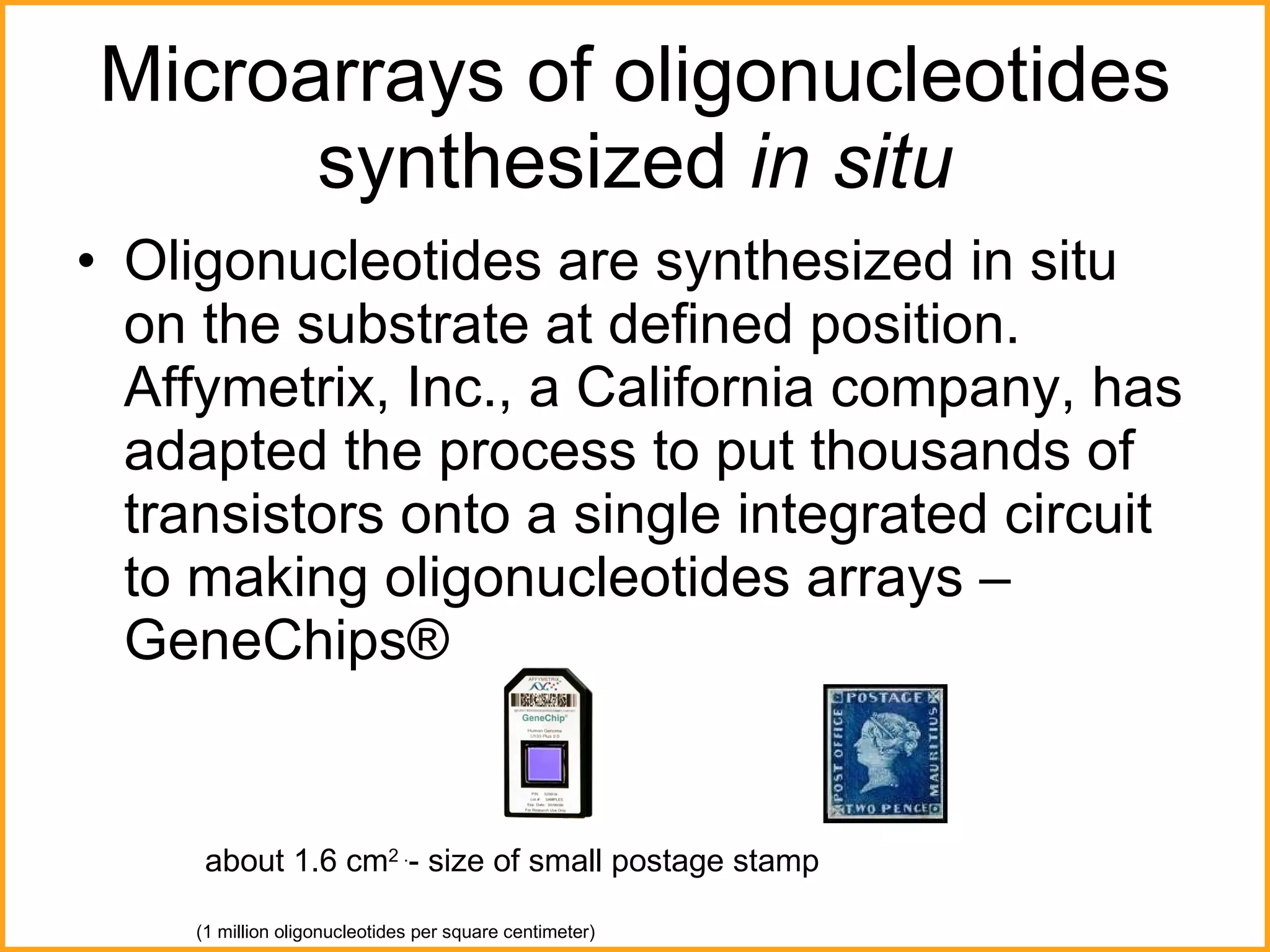
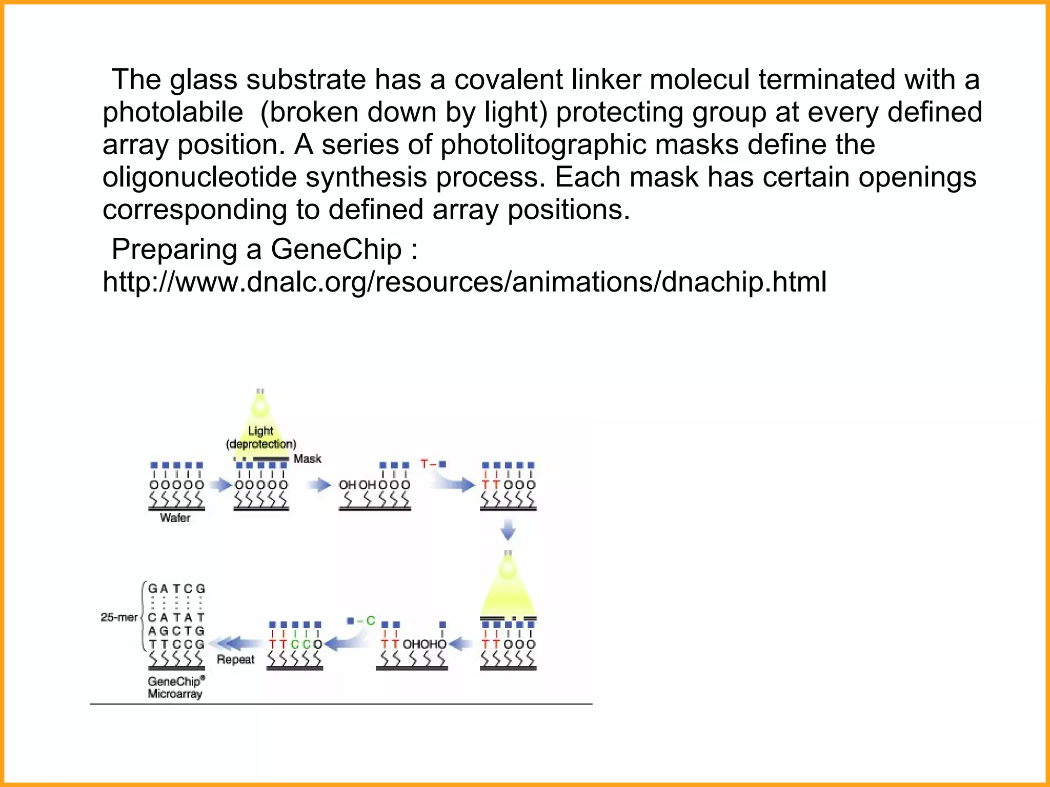
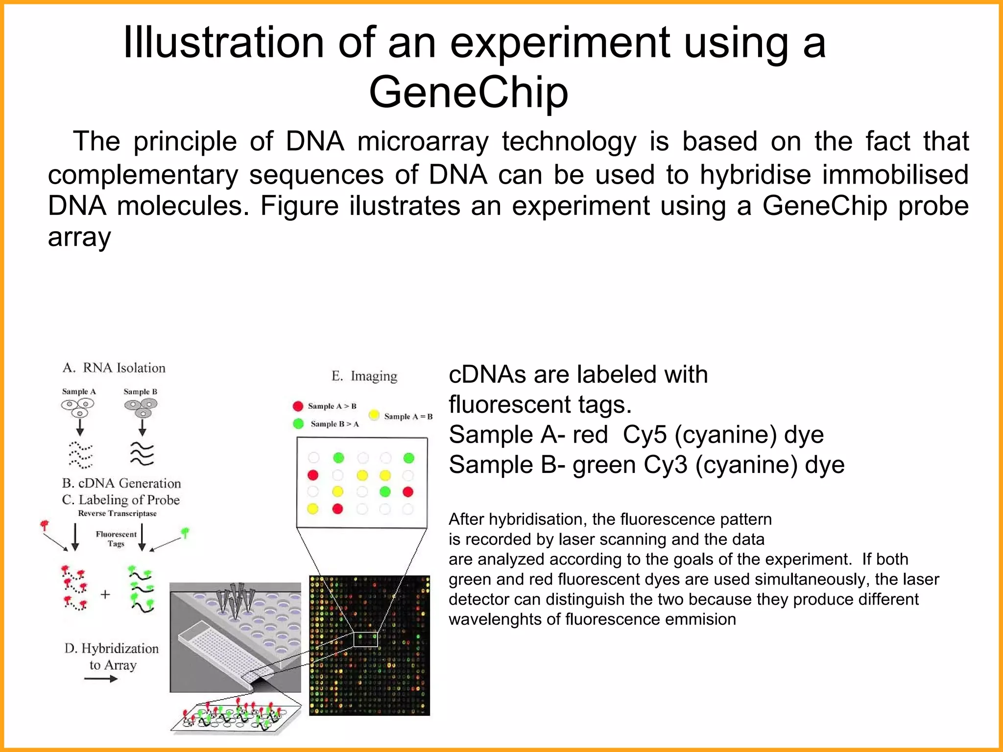
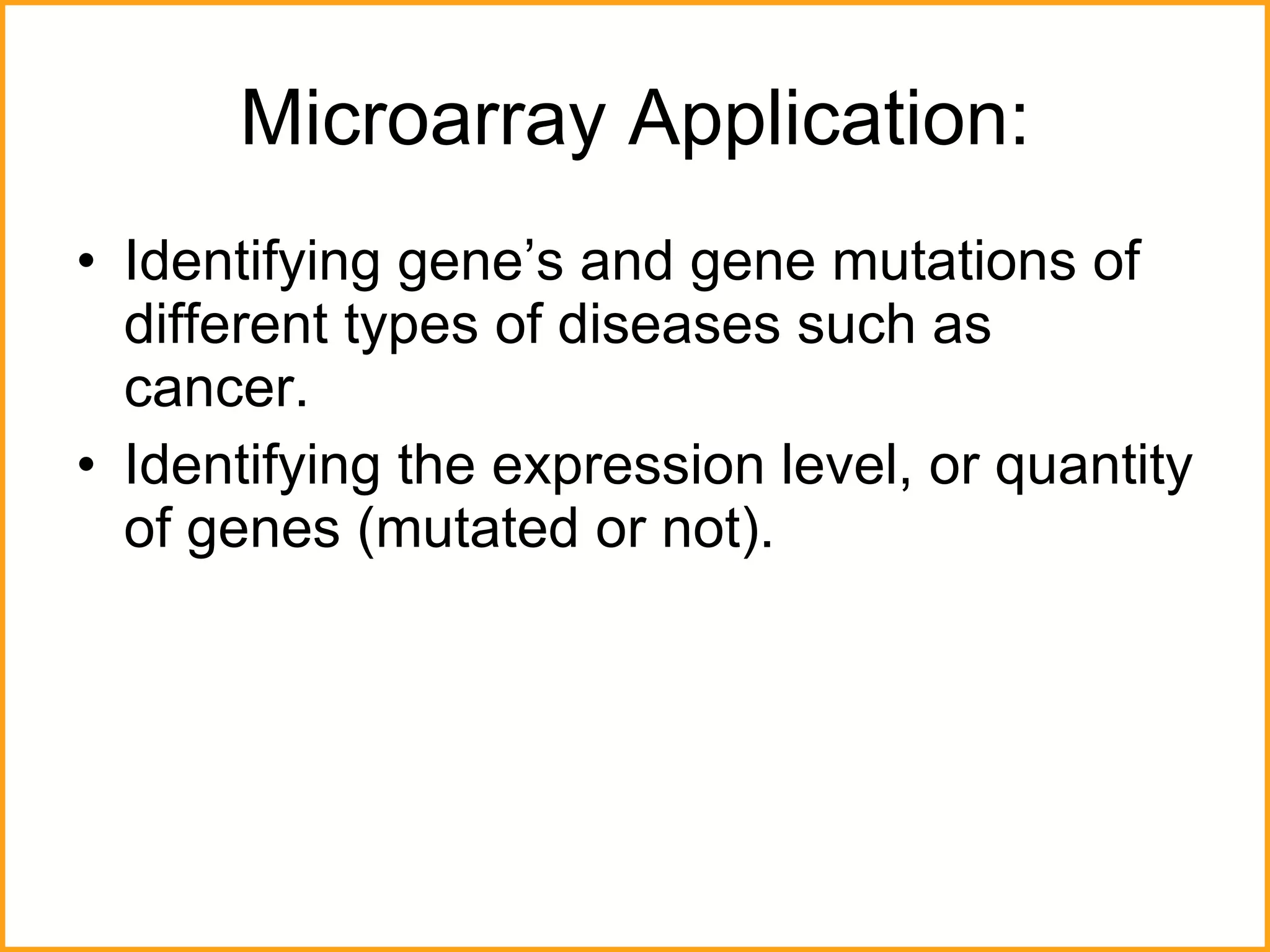
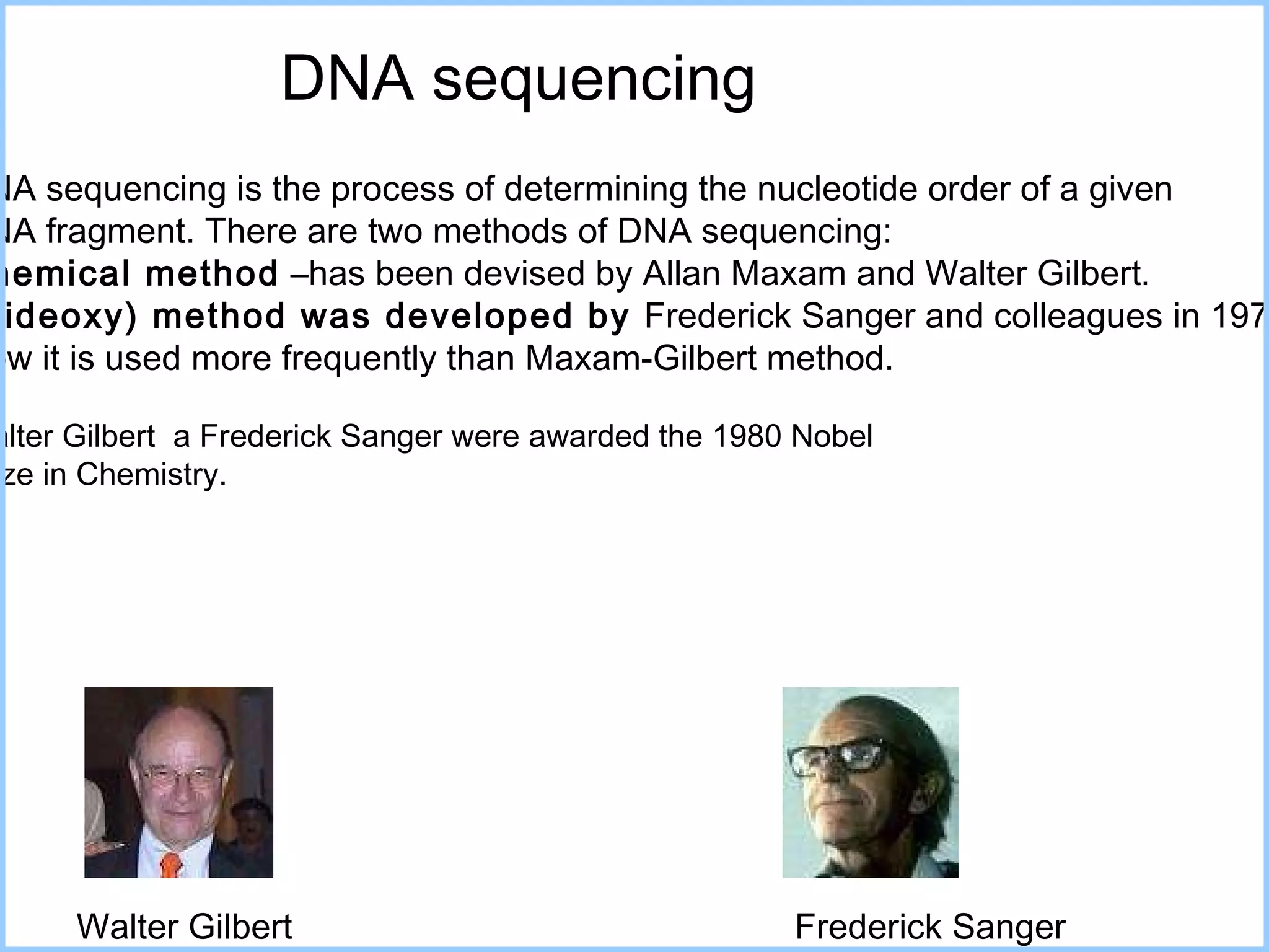
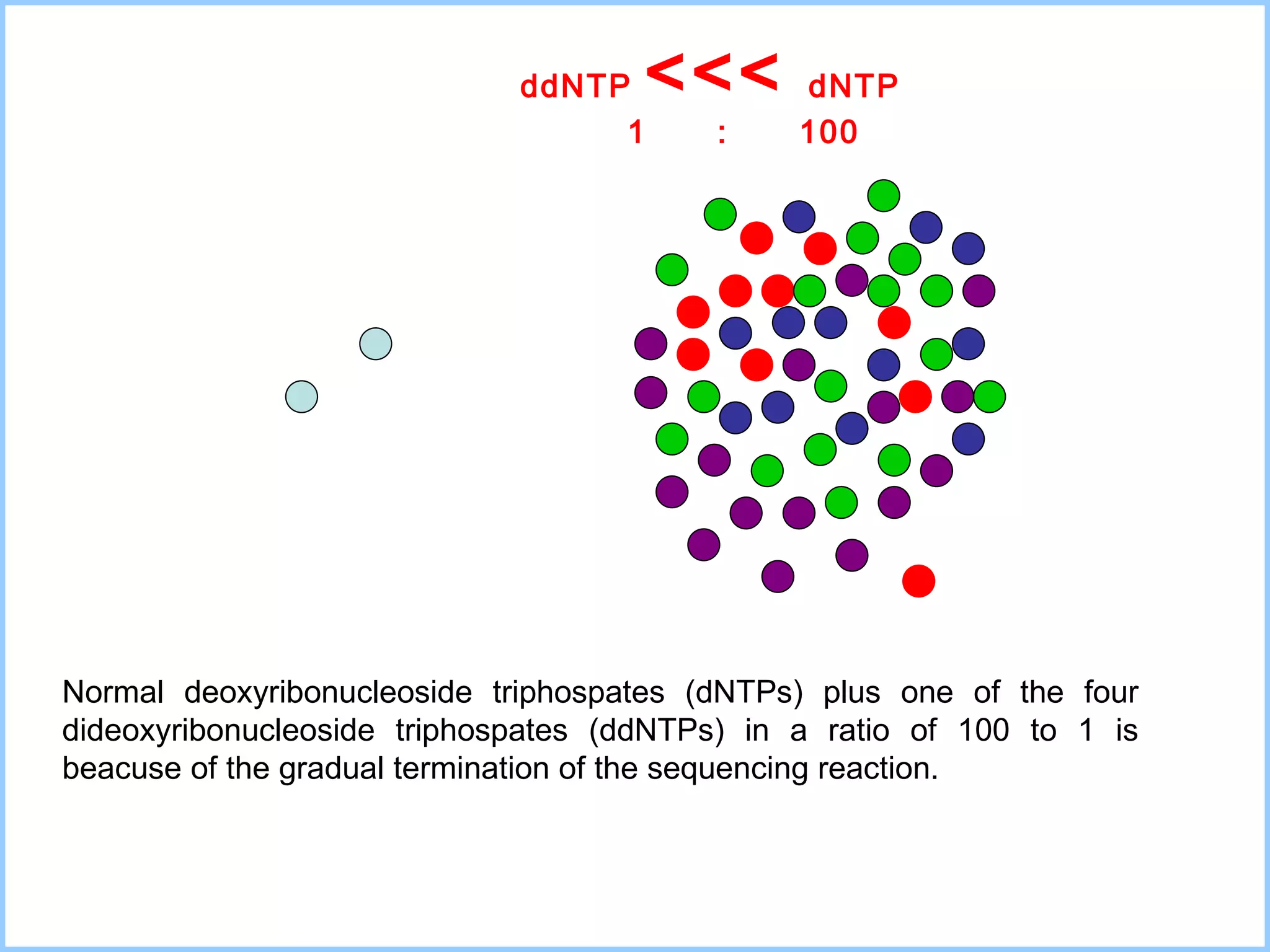
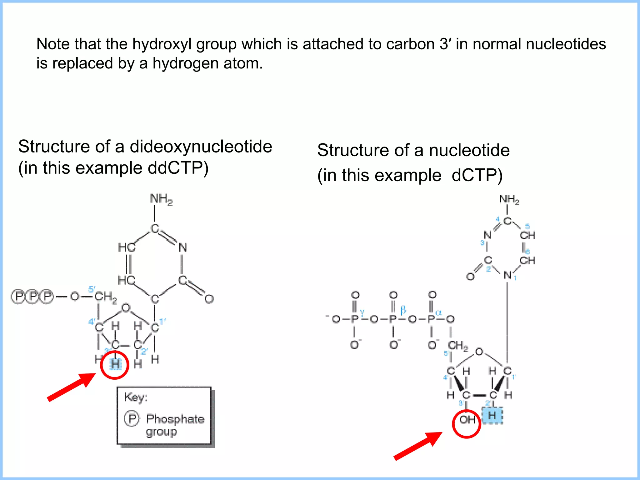
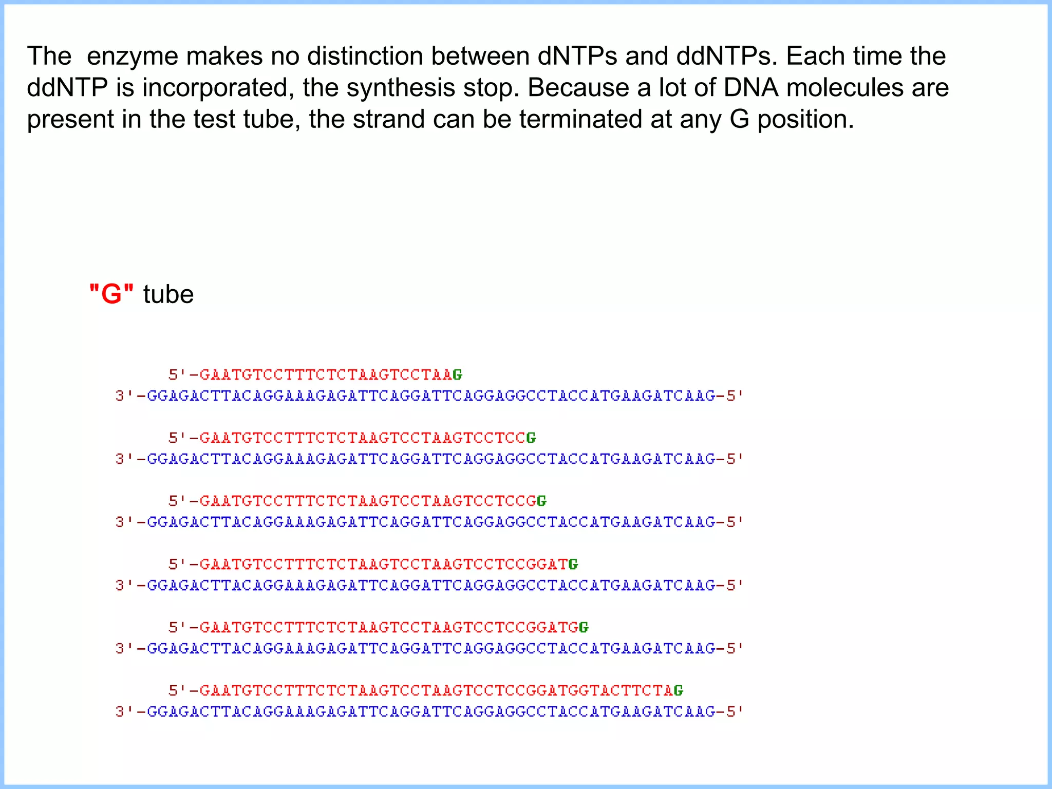
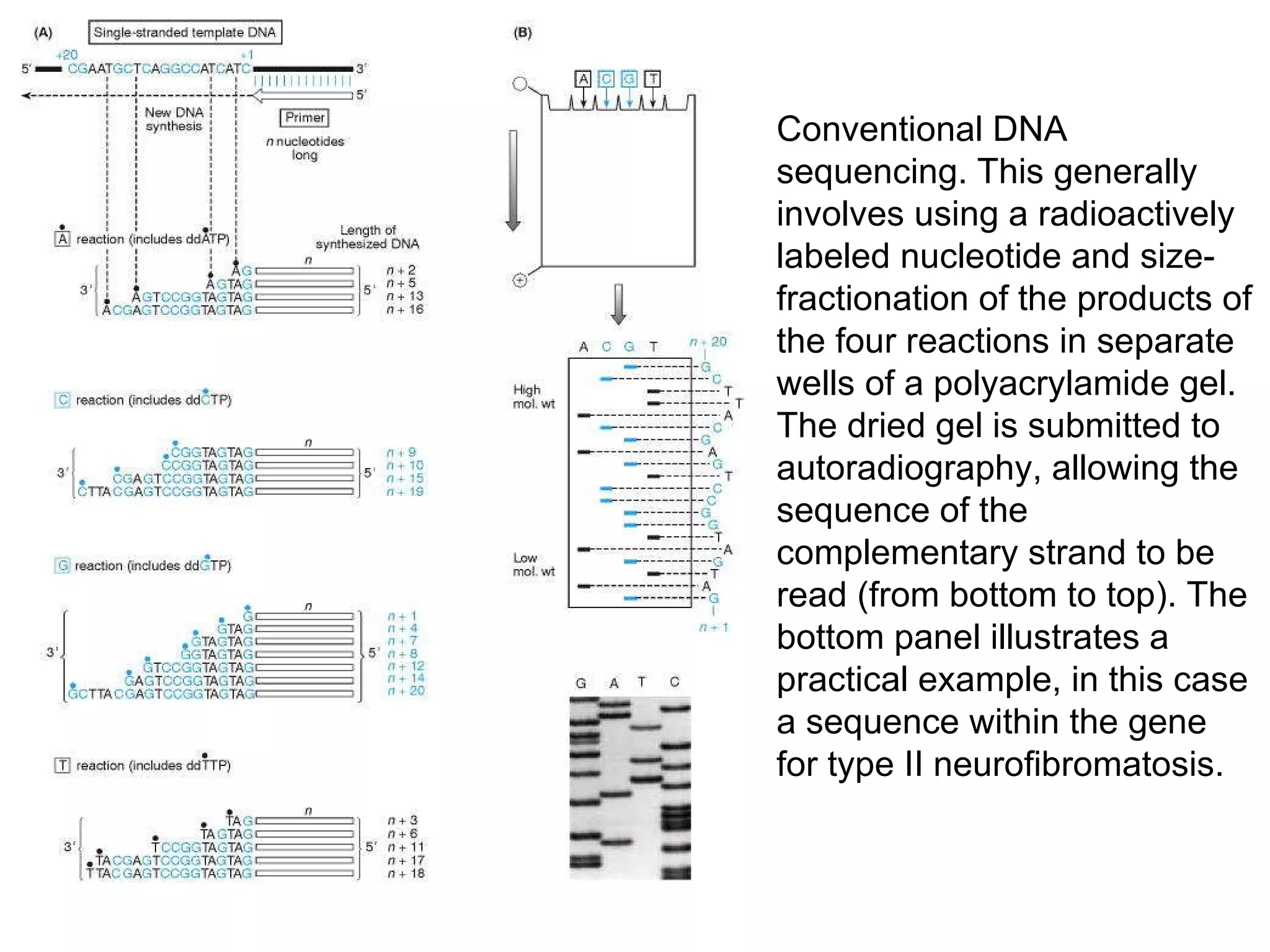
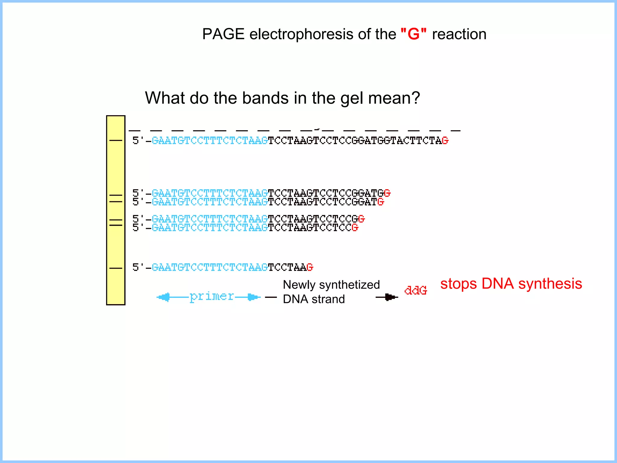
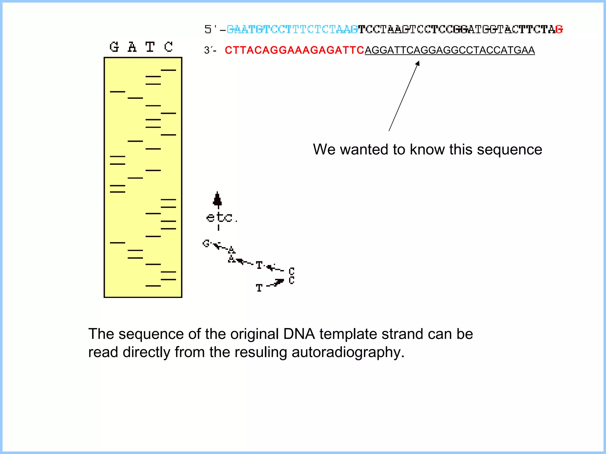
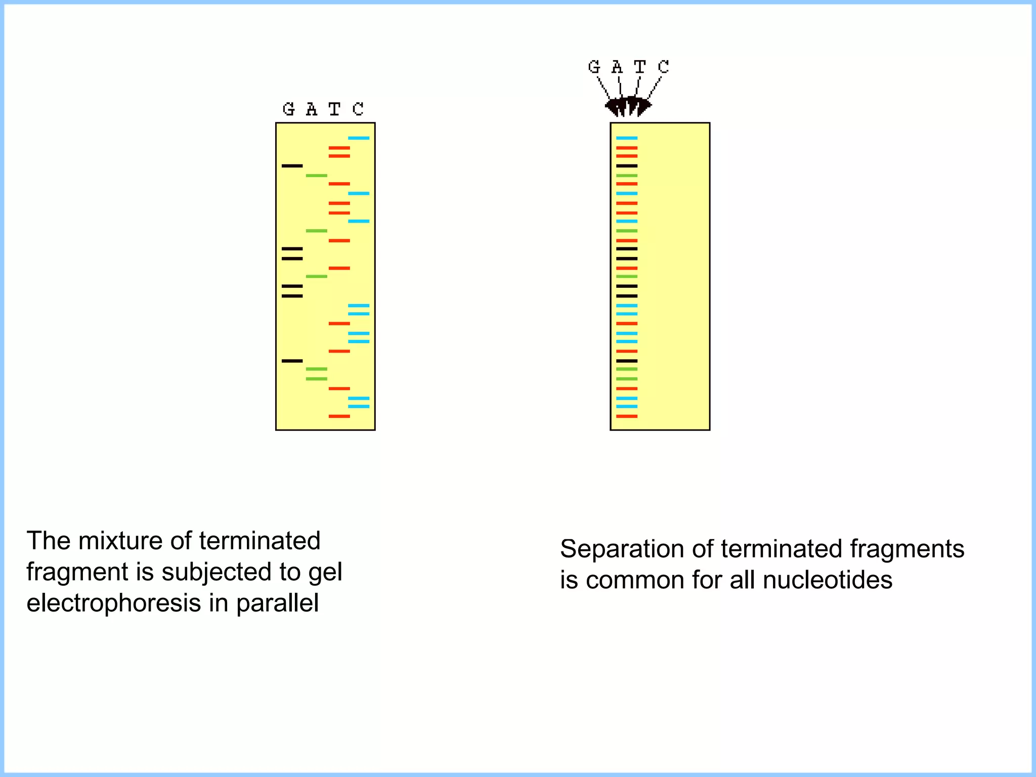
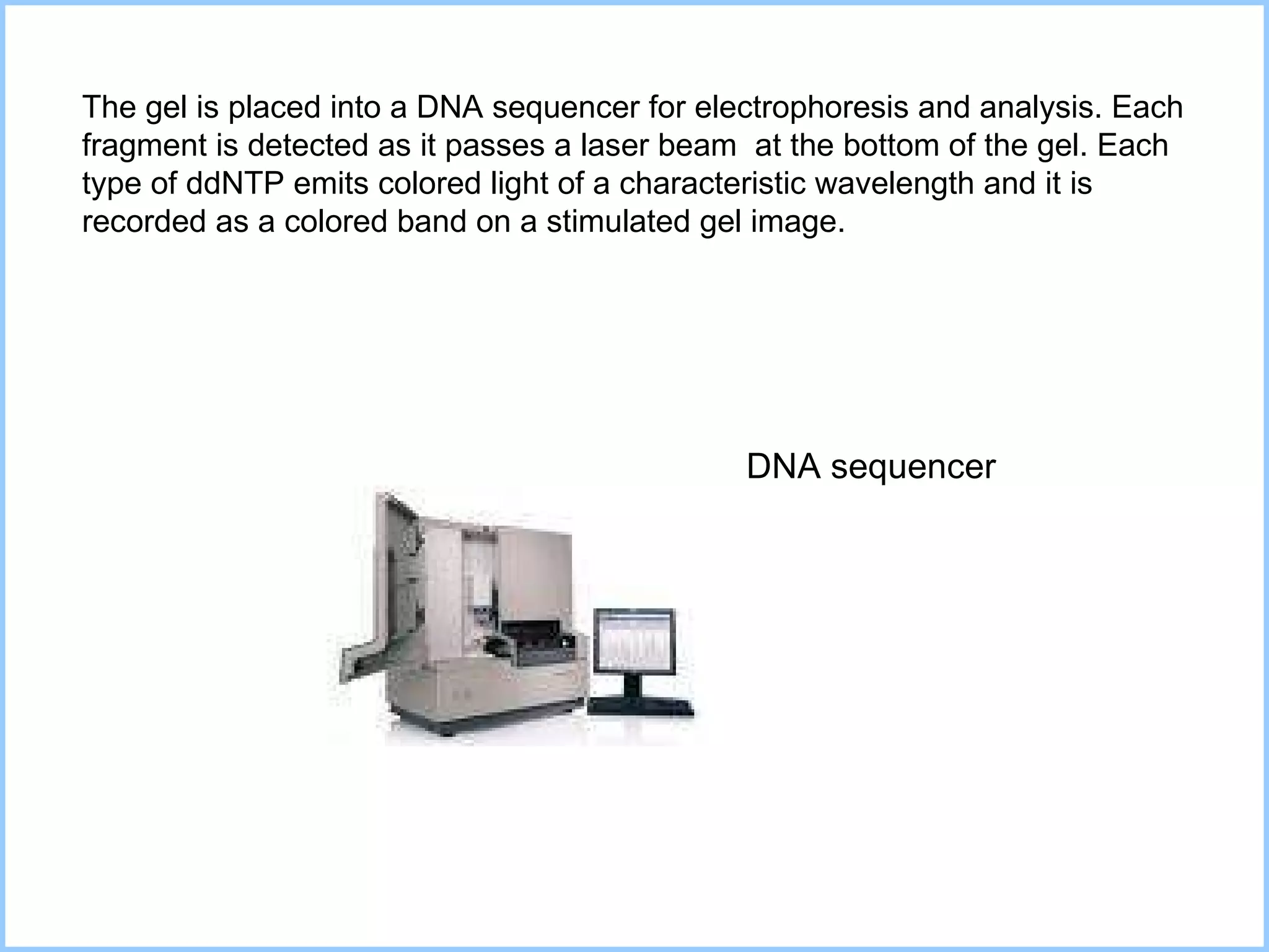
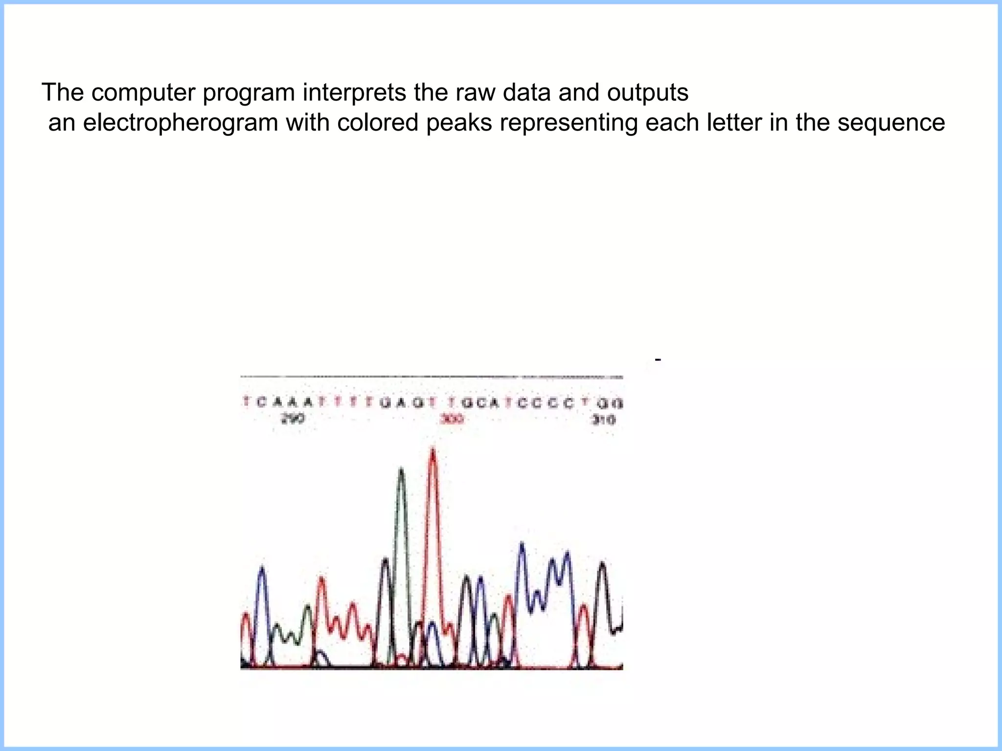
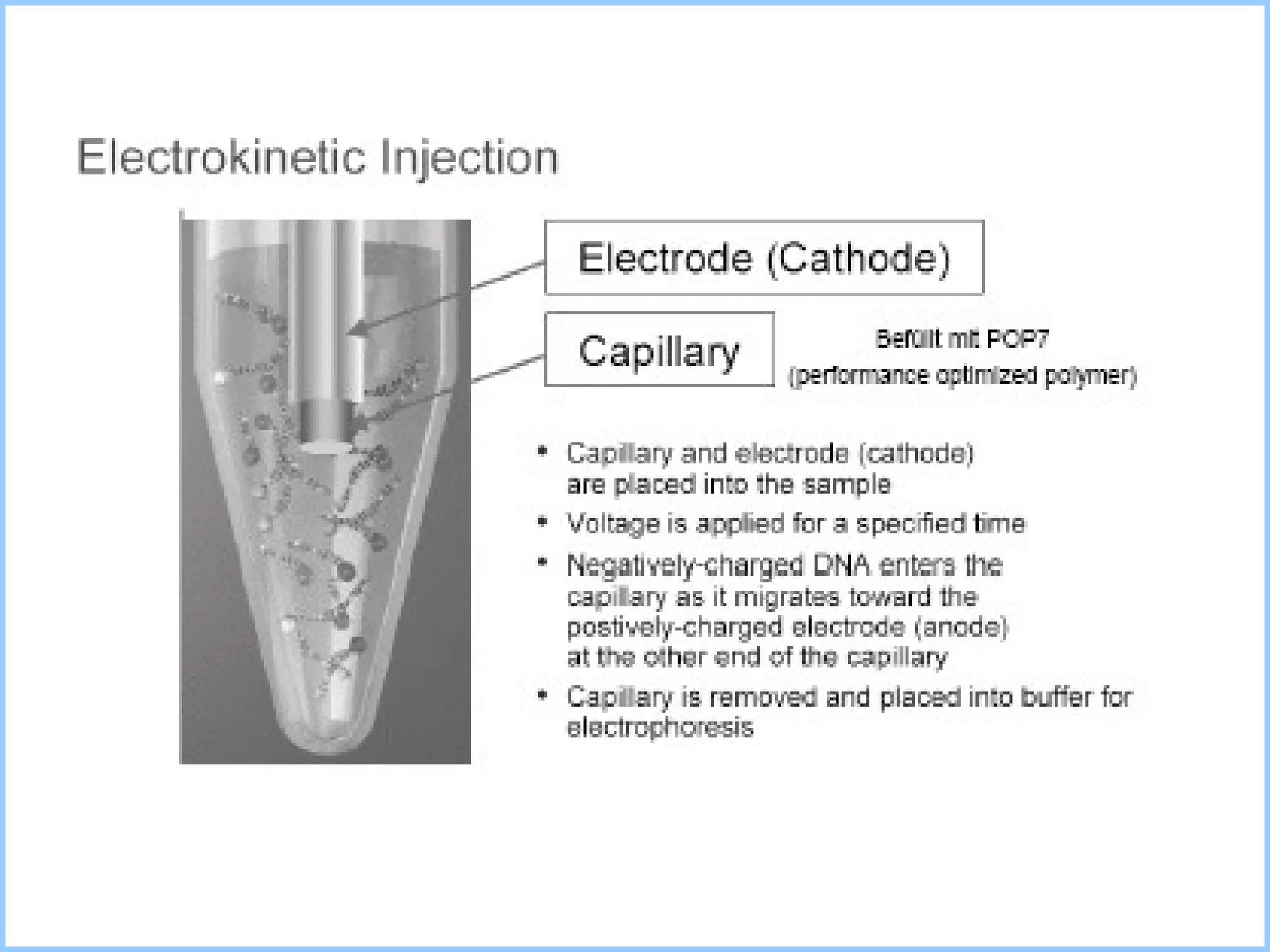
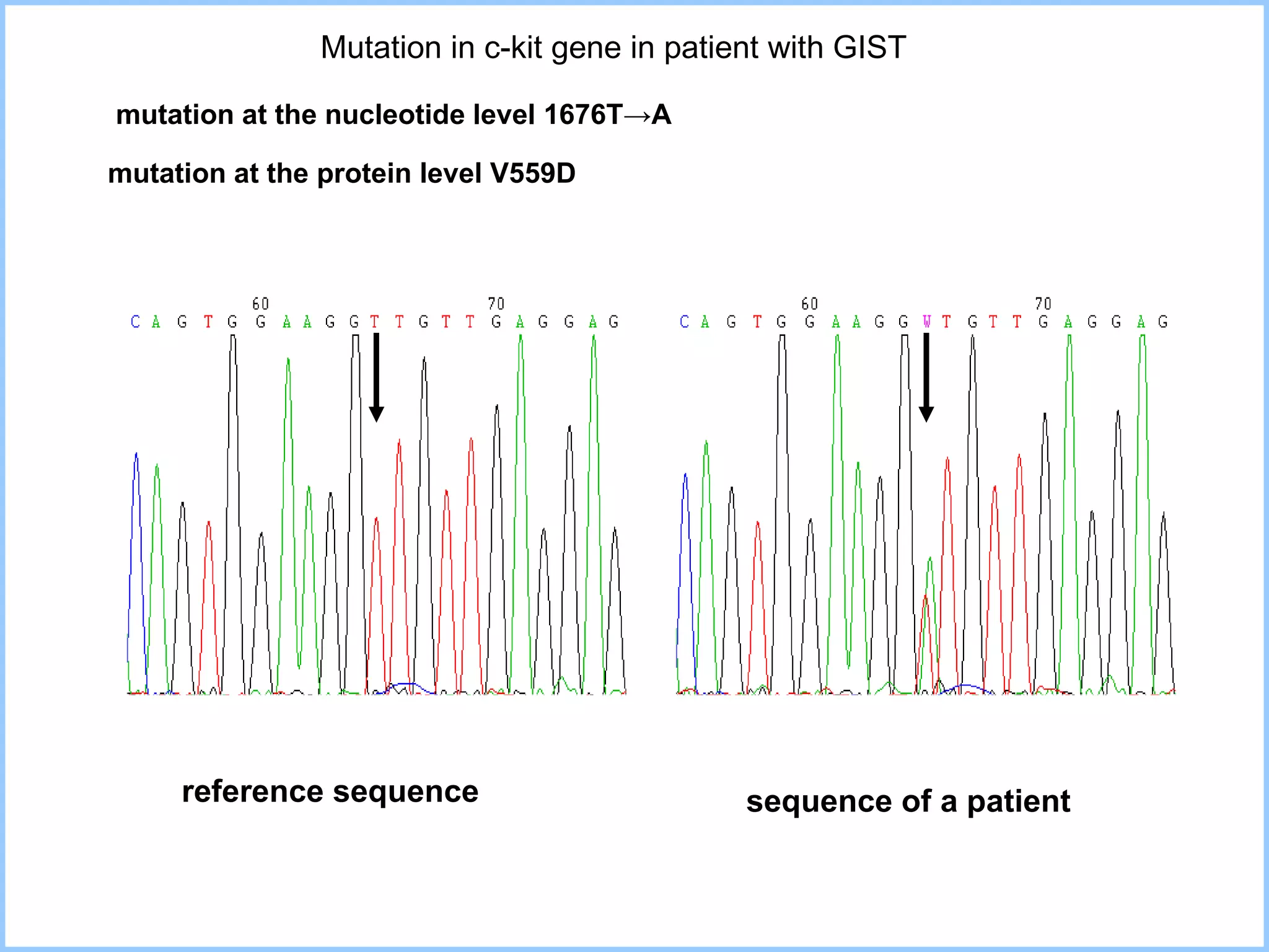
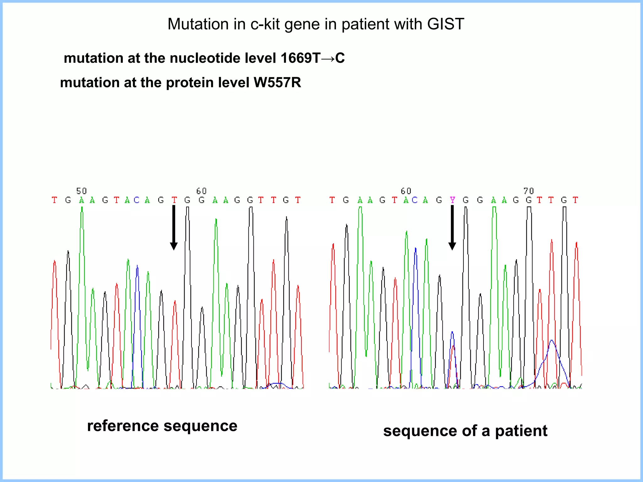
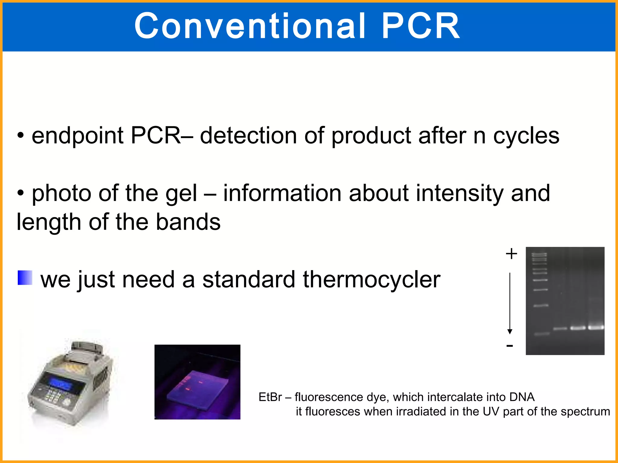
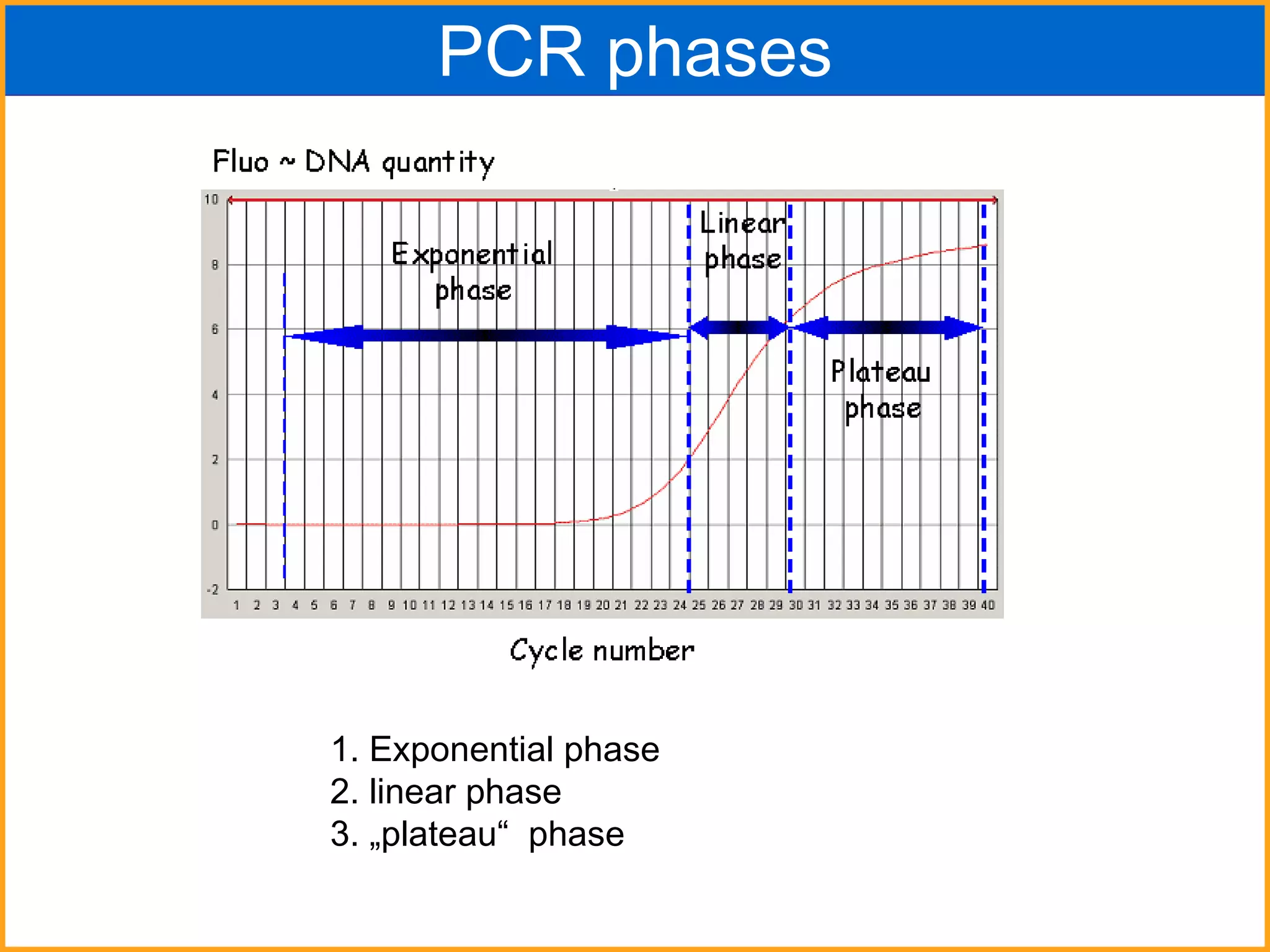
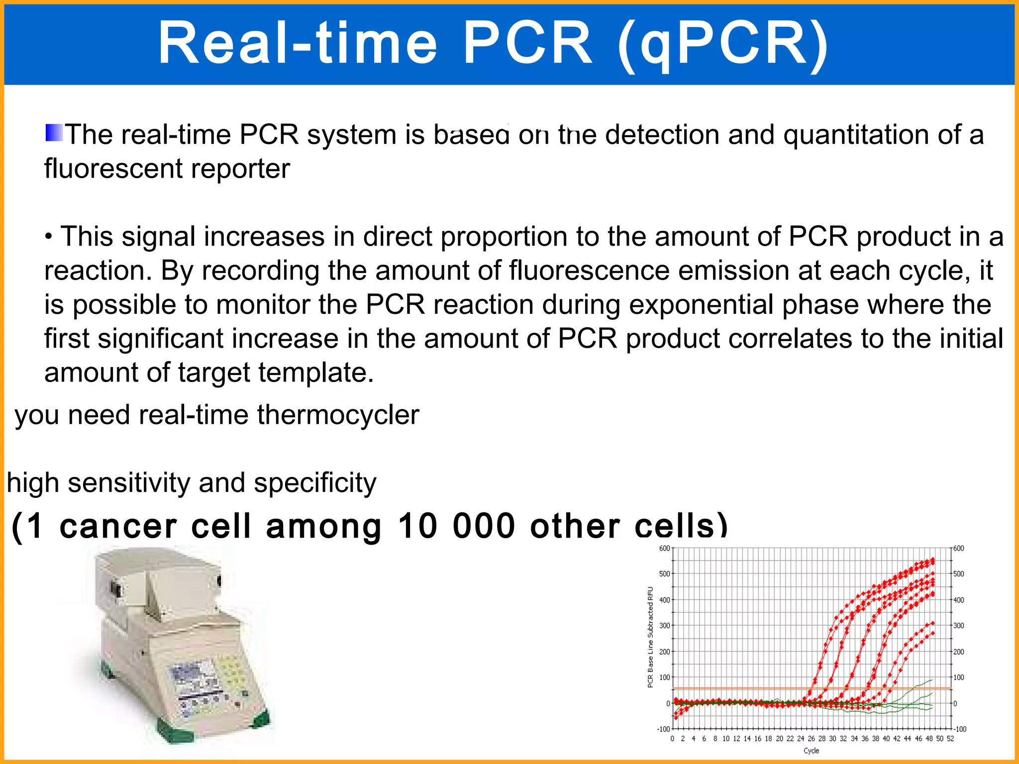
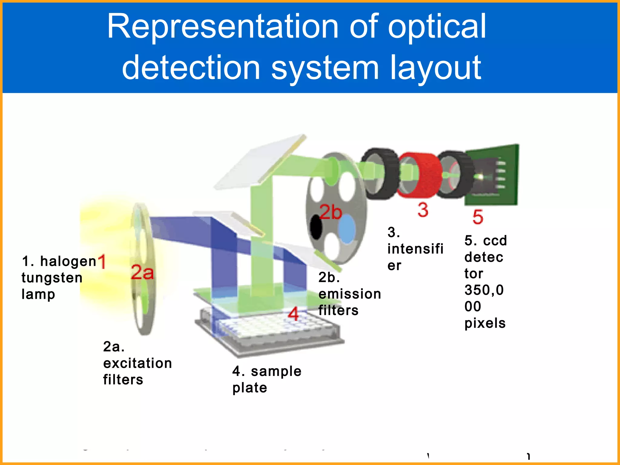
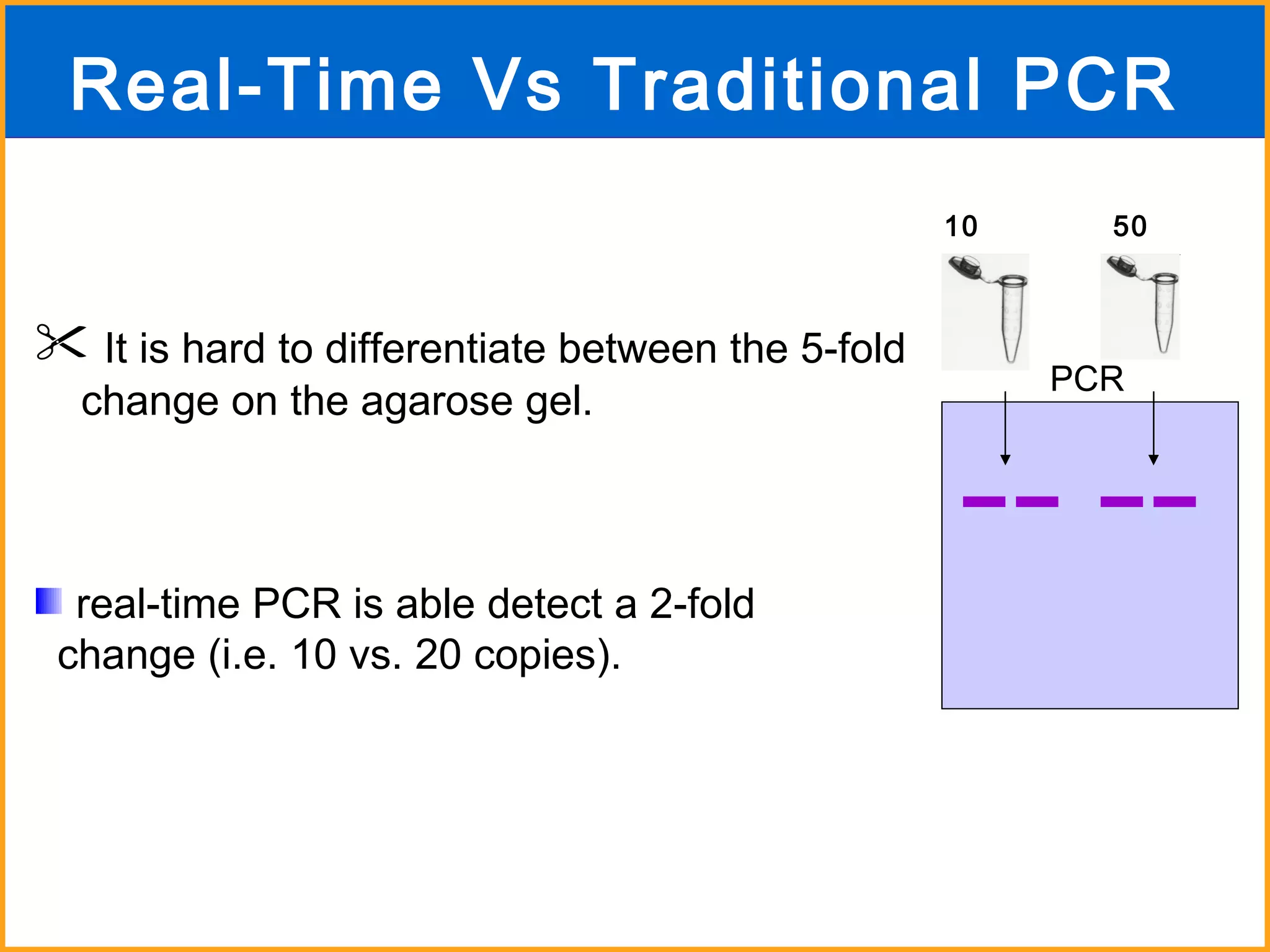
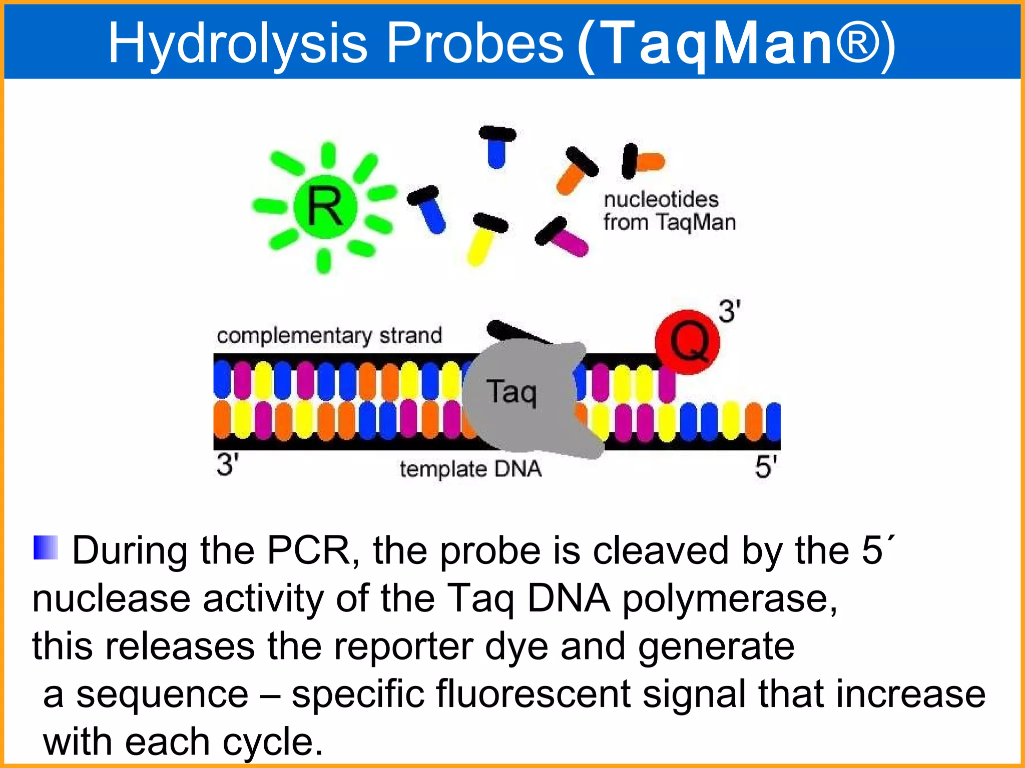
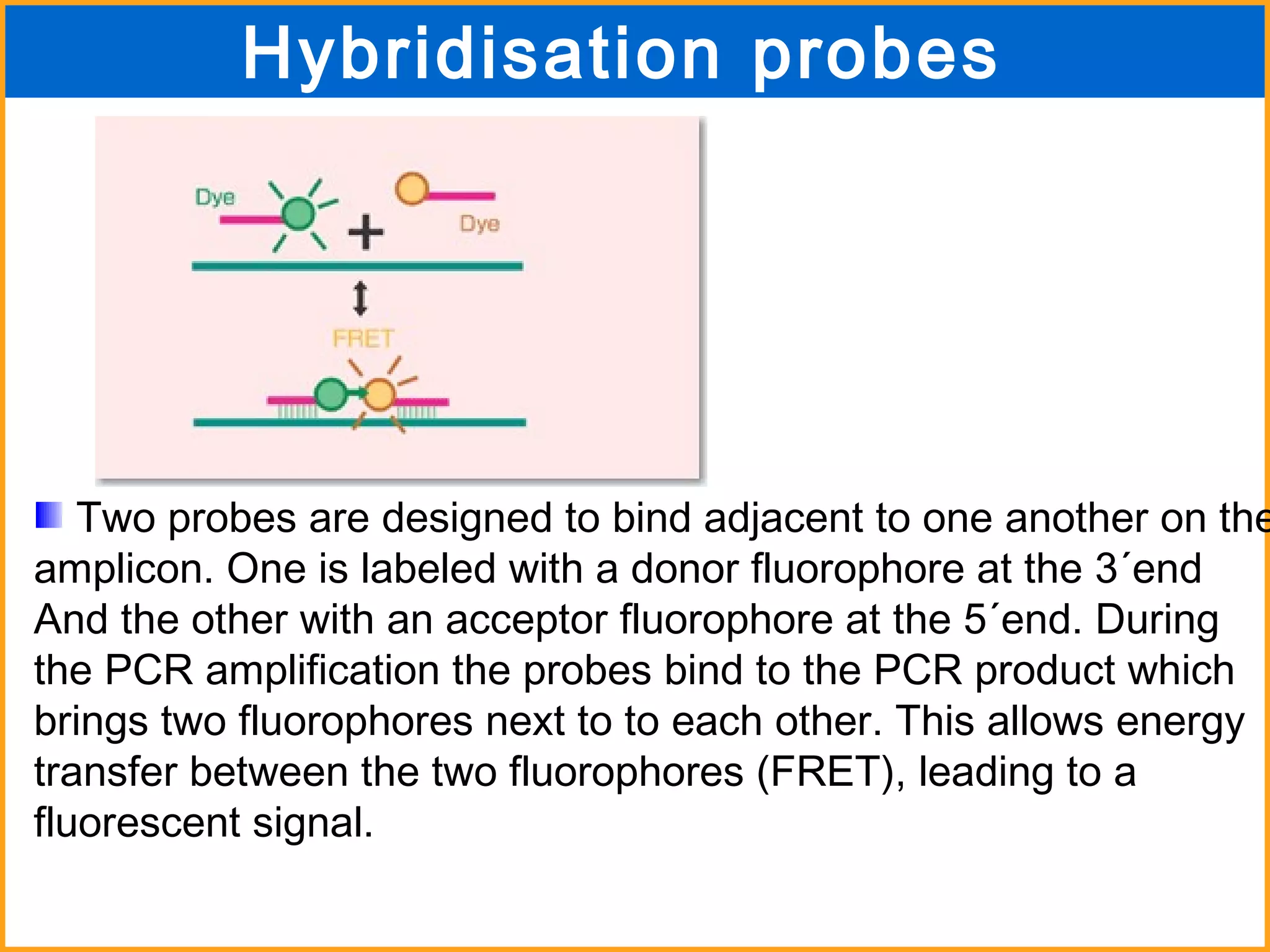
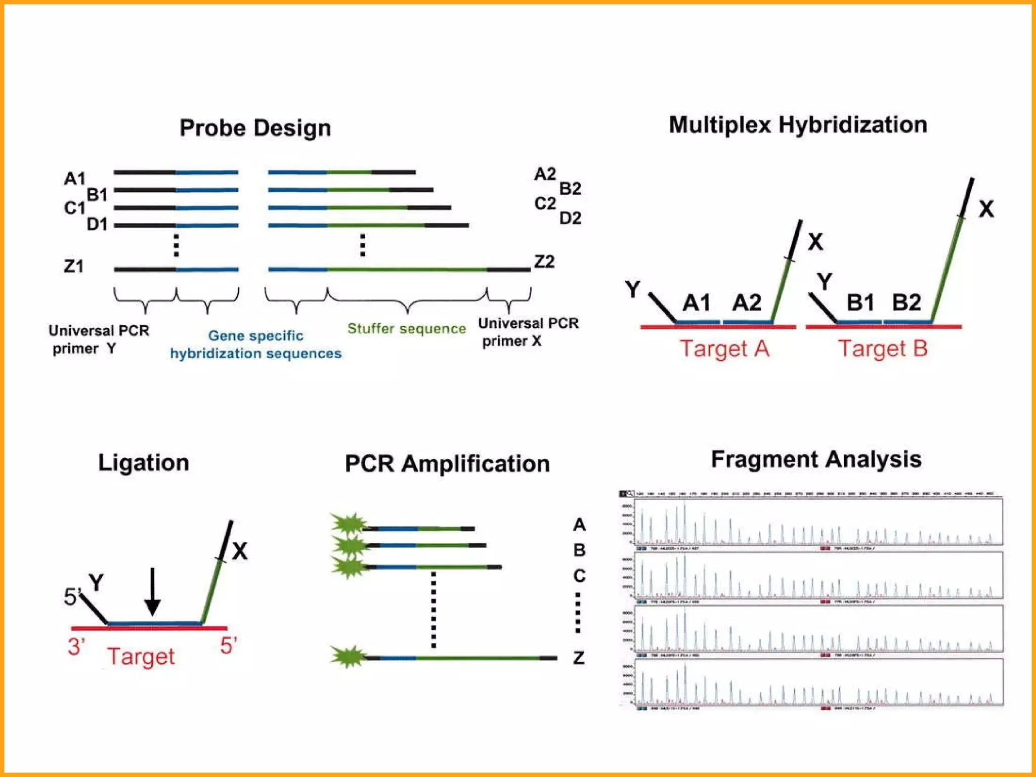
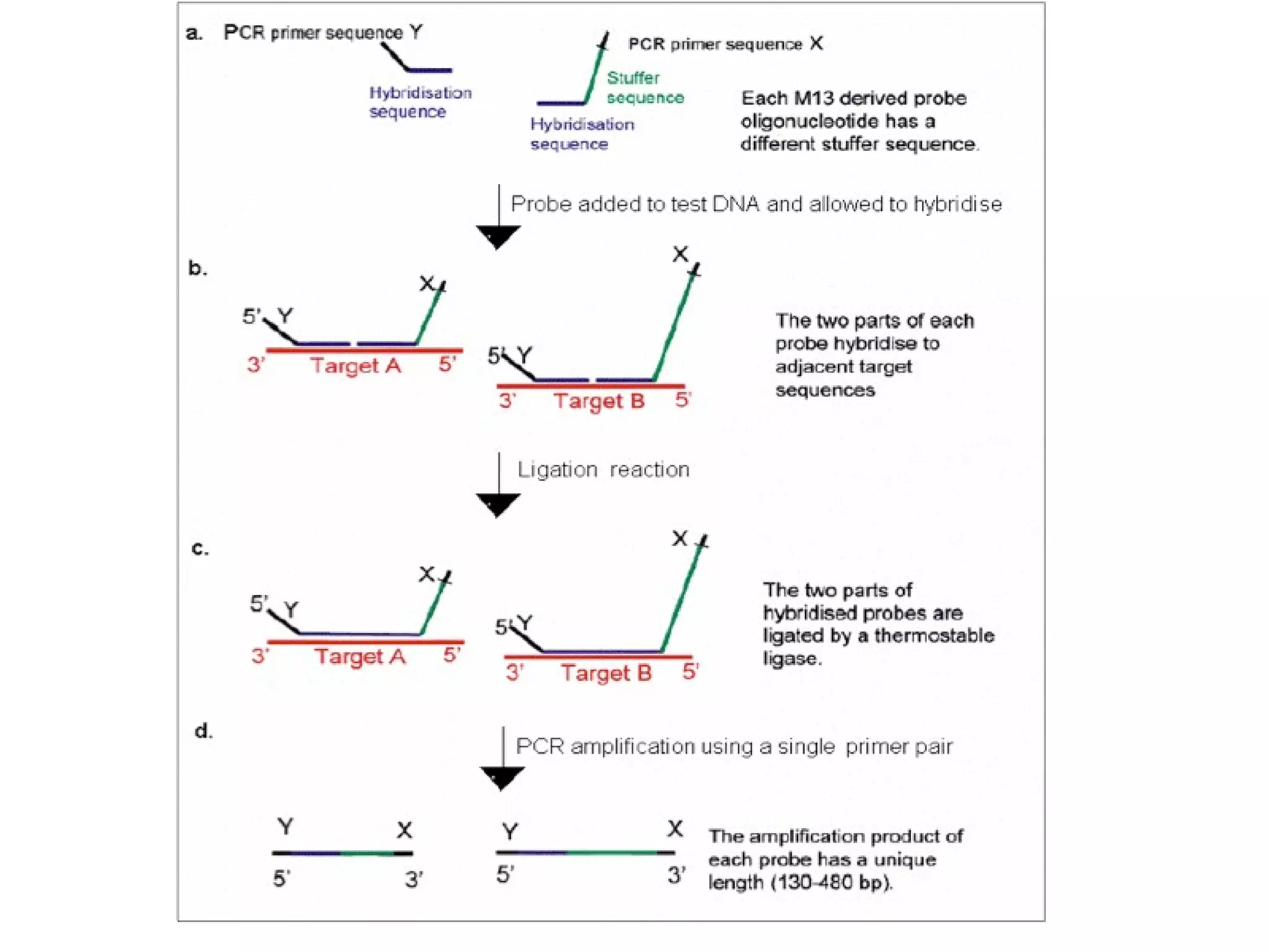
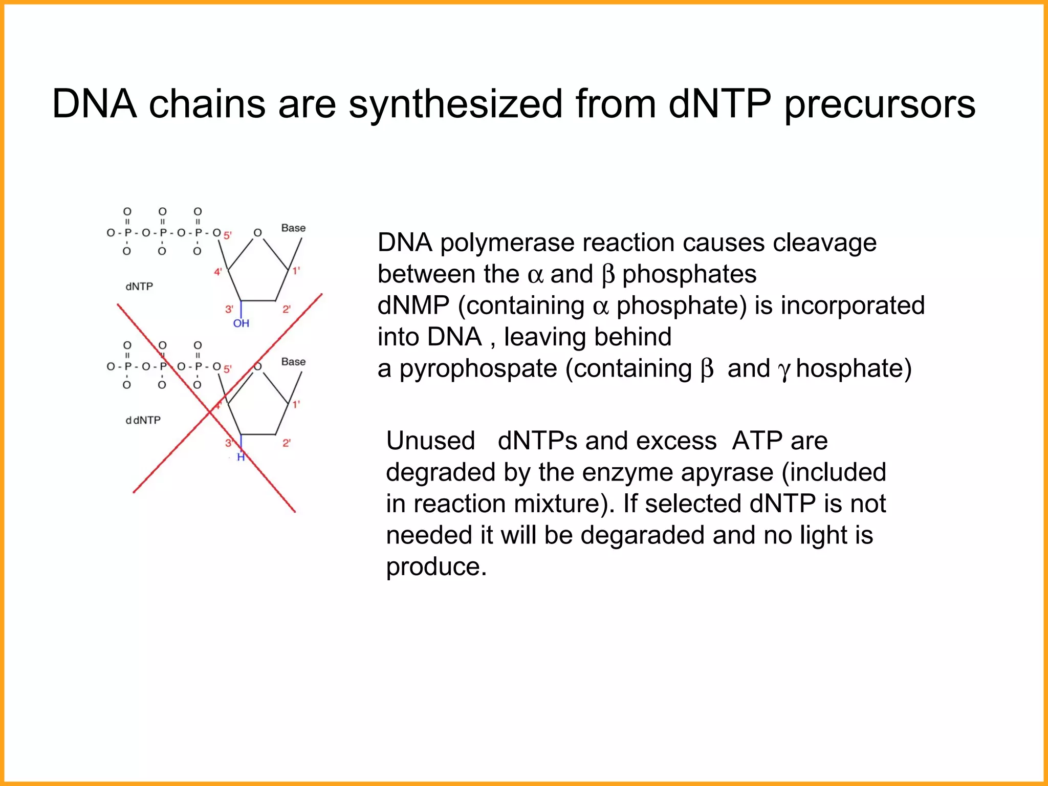
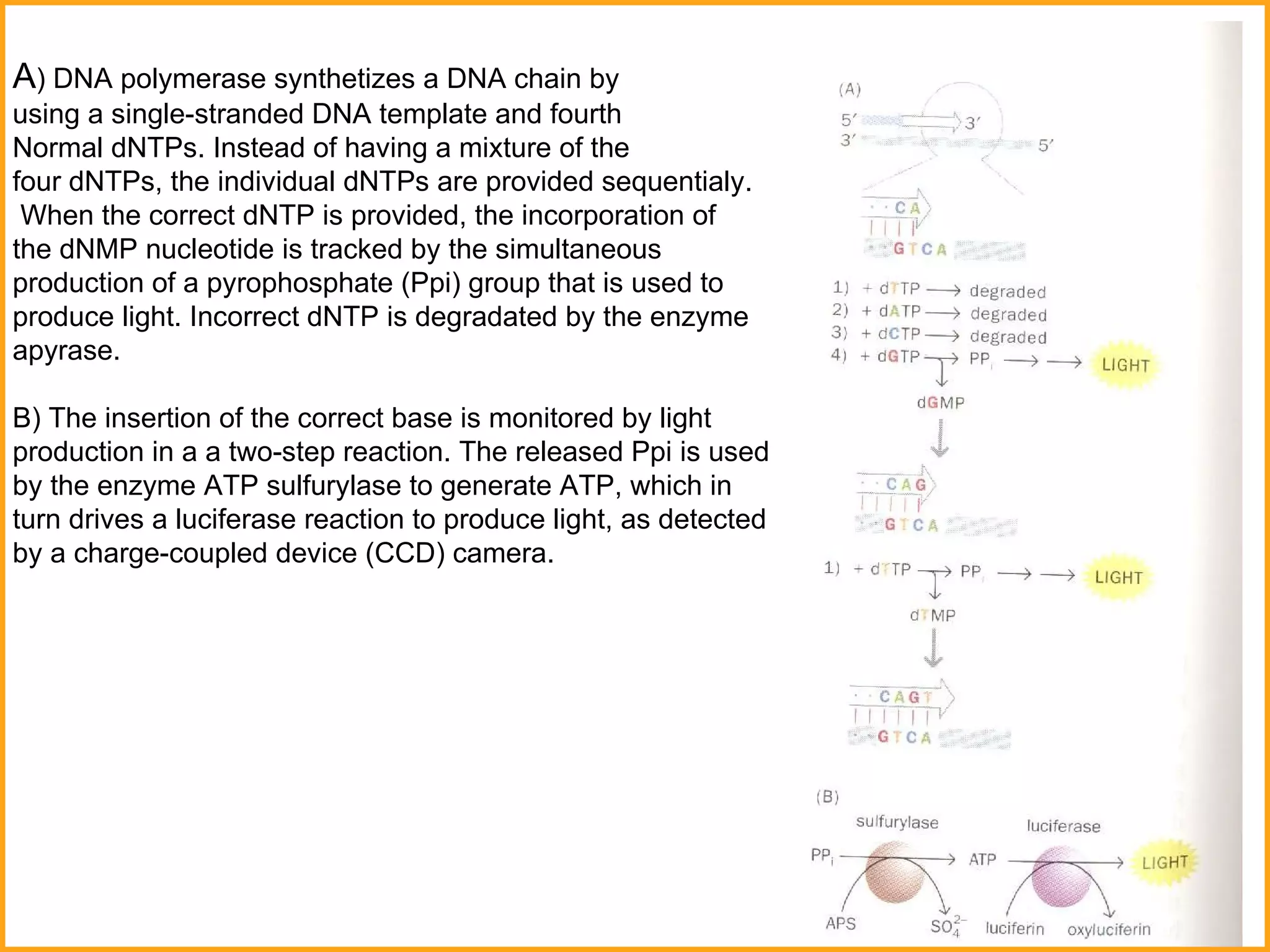
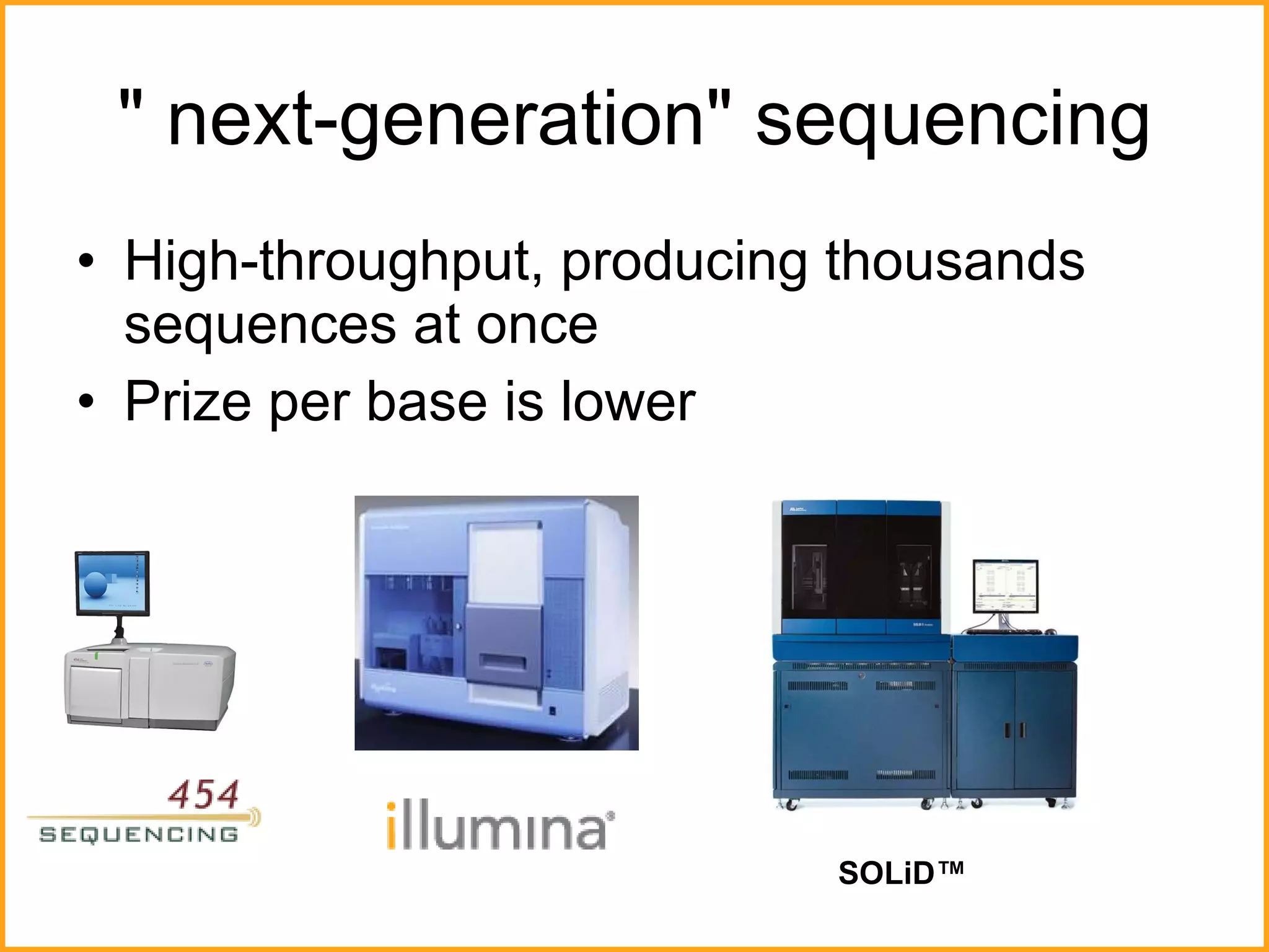
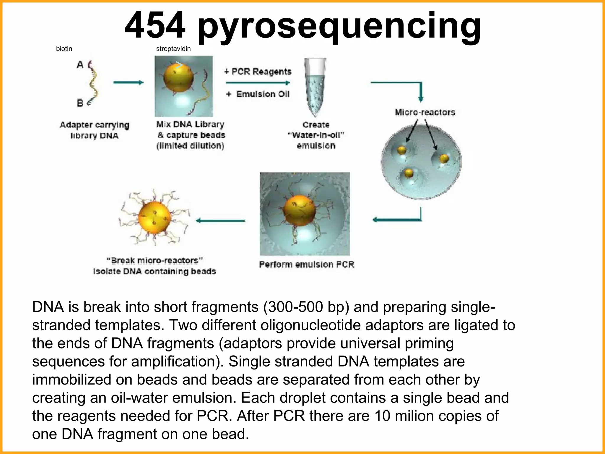
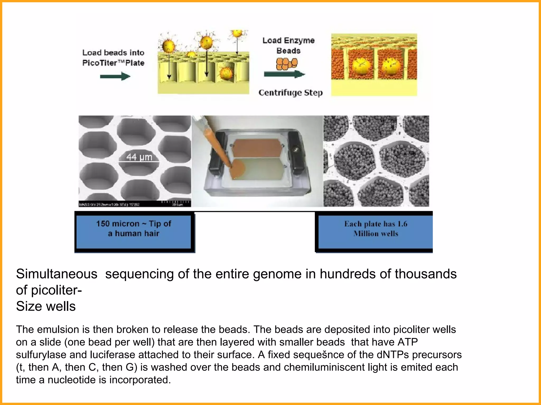
Methods in molecular biology II  By: KHURAM AZIZ
Hybridization of nucleid acids After isolation of the gene from the DNA, we could ask location of the gene on a chromosome  position of the gene on a chromosome if the gene is mutated if the similar gene is in other organisms We could ask these questions by using  Watson-Crick base pairing
Hybridization of nucleid acids Doublestrand DNA High temperature High pH Temperature decrease,  Decreasing The pH Level   Denaturation - double-stranded deoxyribonucleotic acid unwinds and separates into single-stranded strands through the breaking of hydrogen bonding between the bases  DNA pair by hydrogen bonds to a complementary sequence, forming a double - stranded polynucleotide
Nucleic acid hybridization Fundamental tool in molecular biology which  takes advantage of the ability of individual single-stranded nucleic acid molecules to form double-stranded molecules (that is, to  hybridize ) For this to happen , the interacting ss molecules must have a sufficiently high degree of sequence similarity Standard nuclei acid hybridization assays involve using of labeled nucleic acid  probe  to identify related DNA or RNA molecules   (with a significantly high degree of sequence similarity) within a complex mixture of unlabeled nucleic acid molecules, the  target  nucleic acid
Principles of nucleic acid hybridization  Specificity of the interaction between  probe  and  target  comes from  base complementarity  , because both populations are treated in such a way to ensure that all the nucleic acids sequence present are  single stranded (ssDNA)   Thus, if either the probe or the target is initially double-stranded, the individual strands must be separated –  denatured  (heating or alkaline treatment) After mixing single strands of probe with single strands of target, strands with complementary base sequences are allowed to  reassociate (reanneal)
Hybridization of nucleid acids was used for prenatal diagnosis  normal   globin sickle-cell   globin normal protein mutated protein protein patients
Southern blotting
The DNA  probe  is detected by its radioactivity. DNA  probes  detected by chemical or fluorescence methods are also widely used (A) A mixture of either single-stranded RNA  molecules  ( Northern blotting ) or the double-stranded DNA  molecules  created by restriction nuclease treatment ( Southern blotting ) is separated according to length by electrophoresis. (B) A sheet of either nitrocellulose paper or nylon paper is laid over the gel, and the separated RNA or DNA fragments are transferred to the sheet by  blotting . (C) The nitrocellulose sheet is carefully peeled off the gel. (D) The sheet containing the bound  nucleic acids  is placed in a sealed plastic bag together with a buffered salt solution containing a radioactively labeled DNA  probe . The paper sheet is exposed to a labeled DNA  probe  for a prolonged period under conditions favoring  hybridization . (E) The sheet is removed from the bag and washed thoroughly, so that only  probe   molecules  that have hybridized to the RNA or DNA immobilized on the paper remain attached. After  autoradiography , the DNA that has hybridized to the labeled  probe  shows up as bands on the autoradiograph. For  Southern blotting , the strands of the double-stranded DNA  molecules  on the paper must be separated before the  hybridization  process; this is done by exposing the DNA to alkaline denaturing conditions after the gel has been run  Southern blotting
Fluorescence labeling of probes  Direct labeling –  a nucleotide containing an attached labeled group is incorporated. Often such systems involve incorporation of modified nucleotides containing a f luorophore,  a chemical group which can fluorescence when exposed to light of a ceratin wavelenght
DNA microarray (=DNA Chip) Any cells expresses, at any one time, many hundreds or even thousands of genes.  Some of products are expressed at high level (e.g. actin) while others may only  be expressed in a few copies.  Expresion of different sets of genes  Trying to identify these differences is important field of research.  Different stages of maturation tumor cell vs. normal cell
DNA microarray  is a collection of microscopic DNA spots, commonly representing single genes, arrayed on a solid surface   by covalent attachment to a chemical matrix.   There are currently two platforms/types of DNA microarrays  according to the differences in how the nucleid acid samples are generated and delivered to the microarrays:  Microarrays of pre-synthesized nucleid acids Microarrays of oligonucleotides synthesized  in situ Figure 1. Glass complementary DNA (cDNA) microarray produced by using high-speed precision robot. This type of DNA microarray can bear between 10,000 - 20,000 spots (genes) on an area of 3.6 cm2. Each spot represents the product of a specific gene and is generated by depositing a few nano liters of PCR product representing that specific gene usually at concentration of 100-500 µg/ml. The diameter of each spot is also typically 50-150 µm.  Figure 2. Illustration of a DNA GeneChip (Affymetrix).
Microarrays of pre-synthesized nucleid acids T he first type of DNA microarray technology developed   by Patrick Brown and his colleagues at Stanford University . Individual DNA clones or oligonucleotides are spotted at individual locations on the surface of a microscope slide, specified by precise x,y co-ordinates in a miniaturized grid. It  is produced by using a robotic device .  Patrick Brown Robotic spotting for construction of Dna micoarrays Microarray robot Laser scanner http://www.dnalc.org/resources/animations/dnaarray.html
Microarrays of oligonucleotides synthesized  in situ Oligonucleotides are synthesized in situ on the substrate at defined position. Affymetrix, Inc., a California company, has adapted the process to put thousands of transistors onto a single integrated circuit to making oligonucleotides arrays – GeneChips®  about 1.6 cm 2 . - size of small postage stamp (1 million oligonucleotides per square centimeter)
The glass substrate has a covalent linker molecul terminated with a photolabile  (broken down by light) protecting group at every defined array position. A series of photolitographic masks define the oligonucleotide synthesis process. Each mask has certain openings corresponding to defined array positions.  Preparing a GeneChip :  http://www.dnalc.org/resources/animations/dnachip.html
The principle of DNA microarray technology is based on the fact that complementary sequences of DNA can be used to hybridise immobilised DNA molecules.  Figure ilustrates an experiment using a GeneChip probe array Illustration of an experiment using a GeneChip  cDNAs are labeled with  fluorescent tags.  Sample A- red  Cy5 (cyanine) dye Sample B- green Cy3 (cyanine) dye After hybridisation, the fluorescence pattern  is recorded by laser scanning and the data  are analyzed according to the goals of the experiment.  If both green and red fluorescent dyes are used simultaneously, the laser detector can distinguish the two because they produce different wavelenghts of fluorescence emmision
Microarray Application: Identifying gene’s and gene mutations of different types of diseases such as cancer. Identifying the expression level, or quantity of genes (mutated or not).
DNA sequencing  DNA sequencing is the process of determining the nucleotide   order of a given  DNA fragment . There are two methods of DNA sequencing:  Chemical method  –has been devised by Allan Maxam and Walter Gilbert . (Dideoxy) method was developed by  Frederick Sanger and colleagues in 1977. Now it is used more frequently than Maxam-Gilbert method.  Walter Gilbert  a Frederick Sanger  w ere  awarded the 1980 Nobel  Prize in Chemistry .  Walter Gilbert Frederick Sanger
First step is DNA denaturation. So, we obtain single strand DNA.  Labeld synthetic deoxyribonucleotide primer is hybridized to  the single strand of the DNA in the next step. Then, the primer is  elongated in four separate reaction mixtures containing the four  normal deoxyribonucleoside triphospates (dNTPs) plus one of the  four dideoxyribonucleoside triphospates (ddNTPs)  in a ratio of  100 to 1. A ddNTP molecule can add at the position of the  corresponding normal dNTP, but when this occurs, chain  e longation stops because the ddNTP lacks a 3′ hydroxyl. In time, each reaction   mixture will contain a mixture of prematurely  terminated chains ending at every occurrence of the ddNTP .  The oligonucleotide primer is extended using a  T7  DNA  p olymerase  from the  5'- end of the primer.   "G"   tube : all  4  dNTP, ddGTP and DNA polymerase "A„  tube: all  4  dNTP, ddATP and DNA   polymerase "T„  tube: all  4  dNTP, ddTTP and DNA   polymerase  "C„  tube: all  4  dNTP, ddCTP and DNA   polymerase
ddNTP   <<<   dNTP 1  :  100 Normal deoxyribonucleoside triphospates (dNTPs) plus one of the four dideoxyribonucleoside triphospates (ddNTPs)  in a ratio of 100 to 1  is beacuse of the gradual termination of the sequencing reaction.
Structure of a dideoxynucleotide (in this example  ddCTP ) (in this example  dCTP ) N ote that the hydroxyl group which is attached to carbon 3′ in normal nucleotides is replaced by a hydrogen atom. Structure of a nucleotide
The  enzyme makes no distinction between dNTPs and ddNTPs. Each time the ddNTP is incorporated, the synthesis stop. Because a lot of DNA molecules are present in the test tube, the strand can be terminated at any G position.  &quot;G&quot;   tube
Conventional  D NA sequencing. This generally involves using a radioactively labeled nucleotide and size-fractionation of the products of the four reactions in separate wells of a  p olyacrylamide gel. The dried gel is submitted to  au toradiography, allowing the sequence of the  c omplementary strand to be read (from bottom to top). The bottom panel illustrates a practical example, in this case a sequence within the gene for type II neurofibromatosis.
stops DNA synthesis Newly synthetized  DNA strand What do the bands in the gel mean? PAGE electrophoresis of the  &quot;G&quot;   reaction
The sequence of the original DNA template strand can be  read directly from the resuling autoradiography. 3´-  CTTACAGGAAAGAGATTC AGGATTCAGGAGGCCTACCATGAA We wanted to know this sequence
Separation of terminated fragments  is common for all nucleotides The mixture of terminated fragment is subjected to gel electrophoresis in parallel
An alternative to the labelling of the primer is to label the terminators instead, commonly called 'dye terminator sequencing'. The major advantage of this approach is the complete sequencing set can be performed in a single reaction, rather than the four needed with the labeled-primer approach. This is accomplished by labelling each of the dideoxynucleotide chain-terminators with a separate fluorescent dye, which fluoresces at a different wavelength.  M ore commonly now ,  fragments are then size-separated in a narrow glass tube (capillary) filled with a viscous polymer  instead of  electrophoresis in a slab polyacrylamide gel .  The gel is placed into a DNA sequencer for electrophoresis and analysis. Each fragment is detected as it passes a laser beam  at the bottom of the gel. Each type of ddNTP emits colored light of a characteristic wavelength and it is recorded as a colored band on a stimulated gel image.
The gel is placed into a DNA sequencer for electrophoresis and analysis. Each fragment is detected as it passes a laser beam  at the bottom of the gel. Each type of ddNTP emits colored light of a characteristic wavelength and it is recorded as a colored band on a stimulated gel image.  DNA sequencer
The computer program interprets the raw data and outputs an electropherogram with colored peaks representing each letter in the sequence
 
Cappilary Samples Ele ctrode
Separation of the terminated fragments according to their sizes terminated fragment
Automated DNA sequencers
ABI 3100  16-capillary array ABI 310  single capillary Capillary Electrophoresis Instrumentation
GISTs – gastrointestinal stromal tumors mutations in genes c-kit and PDGFRA exon  11 c-kit ,exon 12 PDGFRA  –good  response to imatinib exo n  17   c-kit, exo n  18 PDGFRA  resistent to imatinib
Mutation in c-kit gene in patient with GIST  reference sequence sequence of a patient mutation at the nucleotide level  1676T->A mutation at the protein level V 55 9D
Mutation in c-kit gene in patient with GIST  mutation at the nucleotide level  1669T->C mutation at the protein level W 557R reference sequence sequence of a patient
Real Time PCR Institute of molecular biology
Conventional PCR endpoint PCR– detection of product after n cycles   photo of the gel – information about intensity and length of the bands we just need a standard thermocycler  EtBr – fluorescence dye, which intercalate into DNA it fluoresces when irradiated in the UV part of the spectrum + -
Nowadays it is needed the more precise determination  of the amount of the nucleid acid, which input into a PCR reaction.
PCR phases 1. Exponential phase 2. linear phase 3. „plateau“  phase
The real-time PCR system is based on the detection and quantitation of a fluorescent reporter This signal increases in direct proportion to the amount of PCR product in a reaction. By recording the amount of fluorescence emission at each cycle, it is possible to monitor the PCR reaction during exponential phase where the first significant increase in the amount of PCR product correlates to the initial amount of target template. you need real-time thermocycler high sensitivity and specificity (1 cancer cell among 10 000 other cells) Real-time PCR (qPCR)  PCR
Representation of optical  detection system layout www.biorad.com 2a. excitation filters 2b. emission filters 1. halogen tungsten lamp 4. sample plate 3. intensifier 5. ccd detector 350,000 pixels
 
50 copy 10 copy It is hard to differentiate between the 5-fold change on the agarose gel. real-time PCR is able detect a 2-fold change (i.e. 10 vs. 20 copies). Real-Time Vs Traditional PCR PCR
Quantitation of Gene Expression Overexpression of the receptor tyrosine kinase HER-2/neu  is associated with poor prognosis in patients with breast  and ovarian cancer. Real -Time PCR Applications
Real-Time PCR Applications Patients with non-small cell lung cancer, who has high  EGFR mRNA expression  has better  response to gefitinib and prolonged progression-free survival .  Gefitinib- selective inhibitor of EGF receptor tyrosine kinase Dziadziuszko, R. et al. Clin Cancer Res 2006;12:3078-3084 Copyright ©2006 American Association for Cancer Research
Quantification of viral load in clinical samples (e. g. monitoring of herpes viruses after stem cell transplantation) Quantification of bacterial pathogens as Listeria monocytogenes and Helicobacetr pylory and many others Array Verification Monitoring of minimal residual disease in patients with leukaemias and solid tumors (we can monitor effects of anticancer therapy)  Genotyping ( Factor V Leiden mutation as an aid to diagnosis in the evaluation of patients with suspected thrombophilia )
Detection Systems used in real-time PCR A number of detection systems are available  for real-time PCR. All are based on the generation  of a fluorescent signal that is proportional to the amount  of qPCR product amplified.  Weak band depicted on the upper picture corresponds  to less fluorescence. Stronger band corresponds to high amount of PCR product and more fluorescence intensity.  Band in agarose gel
SYBR ®  Green Non-specific Relatively inexpensive Requires post-pcr dissociation curve analysis ( Tm melting temperature is defined as the temperature at which half of the DNA strands are in the double-helical state and half are in single state. It dependes on the length of the molecule and the specific nucleotide sequence composition. A higher GC content level indicates a higher melting temperature).   Non-specific detection systems
Black line indicate product with lower melting temperature  Tm (81 0 C) than the other products Tm (89  0 C)  Melting curve analysis This peak corresponds to one band in the agarose gel.
SYBR ®  Green is an intercalating dye, which binds only to double strand DNA giving an increase flourescence as the amount of PCR product increases. Only bound SYBR ®  Green is able to flouresce.  SYBR  ®  Green dye
Specific detection systems short oligonucleotide hybridise to the PCR amplicon hydrolysis probes (TaqMan  ®, hybridization probes, molecular beacons,Scorpions TM ) specific expensive
Hydrolysis Probes   (TaqMan ®)   Taqman system makes use of an oligonucleotide  hybridisation probe with both a reporter and  a quencher dye attached. The probe is designed to bind within the amplified PCR fragment.
During the PCR, the probe is cleaved by the 5´  nuclease activity of the Taq DNA polymerase,  this releases the reporter dye and generate a sequence – specific fluorescent signal that increase with each cycle. Hydrolysis Probes   (TaqMan ®)
Hybridisation probes  Two probes are designed to bind adjacent to one another on the amplicon. One is labeled with a donor fluorophore at the 3´end  And the other with an acceptor fluorophore at the 5´end. During the PCR amplification the probes bind to the PCR product which brings two fluorophores next to to each other. This allows energy transfer between the two fluorophores (FRET), leading to a fluorescent signal.
MLPA – multiplex ligation probe assay We can detect long deletions, insertions, duplications and amplifications, known mutations and we could use this method for quantification of mRNA  Principle : uses probes designed to hybridize adjacently to the target sequence. After ligation, the joined probes are amplified and quantified example of using of MLPA : we can detect BRCA1 and BRCA2 mutations in hereditary breast cancers
 
 
Principle of MLPA. For each specific target, a set of two probes was designed that hybridize immediately adjacent to each other on the same target strand. Both probes consist of a short (22–43 nt) target-specific sequence and a universal forward or reverse PCR primer-binding site. In addition, one of the probes contains a so-called stuffer sequence. For each probe, the stuffer part has a specific length (19–364 bp) and sequence. The long probes are M13-derived. The short probes are synthetic. After an overnight hybridization to the target DNA, the two parts of each hybridized probe are joined by a ligation reaction. Next, a PCR is carried out with a single fluorescent-labeled primer pair, which ensures that the relative yield of the PCR products is proportional to the amount of target. The fragment analysis is preferably carried out on an automated capillary sequencer. The multiple fragments can be distinguished based on different length. The peak area value of each product is used to calculate the relative quantity.
Pyrosequencing Dideoxy DNA sequencing - 96 samples at a time ~  30 -60 kb  of sequence  per 3-4 hour electrophoretic run - require electrophoresis Pyrosequencing – is able to monitor incorporation of each nucleotide in the growing DNA chain and to identify which nucleotide was being incorporated at each step.
DNA chains are synthesized from dNTP precursors DNA polymerase reaction causes cleavage between the   and   phosphates dNMP (containing    phosphate) is incorporated into DNA , leaving behind  a pyrophospate (containing   and   hosphate) Unused  dNTPs and excess  ATP are degraded by the enzyme apyrase   (included in reaction mixture). If selected dNTP is not needed it will be degaraded and no light   is produce .
A ) DNA polymerase synthetizes a DNA chain by using a single-stranded DNA template and fourth  Normal dNTPs. Instead of having a mixture of the  four dNTPs, the individual dNTPs are provided sequentialy. When the correct dNTP is provided, the incorporation of the dNMP nucleotide is tracked by the simultaneous  production of a pyrophosphate (Ppi) group that is used to produce light. Incorrect dNTP is degradated by the enzyme apyrase.  B) The insertion of the correct base is monitored by light p roduction in   a a two-step reaction. The released Ppi is used by the enzyme ATP sulfur y lase to generate ATP, which in turn drives a luciferase reaction to produce light, as detected by a charge-coupled device (CCD) camera.
&quot; next-generation&quot; sequencing High-throughput, producing thousands sequences at once Prize per base is lower  SOLiD™
454 pyrosequencing DNA  is break into short fragments (300-500 bp) and preparing single-stranded templates. Two different oligonucleotide adaptors are ligated to the ends of DNA fragments (adaptors provide universal priming sequences for amplification). Single stranded DNA templates are immobilized on beads and beads are separated from each other by creating an oil-water emulsion. Each droplet contains a single bead and the reagents needed for PCR. After PCR there are 10 milion copies of one DNA fragment on one bead.  biotin streptavidin
Simultaneous  sequencing of the entire genome in hundreds of thousands of picoliter-  Size wells The emulsion is then broken to release the beads. The beads are deposited into picoliter wells on a slide (one bead per well) that are then layered with smaller beads  that have ATP sulfurylase and luciferase attached to their surface. A fixed sequešnce of the dNTPs precursors (t, then A, then C, then G) is washed over the beads and chemiluminiscent light is emited each time a nucleotide is incorporated.
Output
Solexa sequencig technology was developed more recently by the Illumina company SOLiD™ System has been developed by the Applied Biosystem company. Also uses an emulsion-based PCR strategy
'3 rd  generation' ('next-next-generation') sequencing  is knocking on the door. It permit the sequencing of single DNA molecules that are not amplified any way (Helicos Bioscience company)  technology Read length Amount per 1 run  Price for 1 kb Sanger 1000 bp 36 KB 10 USD 454 400 -500 bp 0,5GB 0.2 USD Solid 50bp 180GB Illumina 75bp 20GB 0.04 USD

Methods in molecular_biology

  • 1.
    Methods in molecularbiology II By: KHURAM AZIZ
  • 2.
    Hybridization of nucleidacids After isolation of the gene from the DNA, we could ask location of the gene on a chromosome position of the gene on a chromosome if the gene is mutated if the similar gene is in other organisms We could ask these questions by using Watson-Crick base pairing
  • 3.
    Hybridization of nucleidacids Doublestrand DNA High temperature High pH Temperature decrease, Decreasing The pH Level Denaturation - double-stranded deoxyribonucleotic acid unwinds and separates into single-stranded strands through the breaking of hydrogen bonding between the bases DNA pair by hydrogen bonds to a complementary sequence, forming a double - stranded polynucleotide
  • 4.
    Nucleic acid hybridizationFundamental tool in molecular biology which takes advantage of the ability of individual single-stranded nucleic acid molecules to form double-stranded molecules (that is, to hybridize ) For this to happen , the interacting ss molecules must have a sufficiently high degree of sequence similarity Standard nuclei acid hybridization assays involve using of labeled nucleic acid probe to identify related DNA or RNA molecules (with a significantly high degree of sequence similarity) within a complex mixture of unlabeled nucleic acid molecules, the target nucleic acid
  • 5.
    Principles of nucleicacid hybridization Specificity of the interaction between probe and target comes from base complementarity , because both populations are treated in such a way to ensure that all the nucleic acids sequence present are single stranded (ssDNA) Thus, if either the probe or the target is initially double-stranded, the individual strands must be separated – denatured (heating or alkaline treatment) After mixing single strands of probe with single strands of target, strands with complementary base sequences are allowed to reassociate (reanneal)
  • 6.
    Hybridization of nucleidacids was used for prenatal diagnosis normal  globin sickle-cell  globin normal protein mutated protein protein patients
  • 7.
  • 8.
    The DNA probe is detected by its radioactivity. DNA probes detected by chemical or fluorescence methods are also widely used (A) A mixture of either single-stranded RNA molecules ( Northern blotting ) or the double-stranded DNA molecules created by restriction nuclease treatment ( Southern blotting ) is separated according to length by electrophoresis. (B) A sheet of either nitrocellulose paper or nylon paper is laid over the gel, and the separated RNA or DNA fragments are transferred to the sheet by blotting . (C) The nitrocellulose sheet is carefully peeled off the gel. (D) The sheet containing the bound nucleic acids is placed in a sealed plastic bag together with a buffered salt solution containing a radioactively labeled DNA probe . The paper sheet is exposed to a labeled DNA probe for a prolonged period under conditions favoring hybridization . (E) The sheet is removed from the bag and washed thoroughly, so that only probe molecules that have hybridized to the RNA or DNA immobilized on the paper remain attached. After autoradiography , the DNA that has hybridized to the labeled probe shows up as bands on the autoradiograph. For Southern blotting , the strands of the double-stranded DNA molecules on the paper must be separated before the hybridization process; this is done by exposing the DNA to alkaline denaturing conditions after the gel has been run Southern blotting
  • 9.
    Fluorescence labeling ofprobes Direct labeling – a nucleotide containing an attached labeled group is incorporated. Often such systems involve incorporation of modified nucleotides containing a f luorophore, a chemical group which can fluorescence when exposed to light of a ceratin wavelenght
  • 10.
    DNA microarray (=DNAChip) Any cells expresses, at any one time, many hundreds or even thousands of genes. Some of products are expressed at high level (e.g. actin) while others may only be expressed in a few copies. Expresion of different sets of genes Trying to identify these differences is important field of research. Different stages of maturation tumor cell vs. normal cell
  • 11.
    DNA microarray is a collection of microscopic DNA spots, commonly representing single genes, arrayed on a solid surface by covalent attachment to a chemical matrix. There are currently two platforms/types of DNA microarrays according to the differences in how the nucleid acid samples are generated and delivered to the microarrays: Microarrays of pre-synthesized nucleid acids Microarrays of oligonucleotides synthesized in situ Figure 1. Glass complementary DNA (cDNA) microarray produced by using high-speed precision robot. This type of DNA microarray can bear between 10,000 - 20,000 spots (genes) on an area of 3.6 cm2. Each spot represents the product of a specific gene and is generated by depositing a few nano liters of PCR product representing that specific gene usually at concentration of 100-500 µg/ml. The diameter of each spot is also typically 50-150 µm. Figure 2. Illustration of a DNA GeneChip (Affymetrix).
  • 12.
    Microarrays of pre-synthesizednucleid acids T he first type of DNA microarray technology developed by Patrick Brown and his colleagues at Stanford University . Individual DNA clones or oligonucleotides are spotted at individual locations on the surface of a microscope slide, specified by precise x,y co-ordinates in a miniaturized grid. It is produced by using a robotic device . Patrick Brown Robotic spotting for construction of Dna micoarrays Microarray robot Laser scanner http://www.dnalc.org/resources/animations/dnaarray.html
  • 13.
    Microarrays of oligonucleotidessynthesized in situ Oligonucleotides are synthesized in situ on the substrate at defined position. Affymetrix, Inc., a California company, has adapted the process to put thousands of transistors onto a single integrated circuit to making oligonucleotides arrays – GeneChips® about 1.6 cm 2 . - size of small postage stamp (1 million oligonucleotides per square centimeter)
  • 14.
    The glass substratehas a covalent linker molecul terminated with a photolabile (broken down by light) protecting group at every defined array position. A series of photolitographic masks define the oligonucleotide synthesis process. Each mask has certain openings corresponding to defined array positions. Preparing a GeneChip : http://www.dnalc.org/resources/animations/dnachip.html
  • 15.
    The principle ofDNA microarray technology is based on the fact that complementary sequences of DNA can be used to hybridise immobilised DNA molecules. Figure ilustrates an experiment using a GeneChip probe array Illustration of an experiment using a GeneChip cDNAs are labeled with fluorescent tags. Sample A- red Cy5 (cyanine) dye Sample B- green Cy3 (cyanine) dye After hybridisation, the fluorescence pattern is recorded by laser scanning and the data are analyzed according to the goals of the experiment. If both green and red fluorescent dyes are used simultaneously, the laser detector can distinguish the two because they produce different wavelenghts of fluorescence emmision
  • 16.
    Microarray Application: Identifyinggene’s and gene mutations of different types of diseases such as cancer. Identifying the expression level, or quantity of genes (mutated or not).
  • 17.
    DNA sequencing DNA sequencing is the process of determining the nucleotide order of a given DNA fragment . There are two methods of DNA sequencing: Chemical method –has been devised by Allan Maxam and Walter Gilbert . (Dideoxy) method was developed by Frederick Sanger and colleagues in 1977. Now it is used more frequently than Maxam-Gilbert method. Walter Gilbert a Frederick Sanger w ere awarded the 1980 Nobel Prize in Chemistry . Walter Gilbert Frederick Sanger
  • 18.
    First step isDNA denaturation. So, we obtain single strand DNA. Labeld synthetic deoxyribonucleotide primer is hybridized to the single strand of the DNA in the next step. Then, the primer is elongated in four separate reaction mixtures containing the four normal deoxyribonucleoside triphospates (dNTPs) plus one of the four dideoxyribonucleoside triphospates (ddNTPs) in a ratio of 100 to 1. A ddNTP molecule can add at the position of the corresponding normal dNTP, but when this occurs, chain e longation stops because the ddNTP lacks a 3′ hydroxyl. In time, each reaction mixture will contain a mixture of prematurely terminated chains ending at every occurrence of the ddNTP . The oligonucleotide primer is extended using a T7 DNA p olymerase from the 5'- end of the primer. &quot;G&quot; tube : all 4 dNTP, ddGTP and DNA polymerase &quot;A„ tube: all 4 dNTP, ddATP and DNA polymerase &quot;T„ tube: all 4 dNTP, ddTTP and DNA polymerase &quot;C„ tube: all 4 dNTP, ddCTP and DNA polymerase
  • 19.
    ddNTP <<< dNTP 1 : 100 Normal deoxyribonucleoside triphospates (dNTPs) plus one of the four dideoxyribonucleoside triphospates (ddNTPs) in a ratio of 100 to 1 is beacuse of the gradual termination of the sequencing reaction.
  • 20.
    Structure of adideoxynucleotide (in this example ddCTP ) (in this example dCTP ) N ote that the hydroxyl group which is attached to carbon 3′ in normal nucleotides is replaced by a hydrogen atom. Structure of a nucleotide
  • 21.
    The enzymemakes no distinction between dNTPs and ddNTPs. Each time the ddNTP is incorporated, the synthesis stop. Because a lot of DNA molecules are present in the test tube, the strand can be terminated at any G position. &quot;G&quot; tube
  • 22.
    Conventional DNA sequencing. This generally involves using a radioactively labeled nucleotide and size-fractionation of the products of the four reactions in separate wells of a p olyacrylamide gel. The dried gel is submitted to au toradiography, allowing the sequence of the c omplementary strand to be read (from bottom to top). The bottom panel illustrates a practical example, in this case a sequence within the gene for type II neurofibromatosis.
  • 23.
    stops DNA synthesisNewly synthetized DNA strand What do the bands in the gel mean? PAGE electrophoresis of the &quot;G&quot; reaction
  • 24.
    The sequence ofthe original DNA template strand can be read directly from the resuling autoradiography. 3´- CTTACAGGAAAGAGATTC AGGATTCAGGAGGCCTACCATGAA We wanted to know this sequence
  • 25.
    Separation of terminatedfragments is common for all nucleotides The mixture of terminated fragment is subjected to gel electrophoresis in parallel
  • 26.
    An alternative tothe labelling of the primer is to label the terminators instead, commonly called 'dye terminator sequencing'. The major advantage of this approach is the complete sequencing set can be performed in a single reaction, rather than the four needed with the labeled-primer approach. This is accomplished by labelling each of the dideoxynucleotide chain-terminators with a separate fluorescent dye, which fluoresces at a different wavelength. M ore commonly now , fragments are then size-separated in a narrow glass tube (capillary) filled with a viscous polymer instead of electrophoresis in a slab polyacrylamide gel . The gel is placed into a DNA sequencer for electrophoresis and analysis. Each fragment is detected as it passes a laser beam at the bottom of the gel. Each type of ddNTP emits colored light of a characteristic wavelength and it is recorded as a colored band on a stimulated gel image.
  • 27.
    The gel isplaced into a DNA sequencer for electrophoresis and analysis. Each fragment is detected as it passes a laser beam at the bottom of the gel. Each type of ddNTP emits colored light of a characteristic wavelength and it is recorded as a colored band on a stimulated gel image. DNA sequencer
  • 28.
    The computer programinterprets the raw data and outputs an electropherogram with colored peaks representing each letter in the sequence
  • 29.
  • 30.
  • 31.
    Separation of theterminated fragments according to their sizes terminated fragment
  • 32.
  • 33.
    ABI 3100 16-capillary array ABI 310 single capillary Capillary Electrophoresis Instrumentation
  • 34.
    GISTs – gastrointestinalstromal tumors mutations in genes c-kit and PDGFRA exon 11 c-kit ,exon 12 PDGFRA –good response to imatinib exo n 17 c-kit, exo n 18 PDGFRA resistent to imatinib
  • 35.
    Mutation in c-kitgene in patient with GIST reference sequence sequence of a patient mutation at the nucleotide level 1676T->A mutation at the protein level V 55 9D
  • 36.
    Mutation in c-kitgene in patient with GIST mutation at the nucleotide level 1669T->C mutation at the protein level W 557R reference sequence sequence of a patient
  • 37.
    Real Time PCRInstitute of molecular biology
  • 38.
    Conventional PCR endpointPCR– detection of product after n cycles photo of the gel – information about intensity and length of the bands we just need a standard thermocycler EtBr – fluorescence dye, which intercalate into DNA it fluoresces when irradiated in the UV part of the spectrum + -
  • 39.
    Nowadays it isneeded the more precise determination of the amount of the nucleid acid, which input into a PCR reaction.
  • 40.
    PCR phases 1.Exponential phase 2. linear phase 3. „plateau“ phase
  • 41.
    The real-time PCRsystem is based on the detection and quantitation of a fluorescent reporter This signal increases in direct proportion to the amount of PCR product in a reaction. By recording the amount of fluorescence emission at each cycle, it is possible to monitor the PCR reaction during exponential phase where the first significant increase in the amount of PCR product correlates to the initial amount of target template. you need real-time thermocycler high sensitivity and specificity (1 cancer cell among 10 000 other cells) Real-time PCR (qPCR) PCR
  • 42.
    Representation of optical detection system layout www.biorad.com 2a. excitation filters 2b. emission filters 1. halogen tungsten lamp 4. sample plate 3. intensifier 5. ccd detector 350,000 pixels
  • 43.
  • 44.
    50 copy 10copy It is hard to differentiate between the 5-fold change on the agarose gel. real-time PCR is able detect a 2-fold change (i.e. 10 vs. 20 copies). Real-Time Vs Traditional PCR PCR
  • 45.
    Quantitation of GeneExpression Overexpression of the receptor tyrosine kinase HER-2/neu is associated with poor prognosis in patients with breast and ovarian cancer. Real -Time PCR Applications
  • 46.
    Real-Time PCR ApplicationsPatients with non-small cell lung cancer, who has high EGFR mRNA expression has better response to gefitinib and prolonged progression-free survival . Gefitinib- selective inhibitor of EGF receptor tyrosine kinase Dziadziuszko, R. et al. Clin Cancer Res 2006;12:3078-3084 Copyright ©2006 American Association for Cancer Research
  • 47.
    Quantification of viralload in clinical samples (e. g. monitoring of herpes viruses after stem cell transplantation) Quantification of bacterial pathogens as Listeria monocytogenes and Helicobacetr pylory and many others Array Verification Monitoring of minimal residual disease in patients with leukaemias and solid tumors (we can monitor effects of anticancer therapy) Genotyping ( Factor V Leiden mutation as an aid to diagnosis in the evaluation of patients with suspected thrombophilia )
  • 48.
    Detection Systems usedin real-time PCR A number of detection systems are available for real-time PCR. All are based on the generation of a fluorescent signal that is proportional to the amount of qPCR product amplified. Weak band depicted on the upper picture corresponds to less fluorescence. Stronger band corresponds to high amount of PCR product and more fluorescence intensity. Band in agarose gel
  • 49.
    SYBR ® Green Non-specific Relatively inexpensive Requires post-pcr dissociation curve analysis ( Tm melting temperature is defined as the temperature at which half of the DNA strands are in the double-helical state and half are in single state. It dependes on the length of the molecule and the specific nucleotide sequence composition. A higher GC content level indicates a higher melting temperature). Non-specific detection systems
  • 50.
    Black line indicateproduct with lower melting temperature Tm (81 0 C) than the other products Tm (89 0 C) Melting curve analysis This peak corresponds to one band in the agarose gel.
  • 51.
    SYBR ® Green is an intercalating dye, which binds only to double strand DNA giving an increase flourescence as the amount of PCR product increases. Only bound SYBR ® Green is able to flouresce. SYBR ® Green dye
  • 52.
    Specific detection systemsshort oligonucleotide hybridise to the PCR amplicon hydrolysis probes (TaqMan ®, hybridization probes, molecular beacons,Scorpions TM ) specific expensive
  • 53.
    Hydrolysis Probes (TaqMan ®) Taqman system makes use of an oligonucleotide hybridisation probe with both a reporter and a quencher dye attached. The probe is designed to bind within the amplified PCR fragment.
  • 54.
    During the PCR,the probe is cleaved by the 5´ nuclease activity of the Taq DNA polymerase, this releases the reporter dye and generate a sequence – specific fluorescent signal that increase with each cycle. Hydrolysis Probes (TaqMan ®)
  • 55.
    Hybridisation probes Two probes are designed to bind adjacent to one another on the amplicon. One is labeled with a donor fluorophore at the 3´end And the other with an acceptor fluorophore at the 5´end. During the PCR amplification the probes bind to the PCR product which brings two fluorophores next to to each other. This allows energy transfer between the two fluorophores (FRET), leading to a fluorescent signal.
  • 56.
    MLPA – multiplexligation probe assay We can detect long deletions, insertions, duplications and amplifications, known mutations and we could use this method for quantification of mRNA Principle : uses probes designed to hybridize adjacently to the target sequence. After ligation, the joined probes are amplified and quantified example of using of MLPA : we can detect BRCA1 and BRCA2 mutations in hereditary breast cancers
  • 57.
  • 58.
  • 59.
    Principle of MLPA.For each specific target, a set of two probes was designed that hybridize immediately adjacent to each other on the same target strand. Both probes consist of a short (22–43 nt) target-specific sequence and a universal forward or reverse PCR primer-binding site. In addition, one of the probes contains a so-called stuffer sequence. For each probe, the stuffer part has a specific length (19–364 bp) and sequence. The long probes are M13-derived. The short probes are synthetic. After an overnight hybridization to the target DNA, the two parts of each hybridized probe are joined by a ligation reaction. Next, a PCR is carried out with a single fluorescent-labeled primer pair, which ensures that the relative yield of the PCR products is proportional to the amount of target. The fragment analysis is preferably carried out on an automated capillary sequencer. The multiple fragments can be distinguished based on different length. The peak area value of each product is used to calculate the relative quantity.
  • 60.
    Pyrosequencing Dideoxy DNAsequencing - 96 samples at a time ~ 30 -60 kb of sequence per 3-4 hour electrophoretic run - require electrophoresis Pyrosequencing – is able to monitor incorporation of each nucleotide in the growing DNA chain and to identify which nucleotide was being incorporated at each step.
  • 61.
    DNA chains aresynthesized from dNTP precursors DNA polymerase reaction causes cleavage between the  and  phosphates dNMP (containing  phosphate) is incorporated into DNA , leaving behind a pyrophospate (containing  and  hosphate) Unused dNTPs and excess ATP are degraded by the enzyme apyrase (included in reaction mixture). If selected dNTP is not needed it will be degaraded and no light is produce .
  • 62.
    A ) DNApolymerase synthetizes a DNA chain by using a single-stranded DNA template and fourth Normal dNTPs. Instead of having a mixture of the four dNTPs, the individual dNTPs are provided sequentialy. When the correct dNTP is provided, the incorporation of the dNMP nucleotide is tracked by the simultaneous production of a pyrophosphate (Ppi) group that is used to produce light. Incorrect dNTP is degradated by the enzyme apyrase. B) The insertion of the correct base is monitored by light p roduction in a a two-step reaction. The released Ppi is used by the enzyme ATP sulfur y lase to generate ATP, which in turn drives a luciferase reaction to produce light, as detected by a charge-coupled device (CCD) camera.
  • 63.
    &quot; next-generation&quot; sequencingHigh-throughput, producing thousands sequences at once Prize per base is lower SOLiD™
  • 64.
    454 pyrosequencing DNA is break into short fragments (300-500 bp) and preparing single-stranded templates. Two different oligonucleotide adaptors are ligated to the ends of DNA fragments (adaptors provide universal priming sequences for amplification). Single stranded DNA templates are immobilized on beads and beads are separated from each other by creating an oil-water emulsion. Each droplet contains a single bead and the reagents needed for PCR. After PCR there are 10 milion copies of one DNA fragment on one bead. biotin streptavidin
  • 65.
    Simultaneous sequencingof the entire genome in hundreds of thousands of picoliter- Size wells The emulsion is then broken to release the beads. The beads are deposited into picoliter wells on a slide (one bead per well) that are then layered with smaller beads that have ATP sulfurylase and luciferase attached to their surface. A fixed sequešnce of the dNTPs precursors (t, then A, then C, then G) is washed over the beads and chemiluminiscent light is emited each time a nucleotide is incorporated.
  • 66.
  • 67.
    Solexa sequencig technologywas developed more recently by the Illumina company SOLiD™ System has been developed by the Applied Biosystem company. Also uses an emulsion-based PCR strategy
  • 68.
    '3 rd generation' ('next-next-generation') sequencing is knocking on the door. It permit the sequencing of single DNA molecules that are not amplified any way (Helicos Bioscience company) technology Read length Amount per 1 run Price for 1 kb Sanger 1000 bp 36 KB 10 USD 454 400 -500 bp 0,5GB 0.2 USD Solid 50bp 180GB Illumina 75bp 20GB 0.04 USD

Editor's Notes

  • #34 Not too much detail here 4 – 5 dye chemistry used