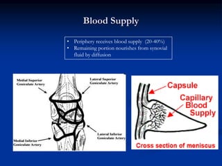
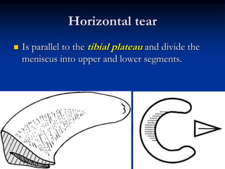
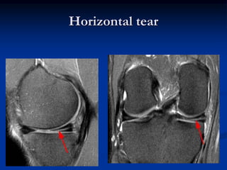
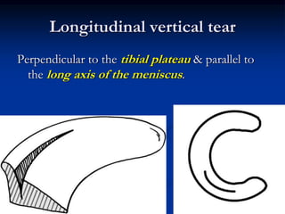
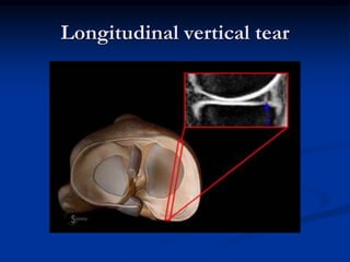
2. Anatomy of the meniscus
Fibrocartilage C-shaped disc
Mesenchymal tissue - appear
in 8 -10 weeks of gestation
Highly cellular and vascular
initially
Type-1 collagen with 75% of
water
Fibers orientation
3. Posterior horn thicker & wider than the
anterior horn in medial meniscus
Lateral meniscus more mobile
Not attached with Tibia at Popliteal hiatus
Insertional Ligaments
1-Transverse Meniscal Ligament
Connects both menisci anteriorly
2- Coronary (meniscotibial) ligaments
Connect medial meniscus to Tibia anterolaterally
3-Meniscofemoral ligament of Wrisberg (70%)
Passes from posterior horn of lateral meniscus to medial femoral
condyle posterior to PCL
4-Meniscofemoral ligament of Humphrey
Passes from posterior horn of lateral meniscus to medial
femoral condyle anterior to PCL
Lateral Medial
4.
5. Biomechanical Functions
Load distribution
Shock absorption
Joint stability
Joint lubrication
Nutrition of articular cartilage
Helps knee locking in extension
Reduce friction between Tibia and Fibula
Chock block
6. Biomechanics
Meniscectomy Meniscus intact
• Limited contact
area
• High contact
stress
6/29/2013 6
Load
Distribution
• More contact
area
• Low contact
stress
7. BIOMECHANICS OF MENISCUS
The contact force of the menisci
on the femur helps guide the femur
anteriorly during flexion
The reaction force of the femur
on the menisci deforms the
menisci posteriorly on the tibial
plateau.
Continuity of peripheral meniscal
rim is very important for load
bearing
Partial meniscectomy still preserve
this function
A radial tear or total menisectomy
massively increase contact stress
and cause OA changes
Femoral
Condyle
Movement
9. Blood Supply
• Periphery receives blood supply (20-40%)
• Remaining portion nourishes from synovial
fluid by diffusion
10. Mechanism of injury
Body rotates with foot
on ground and knee
partially flexed
Repetitive squatting
cause medial meniscal
injury
Trivial injury required
in arthritic knee
Pivoting sports i.e.
soccer, rugby, net ball,
basket ball
12. Unhappy Triad
Also called
Terrible triad
O’Donoghue’s triad
Blown knee
Knee banged on lateral side in semi flexion with foot stable on ground
Injury to
1. Medical collateral ligament
2. ACL
3. Lateral/medial Meniscus
13. Signs & symptoms
Not all meniscal tears are symptomatic
Swelling
Pain and tenderness along joint line - medial or lateral
Pain worse on squatting, kneeling or pivoting
Locking of the knee
Giving way, snaps, clicks, clunks, catches in knee.
Atrophy of quadriceps
Instability of joint
Elastic block at terminal extension
Springy end feel
15. Prone position
Knee flexed to 90 degree and
thigh fixed to the examination
table
compression and rotating
tibial plateau on femoral
condyles
Joint line pain on rotation
16. Thessaly Test
Hold patient’s outstretched hands for support
Ask the patient to stand on normal leg first and ask
to rotate body with knee flexed to 20 degree
Now, ask the patient to stand on affected leg
bend knee to 20 degree and rotate body 3 times
internally and externally
Test Positive if symptoms appear
17. Patient sits with the leg
flexed over the table about
90 degree.
Rotate tibia internally and
externally
Joint line pain confirms test
positive
18. Imaging
Plain Radiographs
Not helpful in meniscal injury
Rule out other bony or joint pathology
Arthrography
Invasive
Accuracy rate 60% - 90%
Largely replaced by MRI
Tibial tunnel enlargement – ACL injury
19. MRI
Non invasive, no ionising radiation
Differentiate between repairable and
non-repairable tears
Average sensitivity:
95% medial and 81% in lateral
Average specificity
88% medial and 95% lateral
Meniscus looks dark due low signal
(high water content).
Picks associated injuries (ACL)
Surgery can be planed ahead
20. Types of meniscal tears according to plane of
cleavage
Meniscal
tears
Vertical Horizontal
Longitudinal Radial
21. Horizontal tear
Is parallel to the tibial plateau and divide the
meniscus into upper and lower segments.
40. Non surgical management
Incomplete tear
Small (5mm) peripheral stable tear
Tears associated with ligamentous injuries (where reconstruction
deferred or contraindicated)
•R-rest
I-ice
C-compression
E-elevation
•NSAIDS
•Physio
•Immobilisation
41. Surgical Management
1. Meniscectomy
By arthrotomy
By arthroscopy
2. Meniscal repair
By arthrotomy
By arthroscopy
3. Meniscal transplantation
Autografts
Allograft
Prosthetic scaffolds
42. Meniscal repair
Arnoczky and Warren -
peripheral zone is repairable
Canine model - fibrin clot
formation in red-red zone
-- scar formation in 10 weeks
Superficial zone cells –
progenitor cells
Factors affecting repair
Location of tear
Location of tear
ACL reconstruction
Age of tear
Age of patient
44. All inside repair (arthroscopic)
Arrows
Darts
Cinch
• Bioabsorbable
material
• Very hard
• Can break
• Can migrate
• Device rubbing can
cause cartilage defect
Second generation delivery system
2 plastic anchors connected with sutures passed by
needle deliver system
Knots tightened outside
Editor's Notes
LOAD DISTRIBUTION
40% load distribution in extension and 80% in knee flexion
Contact area reduced by 75% after menisectomy resulting in increase in peak contact pressure up to 250%
Medial compartment more congruent and less contact stress after menisectomy
SHOCK ABSORPTION
Axial loading extrudes meniscus peripherally. As menisci are attached around and can not move further, these axial loading forces are absorbed as “hoop stresses” (circumferential or wall tension).
Water content is squeezed out of tissue and therefore further engergy is absorbed.
Menisectomy decreases shock absorption by 20%
STABILITY
Menisci work as chock block in ACL deficient knee, inhibit anterior translation (secondary stabiliser)
70% decrease in contact area after meniscectomy
Medial tibial condyl more congruent due to concavity. Less contact stress after menisectomy. More OA after lateral menisectomy
Menisci move few mm forward during knee flexion (lateral move more than medial) and role backwards during knee in extension
Menisci stop anterior and posterior glide of femur on tibia
Take 2 apples, place an apple on table top, it will move freely. Now, cut other apple into 8 pieces and put 2 pieces on table top and put apple on these. The apple will not move. Consider table to –tibial surface and apple femoral condyl.
Apply varus or valgus stress while internally or externall y rotating the knee
Thick body in normal meniscus cause boe-tie sign
In bucket handle body is thin
ARTHROTOMY only done if tear associated with ligamentous injury and osteochondral fracture
OPEN- original technique, suturing under vision, anterior knee excess easy but posterior difficult, injury com peroneal nv
INSIDE OUT: Passing suture arthoscopically , 2 sutures ends tied over outer aspect of knee with small incisions
ALL INSIDE: Arthroscopic repair (1) Arrows (2) Darts (3) Repair devices