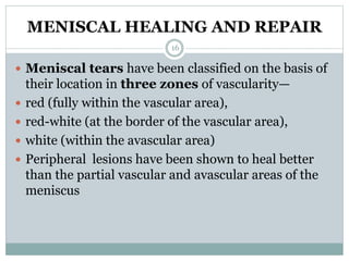
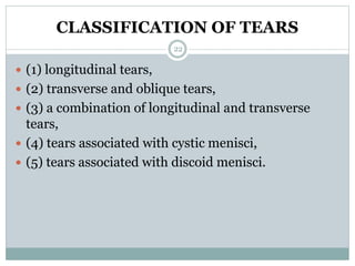
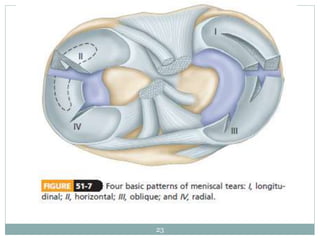
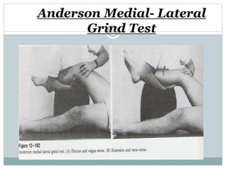
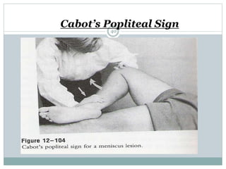
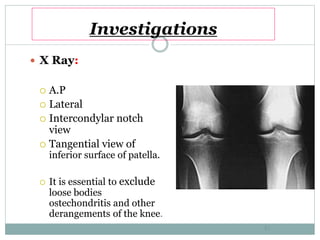
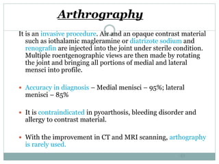
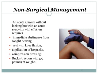
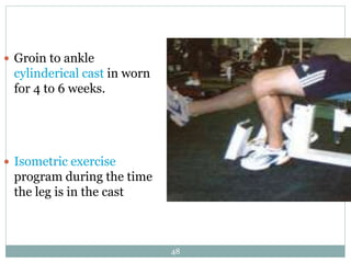
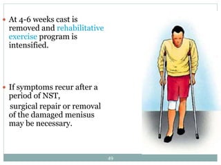
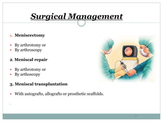
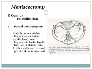
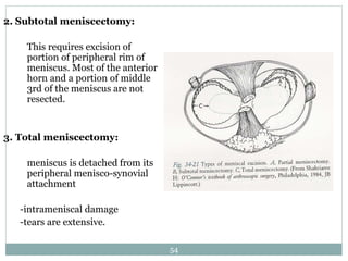
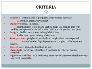
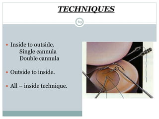
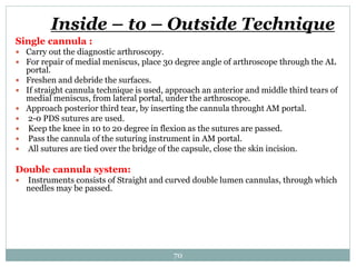
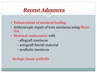
The document discusses meniscal injuries and pathology. It provides information on the anatomy and function of the menisci, as well as types of meniscal tears. The diagnosis of meniscal tears involves taking a history of the injury and examining for symptoms like joint line tenderness, effusion, and a locking sensation. Investigations may include x-rays, MRI, arthrography and arthroscopy. Treatment options discussed include non-surgical management for minor tears and surgical repair or resection for larger tears.
The presentation investigates the following characteristics of the meniscus;
Role of the Meniscus
Material Properties
Structural Limitations / Failure Limits
Mechanism & Treatment of Injuries
Acromioclavicular (AC) joint injury is a term used to describe an injury to the top of the shoulder, where the front of the shoulder blade (acromion) attaches to the collarbone (clavicle).
MCL. LCL.ALL injuries
To understand the relevant anatomy of the side ligaments of the knee
To study the mechanism of injury of each ligament and how to diagnose such injury
To highlight the different treatment options in acute or chronic situations
step by step presentation on ultrasound evaluation of shoulder and knee joints with illustrations of probe positioning.multiple examples of pathologies also added.
These simplified slides by Dr. Sidra Arshad present an overview of the non-respiratory functions of the respiratory tract.
Learning objectives:
1. Enlist the non-respiratory functions of the respiratory tract
2. Briefly explain how these functions are carried out
3. Discuss the significance of dead space
4. Differentiate between minute ventilation and alveolar ventilation
5. Describe the cough and sneeze reflexes
Study Resources:
1. Chapter 39, Guyton and Hall Textbook of Medical Physiology, 14th edition
2. Chapter 34, Ganong’s Review of Medical Physiology, 26th edition
3. Chapter 17, Human Physiology by Lauralee Sherwood, 9th edition
4. Non-respiratory functions of the lungs https://academic.oup.com/bjaed/article/13/3/98/278874
Explore natural remedies for syphilis treatment in Singapore. Discover alternative therapies, herbal remedies, and lifestyle changes that may complement conventional treatments. Learn about holistic approaches to managing syphilis symptoms and supporting overall health.
New Drug Discovery and Development .....NEHA GUPTA
The "New Drug Discovery and Development" process involves the identification, design, testing, and manufacturing of novel pharmaceutical compounds with the aim of introducing new and improved treatments for various medical conditions. This comprehensive endeavor encompasses various stages, including target identification, preclinical studies, clinical trials, regulatory approval, and post-market surveillance. It involves multidisciplinary collaboration among scientists, researchers, clinicians, regulatory experts, and pharmaceutical companies to bring innovative therapies to market and address unmet medical needs.
Tom Selleck Health: A Comprehensive Look at the Iconic Actor’s Wellness Journeygreendigital
Tom Selleck, an enduring figure in Hollywood. has captivated audiences for decades with his rugged charm, iconic moustache. and memorable roles in television and film. From his breakout role as Thomas Magnum in Magnum P.I. to his current portrayal of Frank Reagan in Blue Bloods. Selleck's career has spanned over 50 years. But beyond his professional achievements. fans have often been curious about Tom Selleck Health. especially as he has aged in the public eye.
Follow us on: Pinterest
Introduction
Many have been interested in Tom Selleck health. not only because of his enduring presence on screen but also because of the challenges. and lifestyle choices he has faced and made over the years. This article delves into the various aspects of Tom Selleck health. exploring his fitness regimen, diet, mental health. and the challenges he has encountered as he ages. We'll look at how he maintains his well-being. the health issues he has faced, and his approach to ageing .
Early Life and Career
Childhood and Athletic Beginnings
Tom Selleck was born on January 29, 1945, in Detroit, Michigan, and grew up in Sherman Oaks, California. From an early age, he was involved in sports, particularly basketball. which played a significant role in his physical development. His athletic pursuits continued into college. where he attended the University of Southern California (USC) on a basketball scholarship. This early involvement in sports laid a strong foundation for his physical health and disciplined lifestyle.
Transition to Acting
Selleck's transition from an athlete to an actor came with its physical demands. His first significant role in "Magnum P.I." required him to perform various stunts and maintain a fit appearance. This role, which he played from 1980 to 1988. necessitated a rigorous fitness routine to meet the show's demands. setting the stage for his long-term commitment to health and wellness.
Fitness Regimen
Workout Routine
Tom Selleck health and fitness regimen has evolved. adapting to his changing roles and age. During his "Magnum, P.I." days. Selleck's workouts were intense and focused on building and maintaining muscle mass. His routine included weightlifting, cardiovascular exercises. and specific training for the stunts he performed on the show.
Selleck adjusted his fitness routine as he aged to suit his body's needs. Today, his workouts focus on maintaining flexibility, strength, and cardiovascular health. He incorporates low-impact exercises such as swimming, walking, and light weightlifting. This balanced approach helps him stay fit without putting undue strain on his joints and muscles.
Importance of Flexibility and Mobility
In recent years, Selleck has emphasized the importance of flexibility and mobility in his fitness regimen. Understanding the natural decline in muscle mass and joint flexibility with age. he includes stretching and yoga in his routine. These practices help prevent injuries, improve posture, and maintain mobilit
263778731218 Abortion Clinic /Pills In Harare ,sisternakatoto
263778731218 Abortion Clinic /Pills In Harare ,ABORTION WOMEN’S CLINIC +27730423979 IN women clinic we believe that every woman should be able to make choices in her pregnancy. Our job is to provide compassionate care, safety,affordable and confidential services. That’s why we have won the trust from all generations of women all over the world. we use non surgical method(Abortion pills) to terminate…Dr.LISA +27730423979women Clinic is committed to providing the highest quality of obstetrical and gynecological care to women of all ages. Our dedicated staff aim to treat each patient and her health concerns with compassion and respect.Our dedicated group ABORTION WOMEN’S CLINIC +27730423979 IN women clinic we believe that every woman should be able to make choices in her pregnancy. Our job is to provide compassionate care, safety,affordable and confidential services. That’s why we have won the trust from all generations of women all over the world. we use non surgical method(Abortion pills) to terminate…Dr.LISA +27730423979women Clinic is committed to providing the highest quality of obstetrical and gynecological care to women of all ages. Our dedicated staff aim to treat each patient and her health concerns with compassion and respect.Our dedicated group of receptionists, nurses, and physicians have worked together as a teamof receptionists, nurses, and physicians have worked together as a team wwww.lisywomensclinic.co.za/
Prix Galien International 2024 Forum ProgramLevi Shapiro
June 20, 2024, Prix Galien International and Jerusalem Ethics Forum in ROME. Detailed agenda including panels:
- ADVANCES IN CARDIOLOGY: A NEW PARADIGM IS COMING
- WOMEN’S HEALTH: FERTILITY PRESERVATION
- WHAT’S NEW IN THE TREATMENT OF INFECTIOUS,
ONCOLOGICAL AND INFLAMMATORY SKIN DISEASES?
- ARTIFICIAL INTELLIGENCE AND ETHICS
- GENE THERAPY
- BEYOND BORDERS: GLOBAL INITIATIVES FOR DEMOCRATIZING LIFE SCIENCE TECHNOLOGIES AND PROMOTING ACCESS TO HEALTHCARE
- ETHICAL CHALLENGES IN LIFE SCIENCES
- Prix Galien International Awards Ceremony
NVBDCP.pptx Nation vector borne disease control programSapna Thakur
NVBDCP was launched in 2003-2004 . Vector-Borne Disease: Disease that results from an infection transmitted to humans and other animals by blood-feeding arthropods, such as mosquitoes, ticks, and fleas. Examples of vector-borne diseases include Dengue fever, West Nile Virus, Lyme disease, and malaria.
New Directions in Targeted Therapeutic Approaches for Older Adults With Mantl...i3 Health
i3 Health is pleased to make the speaker slides from this activity available for use as a non-accredited self-study or teaching resource.
This slide deck presented by Dr. Kami Maddocks, Professor-Clinical in the Division of Hematology and
Associate Division Director for Ambulatory Operations
The Ohio State University Comprehensive Cancer Center, will provide insight into new directions in targeted therapeutic approaches for older adults with mantle cell lymphoma.
STATEMENT OF NEED
Mantle cell lymphoma (MCL) is a rare, aggressive B-cell non-Hodgkin lymphoma (NHL) accounting for 5% to 7% of all lymphomas. Its prognosis ranges from indolent disease that does not require treatment for years to very aggressive disease, which is associated with poor survival (Silkenstedt et al, 2021). Typically, MCL is diagnosed at advanced stage and in older patients who cannot tolerate intensive therapy (NCCN, 2022). Although recent advances have slightly increased remission rates, recurrence and relapse remain very common, leading to a median overall survival between 3 and 6 years (LLS, 2021). Though there are several effective options, progress is still needed towards establishing an accepted frontline approach for MCL (Castellino et al, 2022). Treatment selection and management of MCL are complicated by the heterogeneity of prognosis, advanced age and comorbidities of patients, and lack of an established standard approach for treatment, making it vital that clinicians be familiar with the latest research and advances in this area. In this activity chaired by Michael Wang, MD, Professor in the Department of Lymphoma & Myeloma at MD Anderson Cancer Center, expert faculty will discuss prognostic factors informing treatment, the promising results of recent trials in new therapeutic approaches, and the implications of treatment resistance in therapeutic selection for MCL.
Target Audience
Hematology/oncology fellows, attending faculty, and other health care professionals involved in the treatment of patients with mantle cell lymphoma (MCL).
Learning Objectives
1.) Identify clinical and biological prognostic factors that can guide treatment decision making for older adults with MCL
2.) Evaluate emerging data on targeted therapeutic approaches for treatment-naive and relapsed/refractory MCL and their applicability to older adults
3.) Assess mechanisms of resistance to targeted therapies for MCL and their implications for treatment selection
Title: Sense of Taste
Presenter: Dr. Faiza, Assistant Professor of Physiology
Qualifications:
MBBS (Best Graduate, AIMC Lahore)
FCPS Physiology
ICMT, CHPE, DHPE (STMU)
MPH (GC University, Faisalabad)
MBA (Virtual University of Pakistan)
Learning Objectives:
Describe the structure and function of taste buds.
Describe the relationship between the taste threshold and taste index of common substances.
Explain the chemical basis and signal transduction of taste perception for each type of primary taste sensation.
Recognize different abnormalities of taste perception and their causes.
Key Topics:
Significance of Taste Sensation:
Differentiation between pleasant and harmful food
Influence on behavior
Selection of food based on metabolic needs
Receptors of Taste:
Taste buds on the tongue
Influence of sense of smell, texture of food, and pain stimulation (e.g., by pepper)
Primary and Secondary Taste Sensations:
Primary taste sensations: Sweet, Sour, Salty, Bitter, Umami
Chemical basis and signal transduction mechanisms for each taste
Taste Threshold and Index:
Taste threshold values for Sweet (sucrose), Salty (NaCl), Sour (HCl), and Bitter (Quinine)
Taste index relationship: Inversely proportional to taste threshold
Taste Blindness:
Inability to taste certain substances, particularly thiourea compounds
Example: Phenylthiocarbamide
Structure and Function of Taste Buds:
Composition: Epithelial cells, Sustentacular/Supporting cells, Taste cells, Basal cells
Features: Taste pores, Taste hairs/microvilli, and Taste nerve fibers
Location of Taste Buds:
Found in papillae of the tongue (Fungiform, Circumvallate, Foliate)
Also present on the palate, tonsillar pillars, epiglottis, and proximal esophagus
Mechanism of Taste Stimulation:
Interaction of taste substances with receptors on microvilli
Signal transduction pathways for Umami, Sweet, Bitter, Sour, and Salty tastes
Taste Sensitivity and Adaptation:
Decrease in sensitivity with age
Rapid adaptation of taste sensation
Role of Saliva in Taste:
Dissolution of tastants to reach receptors
Washing away the stimulus
Taste Preferences and Aversions:
Mechanisms behind taste preference and aversion
Influence of receptors and neural pathways
Impact of Sensory Nerve Damage:
Degeneration of taste buds if the sensory nerve fiber is cut
Abnormalities of Taste Detection:
Conditions: Ageusia, Hypogeusia, Dysgeusia (parageusia)
Causes: Nerve damage, neurological disorders, infections, poor oral hygiene, adverse drug effects, deficiencies, aging, tobacco use, altered neurotransmitter levels
Neurotransmitters and Taste Threshold:
Effects of serotonin (5-HT) and norepinephrine (NE) on taste sensitivity
Supertasters:
25% of the population with heightened sensitivity to taste, especially bitterness
Increased number of fungiform papillae
Evaluation of antidepressant activity of clitoris ternatea in animals
meniscal injuries
1. D R . A N U B H A V V E R M A
M O D E R A T O R : D R . R A V I K I R A N H G
1 S T M A R C H 2 0 1 6
D E P A R T M E N T O F O R T H O P E D I C S
J S S H O S P I T A L
M Y S O R E
MENISCAL INJURIES AND
PATHOLOGY
1
2. OUTLINE
FUNCTION AND ANATOMY
MENISCAL HEALING AND REPAIR
TEARS OF MENISCI
DIAGNOSIS
INVESTIGATIONS
TREATMENT
2
3. FUNCTION OF MENISCI
JOINT FILLER: compensating for gross incongruity
between femoral and tibial articulating surfaces.
Prevent capsular and synovial impingement
JOINT LUBRICATION: distribute synovial fluid
throughout the joint
STABILITY: flexion to extension, pure hinge to a
gliding/rotary motion
SHOCK ABSORBER: 40 – 60 % of body weight in
standing position
3
4. JOINT FILLER
Following meniscectomy: Flattening of femoral condyl
and formation of osteophytes
Contact area inversely proportional to contact stress
Decreased contact area (Approx 40%), increased contact
stress (100% medial meniscus. 200% lateral meniscus
because of relative convex surface of lateral tibial
plateau)
4
5. STABILITY
Increased joint laxity following meniscectomy.
Insignificant if ligamentous structures intact
ACL deficient knee: increased tibial translation by 58%
after medial meniscectomy (c.f. lateral meniscectomy –
not affixed firmly and does not act as efficient posterior
wedge to prevent translation)
May account for different patterns of meniscal injuries in
ACL deficient knee
5
7. MEDIAL MENISCUS
C shaped, larger in radius than lateral meniscus
Anterior horn: Attached anterior to intercondylar
eminence and to the ACL
Posterior horn: Attached in front of attachment of PCL,
posterior to the intercondylar eminence
Entire peripheral border firmly attached to the medial
capsule and through coronary ligament to the upper
border of tibia
7
8. LATERAL MENISCUS
Smaller, More circular, thicker in periphery, wider in
body and more mobile than medial meniscus
ANTERIOR HORN: attached medially in front of the
intercondylar eminence
POSTERIOR HORN: inserts into the posterior
aspect of the intercondylar eminence and in front of
posterior attachment of medial meniscus.
Attached to both cruciate ligaments and posteriorly
to the medial femoral condyle by either the ligament
of Humphry or the ligament of wrisberg
8
10. LATERAL MENISCUS
Smaller in diameter
Circular
Thicker in periphery
Wider Body
More Mobile
Attached to both ACL/PCL
MEDIAL MENISCUS
Larger in diameter
C shaped
Thinner in periphery
Thinner body
Less mobile
Not attached to ACL/PCL
10
16. MENISCAL HEALING AND REPAIR
Meniscal tears have been classified on the basis of
their location in three zones of vascularity—
red (fully within the vascular area),
red-white (at the border of the vascular area),
white (within the avascular area)
Peripheral lesions have been shown to heal better
than the partial vascular and avascular areas of the
meniscus
16
17. for a meniscus to regenerate, the entire structure
must be resected to expose the vascular synovial
tissue
In subtotal meniscectomy, the excision must
extend to the peripheral vasculature of the meniscus.
Subtotal excisions of the meniscus within the
avascular central half of the meniscus do not show
any regeneration potential.
17
18. MENISCAL TEARS
Mechanism: The menisci follow the tibial condyles
during flexion and extension, but during rotation
they follow the femur and move on the tibia;
consequently, the medial meniscus becomes
distorted.
Its anterior and posterior attachments follow the
tibia, but its intervening part follows the femur; thus
it is likely to be injured during rotation.
18
19. WHY L.M. IS SPARED?
However, the lateral meniscus, because it is firmly
attached to the popliteus muscle and to the ligament
of Wrisberg or of Humphry, follows the lateral
femoral condyle during rotation and therefore is less
likely to be injured.
19
20. During vigorous internal rotation of the femur on the
tibia with the knee in flexion, the femur tends to
force the medial meniscus posteriorly and toward the
center of the joint
The posterior part of the meniscus is forced toward
the center of the joint, is caught between the femur
and the tibia, and is torn longitudinally when the
joint is suddenly extended.
Most common location: posterior horn of
meniscus
Most common type: longitudinal
20
26. DIAGNOSIS
HISTORY: middle aged person who sustains a
weight-bearing twist on the knee or who has pain
after squatting.
The syndromes caused by tears of the menisci can be
divided into two groups:
1. With Locking
2. Without Locking
26
27. THE LOCKING KNEE
Inability to completely extend the knee joint
occurs only with longitudinal tears and is much more
common with bucket-handle tears
Intra articular tumor, an osteocartilaginous loose body,
and other conditions can also cause locking.
False locking :hemorrhage around the posterior part of
the capsule or a collateral ligament with associated
hamstring spasm prevents complete extension of the
knee.
locking may not be recognized unless the injured knee is
compared with the opposite knee, which should exhibit
the 5 to 10 degrees of recurvatum that normally is
present.
27
28. NON LOCKING KNEE
typically gives a history of several episodes of trouble
referable to the knee, often resulting in effusion and
a brief period of disability but no definite locking.
A sensation of “giving way” or snaps, clicks, catches,
or jerks in the knee may be described
IMPORTANT CLUES IN AN INJURED NON
LOCKING KNEE: a sensation of giving way,
effusion, atrophy of the quadriceps, tenderness over
the joint line (or the meniscus), and reproduction of
a click by manipulative maneuvers during the
physical examination.
28
29. GIVING AWAY
a tear in the posterior part of a meniscus, the patient
usually notices this on rotary movements of the knee
and often associates it with a feeling of subluxation
or “the joint jumpin out of place.”
When giving way is a result of other causes, such as
quadriceps weakness, it usually is noticeable
during simple flexion of the knee against resistance,
such as in walking down stairs.
29
30. OTHER FEATURES
EFFUSION: occcurs when vascularised peripheral area
of a meniscus is torn. Mostly it is a hemarthrosis.
MUSCLE ATROPHY: especially of the vastus medialis
JOINT LINE TENDERNESS: most important
physical finding. Localised over the medial or lateral
joint line or over the periphery of the meniscus. The
meniscus itself is without nerve fibers except at its
periphery; therefore, the tenderness or pain is related to
synovitis in the adjacent capsular and synovial tissues.
30
31. THE MC MURRAY TEST
Medial Meniscus: Keeping the knee completely
flexed, the leg is externally rotated as far as possible
and then the knee is slowly extended. As the femur
passes over a tear in the meniscus, a click may be
heard or felt.
Lateral Mensicus: palpating the posterolateral
margin of the joint, internally rotating the leg as far
as possible, and slowly extending the knee while
listening and feeling for a click.
31
33. CLICK: caused by a posterior peripheral tear of the
meniscus and occurs between complete flexion of the
knee and 90 degrees
POPPING: occurs with greater degrees of extension
when it is definitely localized to the joint suggests a
tear of the middle and anterior portions of the
meniscus
The position of the knee when the click occurs thus
may help locate the lesion.
33
34. APLEY’S GRINDING TEST
With the patient prone, the knee is flexed to 90
degrees and the anterior thigh is fixed against the
examining table. The foot and leg are then pulled
upward to distract the joint.
with the knee in the same position, the foot and leg
are pressed downward and rotated as the joint is
slowly flexed and extended
when a meniscus has been torn, popping and pain
localized to the joint line may be noted.
34
41. Investigations
X Ray:
A.P
Lateral
Intercondylar notch
view
Tangential view of
inferior surface of patella.
It is essential to exclude
loose bodies
ostechondritis and other
derangements of the knee.
41
42. Arthrography
It is an invasive procedure. Air and an opaque contrast material
such as iothalamic magleramine or diatrizote sodium and
renografin are injected into the joint under sterile condition.
Multiple roentgenographic views are then made by rotating
the joint and bringing all portions of medial and lateral
mensci into profile.
Accuracy in diagnosis – Medial menisci – 95%; lateral
menisci – 85%
It is contraindicated in pyoarthosis, bleeding disorder and
allergy to contrast material.
With the improvement in CT and MRI scanning, arthography
is rarely used.
42
43. Arthroscopy
• It has an accuracy of 98% for
medial meniscus & 90% for
lateral meniscus.
43
44. MRI
Grading:
Grade I Tear of the menisus has
increased signal in the meniscal substance.
Grade II Involves a more pronounced
and frequently linear signal that does not
break the surface of the menisus.
Grade III Signal that traverses through
the meniscal surface.
Grade IV There is extension of tear
through both tibial and femoral surfaces of
the menisus.
Grade I and II changes appear
normal on arthoscopic evaluation.
44
46. Non-Surgical Management
Indications:
Partial thickness splits.
full thickness oblique or vertical tears less than
5mm, if stable
Short radial tears.
Degenerative tears in OA, without mechanical
symptoms.
Stable tears with inability to displace the central
portion, by greater than 3mm.
Contra indications:
Chronic tears with superimposed acute injury.
In a locked knee caused by bucket handle tear of
meniscus.
46
47. Non-Surgical Management
An acute episode without
locking but with an acute
synovitis with effusion
requires
immediate abstinence from
weight bearing,
rest with knee flexion,
application of ice packs,
compression dressing,
Buck’s traction with 5-7
pounds of weight.
47
48. Groin to ankle
cylinderical cast in worn
for 4 to 6 weeks.
Isometric exercise
program during the time
the leg is in the cast
48
49. At 4-6 weeks cast is
removed and rehabilitative
exercise program is
intensified.
If symptoms recur after a
period of NST,
surgical repair or removal
of the damaged menisus
may be necessary.
49
50. Surgical Management
1. Meniscectomy
By arthrotomy or
By arthroscopy
2. Meniscal repair
By arthrotomy or
By arthoscopy
3. Meniscal transplantation
With autografts, allografts or prosthetic scaffolds.
.
50
51. General Principles
Partial meniscectomy is always preferable to subtotal or total
meniscectomy.
The objective is to remove the torn, mobile meniscal fragment and
contour the peripheral rim, leaving a balanced, stable rim of meniscal
tissue.
Pneumatic tourniquet to be used to avoid constant sponging which
prolongs and damages the joint surfaces. Before wound closure
tourniquet to be released and bleeding vessels are ligated or
electrocauterized.
The knee should be examined carefully for stability after the patient is
anesthetized.
The anterior compartment of knee should be explored first, then the
posteomedial and lastly the lateral compartment should be explored.
The condition of the synovial membrane, articular surfaces, medial and
lateral menisci and ligaments should be noted.
51
52. Objective of the Treatment
to remove the torn
mobile meniscal
fragment
contour the peripheral
rim leaving a balance
stable rim of meniscal
tissue.
No standard technique
can be used in every
case.
52
53. Meniscectomy
O Connor
classification
1. Partial meniscectomy:
Only the loose unstable
fragments are excised;
e.g: displaced inner
fragments in bucket handle
tear, flap in oblique tears.
In this a stable and balanced
peripheral rim is preserved.
53
54. 2. Subtotal meniscectomy:
This requires excision of
portion of peripheral rim of
meniscus. Most of the anterior
horn and a portion of middle
3rd of the meniscus are not
resected.
3. Total meniscectomy:
meniscus is detached from its
peripheral menisco-synovial
attachment
-intrameniscal damage
-tears are extensive.
54
55. Partial Total Meniscectomy ?
Deciding factors
Location of tear
Length
Pattern
Stability
Condition of whole meniscus
55
56. Advantages of Partial Over Total
Shorter operating time
Faster recovery
Better post operative function
Better self assessment of outcome
56
57. POST MENISCECTOMY REHAB
PROTOCOL
A compression bandage is
applied to the knee.
Knee is immobilized for 5-7
days. Then it is discontinued.
Ice is applied over the knee
and limb is elevated for 24-
48 hours postoperatively.
57
58. Quadriceps exercises are started
on 2nd day onwards, SLR
isometric quadriceps exercises
are carried out on every hour
when the patient is awake.
When the good muscular control
is achieved patient is allowed to
walk with crutches and with
partial weight bearing.
The sutures are removed at 2
weeks and gentle resistive
exercises are begun.
58
59. Open Excision of Medial Meniscus
Using anteromedial incision
Begin the incision just medial to patella, 5 cm distally
parallel with patella and patellar tendon. Incise the
fascia and capsule 0.5 cm medial to the edge of patellar
tendon .
Grasp the synovium, make a small opening through it
into the joint. Mobilize the anterior third segment of
meniscus.
Grasp the anterior segment with martin clamp.
Free the middle third of the meniscus at its
periphery.
Mobilize the posterior third of meniscus.
Displace the meniscus into the intercondylar notch,
leave a stable balanced menisceal rim.
Close the incision, evert the cut edge of synovium.
Close fascia, extensor aponeurosis and capsule in one
layer.
59
60. Open Excision of Lateral
Meniscus
Anterolateral incision : begin the incision
at the level at the middle portion of the
patella extend it distally to the upper tibial
surface incise the anterolateral capsule
and synovium.
Free the anterior third of lateral meniscus,
and grasp it with martin grasper
Maintain traction on free anterior
segment.
Flex the knee, place the foot on opposite
knee and apply varus strain.
By continued gentle traction, posterior
third of meniscus is separated, and
complete lateral meniscus is excised. Close
the capsule with intermediate sutures.
60
61. Arthroscopic Meniscectomy
Longitudinal tears:
30 degree oblique viewing arthroscope is inserted through an AL
portal.
Probe is placed through the AM portal.
Horizontal tear:
30 degree oblique viewing arthroscope is used through AL portal.
Superior and inferior leaves of the tear is removed with basket
forceps. Peripheral rim is trimmed and contoured.
Oblique tears:
Three portal procedures is adopted. Small posteriorly based
oblique tears are usually removed by morcellation of the flaps
with basket forceps or motorized cutter, trimmer instruments.
Large posterior or oblique tears are removed intact enbloc.
Anterior oblique tears are removed by triangulation technique.
61
62. Three Portal Technique
It is used excision of large
complete intrameniscal
tears of posterior horn.
Arthoscope, grasping
instrument and cutting
instruments are used
through the three portals.
Arthroscope placed in AL
portal. Probe the posterior
limits of displaced bucket
handle through AM portal.
.
62
63. Through AM portal anterior
horn attachment of the
meniscus is released.
Grasping clamp is placed
through the AM portal to
grasp the anterior horn and
it is removed.
Now probe is used through
AM portal to check the
stability of the remaining
rim and look for any tears.
Basket forceps or motorized
shaver are introduced
through AM portal to
smoothen the remaining rim.
63
64. Complications after Meniscectomy
1. Post operative haemarthrosis.
2. Chronic synovitis.
3. Svnovial fistulae.
4. Painful neuromas of the branches of the
infrapatellar portion of saphenous nerve.
5. Thrombophlebitis – suggested by postoperative
pain and swelling in the calf and distal extremity
with low-grade fever.
6. Postoperative infection – increasing effusion, pain
and fever beginning 2 to 3 days after surgery
indicates the onset of pyarthrosis.
64
65. 7. Reflex sympathetic dystrophy.
8. Retained meniscal fragment.
9. Capsular and ligamentous laxity.
10. Late changes degenerative changes with
in the joint. Fairbank described three
changes.
a. Narrowing of joint space.
b. Flattening of peripheral half of the articular
surface of condyle.
c. Development of anteroposterior ridge that
projected distally from the margin of
femoral condyle.
65
67. Arthroscopic Meniscal Tear Repair
Consists of 3 important steps:
1. Appropriate patient selection – should have
documented tear that is able to heal.
2. Tear debridement and local synovial, meniscal and
capsular ablation to stimulate a proliferative
fibroblastic healing response.
3. Suture placement to reduce and stabilize the
meniscus.
67
68. CRITERIA
Location : within 3 mm of periphery are presumed vascular.
More than 5mm are avascular.
Stability : partial thickness.
Full thickness- oblique and vertical tears less than 10 mm with
inability to displace the central portion with a probe greater than 3mm.
Length : Stable tear <10mm in length left alone.
Radial tear <5mm in length left alone.
Tear pattern : peripheral , vertical and longitudinal tears repaired.
Bucket handle, flap, degenerative, complex, radial tears are
excised.
Patient age : should be less than 50 yrs.
Chronicity : Acute tears less than 8 weeks old have better healing
potential.
Ligament stability : ACL deficiency must also be corrected simultaneously
to prevent instability.
68
69. TECHNIQUES
Inside to outside.
Single cannula
Double cannula
Outside to inside.
All – inside technique.
69
70. Inside – to – Outside Technique
Single cannula :
Carry out the diagnostic arthroscopy.
For repair of medial meniscus, place 30 degree angle of arthroscope through the AL
portal.
Freshen and debride the surfaces.
If straight cannula technique is used, approach an anterior and middle third tears of
medial meniscus, from lateral portal, under the arthroscope.
Approach posterior third tear, by inserting the cannula throught AM portal.
2-0 PDS sutures are used.
Keep the knee in 10 to 20 degree in flexion as the sutures are passed.
Pass the cannula of the suturing instrument in AM portal.
All sutures are tied over the bridge of the capsule, close the skin incision.
Double cannula system:
Instruments consists of Straight and curved double lumen cannulas, through which
needles may be passed.
70
71. Outside – to – Inside Technique
In this method suture is introduced through the spinal
needle i.e. Inserted from outside to inside.
This technique is safe approach to posterior horn.
Technique is same for both menisci.
For large peripheral lesions of medial meniscus, such as
bucket handle tears, combination of inside to outside
and outside to inside methods can be used.
71
72. All Inside Technique
Morgan described, this technique for repair of posterior horn.
Advantages:
It allows placement of vertical sutures.
Smaller incision can be used.
Disadvantages:
Need for special instrumentation.
Difficulty with intraarticular knot tieing.
All inside technique, can be performed by using commercial
available T – fix sutures.
72
73. Advantages of Open Technique
1. Vertically oriented sutures are easy to do by open arthrotomy.
It is more secure than more horizontally oriented suturing by
arthoscope techniques.
2. In repair of posterior horn peripheral tears by open arthotomy
technique, posteromedial or posterolateral capsular
reconstruction can be done concurrently.
3. Immobilization required is the same for both open and
arthroscopic technique.
73
74. 1. Small incisions .
2. Short stay.
3. Early mobilization.
4. All corners of the joint can be visualised.
5. Cosmeticaly very minimal scar.
6. Cost effective.
7. Patient is comfortable.
8. Less infection.
9. Less joint stiffness.
10. Morbidity is less.
Advantages of arthroscopic
technique
74
76. After Treatment
Knee is placed in a hinged brace and immediate
range of motion from 0-90 degrees is permitted.
Touchdown weight bearing is permitted
immediately, and
Full weight bearing is permitted at 6 weeks when
the brace and crutches are discarded.
No sports are allowed for 3 months.
If tear is large crutches are discarded at 8 weeks. No
sports are allowed for 6 months.
76
77. Exercises after Injury to the Meniscus
Are designed to build up the quadriceps and
hamstring muscles and increase flexibility and
strength:
Warming up the joint by riding a stationary bicycle,
then straightening and raising the leg (but avoiding
straightening the leg too much).
Extending the leg while sitting (a weight may be
worn on the ankle for this exercise).
Raising the leg while lying on the stomach.
Exercising in a pool.
77
83. Recent Advances
Enhancement of meniscal healing.
Arthroscopic repair of torn meniscus using fibrin
clot.
Meniscal replacement with
- allograft meniscus
- autograft fascial material
- synthetic meniscus
Biologic tissue scaffolds
83
84. Enhancement of Meniscal Healing
Vascular access channels:
creating access of peripheral vessels to avascular region, by a
channel (trephination) allows avascular portion of the
meniscus to heal throught the proliferation of the fibrous
scar.
Synovial abrasion :
encourages vascular extension to avascular regions via.,
formation of vascular synovial pannus.
Exogenous fibrin clot :
a clot precipitated on a sterile glass surface, and placed
within the defect within the vascular zone can promote
healing.
84
85. Arthroscopic Repair of Torn Meniscus
using Fibrin Clot
The fibrin clot appears to act as a chemotoctic and mitogenic stimulus
for reparative cells and provide scaffolding for reparative process.
Arnocky and Warren reported the injection of exogenous fibrin clot
obtained form the patients coagulated blood as promoting improving
meniscal healing. Exogenous fibrin clot is injected with a blunt needle in
the stem of the tear. 1 to 2ml of clot was sufficient to fill an average defect.
When gaps are big, a facial sheath was used to cover these defects and
the exogenous fibrin clot was injected under the cover of sheath i.e., for
complex tears.
Repairs of tears less than 2 months from the time of injury to surgery
result higher healing rates than those of more chronic tears.
Isolated repairs heal significantly better with exogenous fibrin clot
injection.
85
86. Meniscal Replacement
Attempts at meniscal replacement with allograft
menisci, autograft fascial material and synthetic
menisci scaffold are in various stage of study.
Investigation studies of biological tissue scaffolds are
in progress. These grafts may provide a more
acceptable meniscal replacement in the future.
As technology and the biomechanics and physiology
of menisci tissue are better understood. These
techniques may become more popular.
86
87. Allograft Meniscal Transplantation
Aim:
To prevent degenerative changes, in the post meniscectomy
patient.
Indications:
Patient less than 45 yrs age, with pain and discomfort associated
with early OA, without ACL deficiency or significant
malalignment.
Contraindications:
Age more than 60 yrs.
Bony architectural changes.
Prior infection.
Significant malalignment.
Instability.
87
88. Graft Preservation Technique:
Fresh freezing.
Cryo preservation.
Freeze dried.
Secondary sterilization with radiation less than 2.5 M Rad.
Steps:
Graft preparation.
Tunnel placement.
Graft insertion.
Graft fixation.
After Treatment:
Limb placed in long leg hinged knee brace.
Range of movement from 0 to 90 degree begin immediatiely.
Partial weight bearing with brace for first 6 weeks.
Brace removed at 6 weeks.
Full weight bearing started.
Deep flexion avoided for 6 months.
88
89. Bio- absorbable Implants
Poly glycolic acid
Poly levo lactic acid
Raecemic poly lactic acid
Poly dexanone.
All these materials degrade into co2 and
water.
Devices include Anchors,Arrows,
screws,staplers.
89
90. Experimental Studies
Angiogenin, a potent blood vessel inducing
protein- a 123 AA protein
Implantation into the experimentally injured
menisci in rats induces neo vascularisation of
meniscus
90
91. References
Campbell’s operative orthopaedics. Vol. 3, 11th
Edition.
Orthopaedics principles and their applications.
6th Edition Turek.
Mercer’s Orthopaedic Surgery, 10th Edition.
Rockwood and Green’s Fractures in Adults. Vol 2.
7th Edition.
Techniques in Therapeutic Arthroscopy by J.
Serge Parisien.
Athletic Injuries and Rehabilitation by James.
David and William .
JBJS.
Current Orthopedic Diagnosis and Treatment.
91