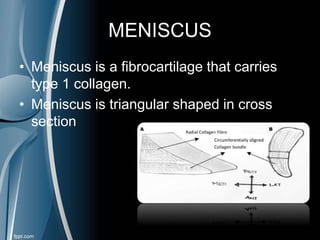
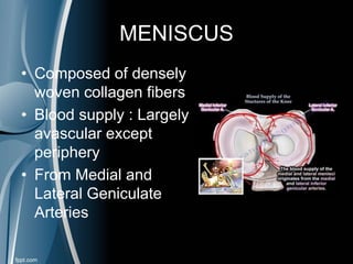
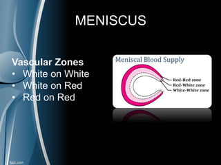
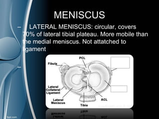
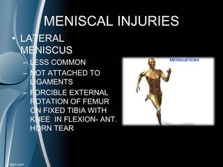
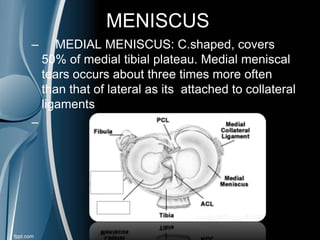
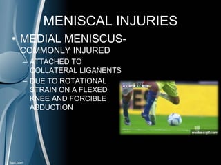
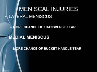
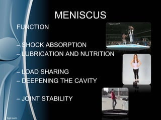
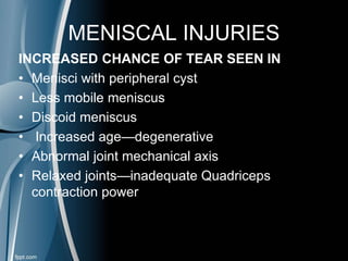
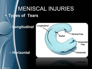
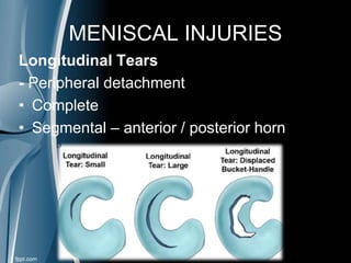
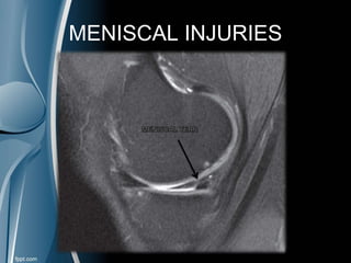
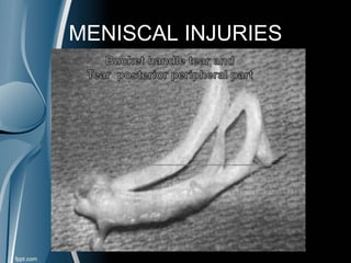
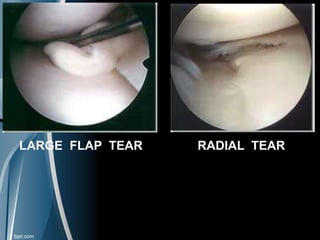
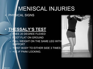
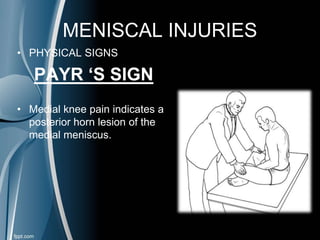
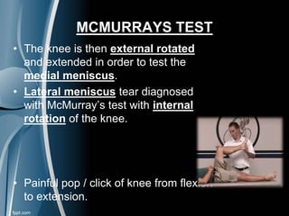
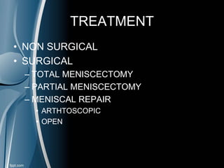
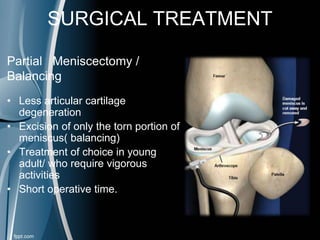
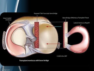
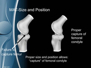
Meniscal injuries
MENISCUS
• Wedge shaped
• Fibro cartilaginous
• Triangular on CS
• Cover ½- 2/3 of surface
• Lateral – Shorter
• Small O shaped
• Medial- longer C shaped
MENISCUS
• Meniscus is a fibrocartilage that carries
type 1 collagen.
• Meniscus is triangular shaped in cross
section
MENISCUS
• Composed of densely
woven collagen fibers
• Blood supply : Largely
avascular except
periphery
• From Medial and
Lateral Geniculate
Arteries
MENISCUS
Vascular Zones
• White on White
• White on Red
• Red on Red
MENISCUS
– LATERAL MENISCUS: circular, covers
70% of lateral tibial plateau. More mobile than
the medial meniscus. Not attatched to
ligament
–
MENISCAL INJURIES
• LATERAL
MENISCUS
– LESS COMMON
– NOT ATTACHED TO
LIGAMENTS
– FORCIBLE EXTERNAL
ROTATION OF FEMUR
ON FIXED TIBIA WITH
KNEE IN FLEXION- ANT.
HORN TEAR
MENISCUS
– MEDIAL MENISCUS: C.shaped, covers
50% of medial tibial plateau. Medial meniscal
tears occurs about three times more often
than that of lateral as its attached to collateral
ligamentsniscus.
–
MENISCAL INJURIES
• MEDIAL MENISCUS-
COMMONLY INJURED
– ATTACHED TO
COLLATERAL LIGANENTS
– DUE TO ROTATIONAL
STRAIN ON A FLEXED
KNEE AND FORCIBLE
ABDUCTION
MENISCUS
• Common location –post horn
• Length, depth and position of tear
depend on the position of the
meniscus in relation to femur and
tibia
MENISCAL INJURIES
• LATERAL MENISCUS
– MORE CHANCE OF TRANSVERSE TEAR
• MEDIAL MENISCUS
– MORE CHANCE OF BUCKET HANDLE TEAR
MENISCUS
FUNCTION
– SHOCK ABSORPTION
– LUBRICATION AND NUTRITION
– LOAD SHARING
– DEEPENING THE CAVITY
– JOINT STABILITY
MENISCAL INJURIES
INCREASED CHANCE OF TEAR SEEN IN
• Menisci with peripheral cyst
• Less mobile meniscus
• Discoid meniscus
• Increased age—degenerative
• Abnormal joint mechanical axis
• Relaxed joints—inadequate Quadriceps
contraction power
MENISCAL INJURIES
• Types of Tears
– Longitudinal
– Horizontal
MENISCAL INJURIES
Longitudinal Tears
- Peripheral detachment
• Complete
• Segmental – anterior / posterior horn
MENISCAL INJURIES
MENISCAL INJURIES
LARGE FLAP TEAR RADIAL TEAR
MENISCAL INJURIES
Horizontal Tears
• Transverse/radial/ oblique
• Affects both medial and lateral meniscus
• More common in lateral meniscus
• Ant/ middle/ post segments
• More in junction of ant. &middle 3rd
MENISCAL INJURIES
TEARS ASSOCIATED WITH
CYSTIC DEGENERATION
• Trauma  degeneration and secondary
mucinous and cystic changes in the periphery
of the meniscus that leads to less mobile
meniscus and more susceptible to tearing
MENISCAL INJURIES
• TEARS ASSOCIATED WITH CONGENITAL
ANOMALIES
– DISCOID MENISCUS
• THICKER THAN NORMAL
• OVAL OR DISC SHAPED
• MORE PRONE TO INJURY
• PAIN , SWELLING, STIFFNESS(+)
• INABILTY TO EXTEND KNEE
• IF ASYMPTOMATIC , NO TREATMENT NEEDED
MENISCAL INJURIES
• CLINICAL DIAGNOSIS
• HISTORY
– MAY BE SYMPTOMATIC
– H/O TRAUMA
– IMMEDIATE AND SEVERE PAIN
– SWELLING
• ISOLATED TEAR
• HEAMARTHROSIS WITH LIGANMENT INJURY
MENISCAL INJURIES
• CLINICAL DIAGNOSIS
• GIVING AWAY
• LOCKING
MENISCAL INJURIES
• PHYSICAL SIGNS
• EFFUSION
• QUADRICEPS WASTING
• JOINT LINE TENDERNESS
• LIMITATION OF MOVEMENTS
MENISCAL INJURIES
• PHYSICAL SIGNS
• BOHLER’S SIGN
• VALGUS STRESS TEST
– MEDIAL COLLATERAL LIG.
• VARUS STRESS TEST
– LATERAL COLLATEAL LIG.
MENISCAL INJURIES
• PHYSICAL SIGNS
• THESSALY’S TEST
– KNEE 20 DEGREE FLEXED
– FOOT FLAT ON GROUND
– FULL WEIGHT ON THE SAME LEG WITH
SUPPORT
– TWIST BODY TO EITHER SIDE 3 TIMES
– +VE IF PAIN/ LOCKING.
MENISCAL INJURIES
• PHYSICAL SIGNS
PAYR ‘S SIGN
• Medial knee pain indicates a
posterior horn lesion of the
medial meniscus.
MENISCUS
• SPECIAL TEST
• MC MURRAY TEST
• APLEY’S GRINDING TEST
• STIENMANN TEST
• CHILDRESS TEST
MCMURRAYS TEST
• The knee is then external rotated
and extended in order to test the
medial meniscus.
• Lateral meniscus tear diagnosed
with McMurray’s test with internal
rotation of the knee.
• Painful pop / click of knee from flexion
to extension.
APLEY’S DISTRACTION-COMPRESSION TEST
• Make the patient in prone ,stabilize thigh with your knee ,
pull leg up in 90 degree flexion
• Then, rotate foot internally and externally: if painful ,
abandon test.
• If no pain , perform in compression, if pain is elicited on
medial side, MCL injury, If lateral ,LCL injury.
• This test is used
to evaluate the
collateral
integrity of knee
MENISCUS
• SPECIAL TEST
STIENMANN TEST
MENISCUS
• SPECIAL TEST
CHILDRESS TEST
• IN SQUATING POSITION
• ASK PATIENT TO MOVE
FORWARD , BACKWARDS
AND BOTH SIDES
• LOOK FOR PAIN
MENISCAL TEAR
INVESTIGATIONS
• XRAYS- Usually Normal
• ARTHROGRAPHY
• ARTHNROSCOPY
• MRI- Reliable and Confirmatory than
arthroscopy
MRI or Arthroscopy?
• False positive with MRI
• Increased signal intensity with in the
post 3rd of med. Meniscus due to central
myxoid degeneration.
• Appropriate use of MRI can reduce the no.
of diagnostic arthroscopies.
TREATMENT
• NON SURGICAL
• SURGICAL
– TOTAL MENISCECTOMY
– PARTIAL MENISCECTOMY
– MENISCAL REPAIR
• ARTHTOSCOPIC
• OPEN
TREATMENT DEPENDS ON
• Type of tear
• Stability
• Site of tear
• Associated injuries
• Age
• Compliance
NON SURGICAL TREATEMENT
• Indications
1. Incomplete meniscal tear
2. A small stable peripheral tear (5mm) without any
other injuries
• Healing in 3-6 weeks
• Limb to be protected – cylinder cast or knee
immobilizer
MENISCAL TEAR
• SURGICAL TREATMENT
SURGICAL TREATMENT
Total meniscectomy
1.It is justified only when meniscus
is irreparably torn
2.To be avoided as much as possible
3.Late degenerative changes after Total
Meniscectomy  Fairbank’s triad
1. Development of an AP ridge that
project distally from margin of femoral
condyle
2. Flattening of the peripheral half of
the articular surface of the condyle
3. Narrowing of the joint space
More in lat meniscus and More in
older people
SURGICAL TREATMENT
Partial Meniscectomy /
Balancing
• Less articular cartilage
degeneration
• Excision of only the torn portion of
meniscus( balancing)
• Treatment of choice in young
adult/ who require vigorous
activities
• Short operative time.
POST OPERATIVE
• Comp. Bandage
• Knee immobilised in ext . for 1 week
• Quadriceps exercises on next day.
• Crutch walking with partial weight bearing
on next day
• Isometric exercises continued till 90* of
flexion
• Then, progressive resistance exercises
COMPLICATIONS
• Haemarthrosis
• Chronic synovitis
• Synovial fistulae
• Painful neuroma
• Infections
• Late Degenerative Arthritis
REGENERATION OF MENISCI
AFTER EXCISION
• After complete meniscectomy – fibrous
regeneration with in 6 weeks to 3 months
• Thinner and narrower than normal
meniscus
• Decreased surface area and mobility
• Tears are rare
MENISCAL REPAIR
• Depends on
- location of the tear,
- its morphology
- patient factors
• Age – young patient, better outcome
• can be open or Arthroscopic.
LOCATION OF REPAIR
• Peripheral tear- red on red region
• Also on red on white region
• Size <1-2 cms
• Appearance- vertical longitudinal tear is
ideal.
Meniscal repair- contraindications:
• > 3 cm Tear
• Transverse tear even in periphery
• Flap tear, radial tear, vertical tear with
secondary lesions
• Ligament instability.
Open repair
• For post 1/3 tear, no more than 2 mm
from the menisco – Synovial junction
• Advantage:
- More precise suture placement
-Sutures placed vertically through
meniscus
- Better preparation of site
Arthroscopic repair
• Techniques
* Inside To Outside
* Outside To Inside
* All Inside
Arthroscopic Examination of Knee
Techniques
• Inside out
• Use zone specific canulas to pass sutures
through joint and across the tear.
• Sutures attached to flexible needle.
• Small post incision.
• Gold standard repairable techniques for
medial and lateral menisci
Techniques
• Outside in
• Sutures passed
percutaneously
across the tear
through spinal needle
and retrieved intra
articularly.
• Used for repair in
anterior horn and
mid-third tears
Techniques
• All inside
• For repair of post. Horn peripheral
tear.
• Allows placement of vertical sutures.
• Smaller incision
Arthroscopy Disadvantages:
• Need for second incision
• Potential for needle stick injuries
• Neurovascular complications
• Prolonged learning curve
• Specific instrumentation
• Difficulty in intraarticular Knot tying
• No long term clinical studies
• Prolonged immobilization
Suture Materials
• Poly glycolic acid
• Poly levo lactic acid
• Raecemic poly lactic acid
• Poly dexanone.
Meniscal transplant
• Allograft implantation: Transplant issues
 Poor fixation technique
 Difficulties with finding a donor of correct
size
• Recent Options: SIS and CMI
• Indications
 Patients that have had meniscectomy
 Patients with pain and discomfort associated with
osteoarthritis
 Patients undergoing ACL reconstruction for increased
stability
 Athletes for determent of osteoarthritis development
• Preservation techniques
 Fresh
 Freeze-Dried
 Cryo-preserved
Meniscal Allograft Transplants (MAT)
• Bone Bridge in Slot
• Loose bridge in slot allows
condyle capture to aid in
optimal anteroposterior
placement
• Guide prepares slot parallel
to tibial slope
• Fixation independent of
bridge placement
J Farr et al 2004
MAT-Bone Bridge
Failure to
capture femur
Proper
capture of
femoral
condyle
Proper size and position allows:
“capture” of femoral condyle
MAT-Size and Position
• Availability
• Reproducibility/Accuracy of Sizing
• Lack of control over final cellular and extracellular matrix
composition/ mechanical properties
• Potential for disease transmission
• Variation of Surgical Technique/ Placement
MAT problems
• Transplant is immunopriveliged -No immunosuppressants
• Avascular meniscus has been frozen-Decreased viability
• Inflammatory response
• Disease transmission risk (eg. HIV risk 1/Million)
• Meniscus may or may not be fully repopulated by patient
MAT-problems
• Manipulated Meniscal Allograft
• Collagen Meniscal Implant (CMI)
• Small Intestine Submucosal (SIS) Implant
• Adult Mesenchymal Stem Cell Implants
• Tissue Engineered Meniscus: Scaffolds + Cells0
• Tissue Engineered Meniscus: Cell-based Therapies
• Tissue Engineered Meniscus: Gene Therapies
Meniscal Substitutes
THE END

Meniscal Injuries

  • 1.
  • 2.
    MENISCUS • Wedge shaped •Fibro cartilaginous • Triangular on CS • Cover ½- 2/3 of surface • Lateral – Shorter • Small O shaped • Medial- longer C shaped
  • 3.
    MENISCUS • Meniscus isa fibrocartilage that carries type 1 collagen. • Meniscus is triangular shaped in cross section
  • 4.
    MENISCUS • Composed ofdensely woven collagen fibers • Blood supply : Largely avascular except periphery • From Medial and Lateral Geniculate Arteries
  • 5.
    MENISCUS Vascular Zones • Whiteon White • White on Red • Red on Red
  • 6.
    MENISCUS – LATERAL MENISCUS:circular, covers 70% of lateral tibial plateau. More mobile than the medial meniscus. Not attatched to ligament –
  • 7.
    MENISCAL INJURIES • LATERAL MENISCUS –LESS COMMON – NOT ATTACHED TO LIGAMENTS – FORCIBLE EXTERNAL ROTATION OF FEMUR ON FIXED TIBIA WITH KNEE IN FLEXION- ANT. HORN TEAR
  • 8.
    MENISCUS – MEDIAL MENISCUS:C.shaped, covers 50% of medial tibial plateau. Medial meniscal tears occurs about three times more often than that of lateral as its attached to collateral ligamentsniscus. –
  • 9.
    MENISCAL INJURIES • MEDIALMENISCUS- COMMONLY INJURED – ATTACHED TO COLLATERAL LIGANENTS – DUE TO ROTATIONAL STRAIN ON A FLEXED KNEE AND FORCIBLE ABDUCTION
  • 10.
    MENISCUS • Common location–post horn • Length, depth and position of tear depend on the position of the meniscus in relation to femur and tibia
  • 11.
    MENISCAL INJURIES • LATERALMENISCUS – MORE CHANCE OF TRANSVERSE TEAR • MEDIAL MENISCUS – MORE CHANCE OF BUCKET HANDLE TEAR
  • 12.
    MENISCUS FUNCTION – SHOCK ABSORPTION –LUBRICATION AND NUTRITION – LOAD SHARING – DEEPENING THE CAVITY – JOINT STABILITY
  • 13.
    MENISCAL INJURIES INCREASED CHANCEOF TEAR SEEN IN • Menisci with peripheral cyst • Less mobile meniscus • Discoid meniscus • Increased age—degenerative • Abnormal joint mechanical axis • Relaxed joints—inadequate Quadriceps contraction power
  • 14.
    MENISCAL INJURIES • Typesof Tears – Longitudinal – Horizontal
  • 15.
    MENISCAL INJURIES Longitudinal Tears -Peripheral detachment • Complete • Segmental – anterior / posterior horn
  • 16.
  • 17.
  • 18.
    LARGE FLAP TEARRADIAL TEAR
  • 19.
    MENISCAL INJURIES Horizontal Tears •Transverse/radial/ oblique • Affects both medial and lateral meniscus • More common in lateral meniscus • Ant/ middle/ post segments • More in junction of ant. &middle 3rd
  • 20.
  • 21.
    TEARS ASSOCIATED WITH CYSTICDEGENERATION • Trauma  degeneration and secondary mucinous and cystic changes in the periphery of the meniscus that leads to less mobile meniscus and more susceptible to tearing
  • 22.
    MENISCAL INJURIES • TEARSASSOCIATED WITH CONGENITAL ANOMALIES – DISCOID MENISCUS • THICKER THAN NORMAL • OVAL OR DISC SHAPED • MORE PRONE TO INJURY • PAIN , SWELLING, STIFFNESS(+) • INABILTY TO EXTEND KNEE • IF ASYMPTOMATIC , NO TREATMENT NEEDED
  • 23.
    MENISCAL INJURIES • CLINICALDIAGNOSIS • HISTORY – MAY BE SYMPTOMATIC – H/O TRAUMA – IMMEDIATE AND SEVERE PAIN – SWELLING • ISOLATED TEAR • HEAMARTHROSIS WITH LIGANMENT INJURY
  • 24.
    MENISCAL INJURIES • CLINICALDIAGNOSIS • GIVING AWAY • LOCKING
  • 25.
    MENISCAL INJURIES • PHYSICALSIGNS • EFFUSION • QUADRICEPS WASTING • JOINT LINE TENDERNESS • LIMITATION OF MOVEMENTS
  • 26.
    MENISCAL INJURIES • PHYSICALSIGNS • BOHLER’S SIGN • VALGUS STRESS TEST – MEDIAL COLLATERAL LIG. • VARUS STRESS TEST – LATERAL COLLATEAL LIG.
  • 27.
    MENISCAL INJURIES • PHYSICALSIGNS • THESSALY’S TEST – KNEE 20 DEGREE FLEXED – FOOT FLAT ON GROUND – FULL WEIGHT ON THE SAME LEG WITH SUPPORT – TWIST BODY TO EITHER SIDE 3 TIMES – +VE IF PAIN/ LOCKING.
  • 28.
    MENISCAL INJURIES • PHYSICALSIGNS PAYR ‘S SIGN • Medial knee pain indicates a posterior horn lesion of the medial meniscus.
  • 29.
    MENISCUS • SPECIAL TEST •MC MURRAY TEST • APLEY’S GRINDING TEST • STIENMANN TEST • CHILDRESS TEST
  • 30.
    MCMURRAYS TEST • Theknee is then external rotated and extended in order to test the medial meniscus. • Lateral meniscus tear diagnosed with McMurray’s test with internal rotation of the knee. • Painful pop / click of knee from flexion to extension.
  • 31.
    APLEY’S DISTRACTION-COMPRESSION TEST •Make the patient in prone ,stabilize thigh with your knee , pull leg up in 90 degree flexion • Then, rotate foot internally and externally: if painful , abandon test. • If no pain , perform in compression, if pain is elicited on medial side, MCL injury, If lateral ,LCL injury. • This test is used to evaluate the collateral integrity of knee
  • 32.
  • 33.
    MENISCUS • SPECIAL TEST CHILDRESSTEST • IN SQUATING POSITION • ASK PATIENT TO MOVE FORWARD , BACKWARDS AND BOTH SIDES • LOOK FOR PAIN
  • 34.
    MENISCAL TEAR INVESTIGATIONS • XRAYS-Usually Normal • ARTHROGRAPHY • ARTHNROSCOPY • MRI- Reliable and Confirmatory than arthroscopy
  • 37.
    MRI or Arthroscopy? •False positive with MRI • Increased signal intensity with in the post 3rd of med. Meniscus due to central myxoid degeneration. • Appropriate use of MRI can reduce the no. of diagnostic arthroscopies.
  • 38.
    TREATMENT • NON SURGICAL •SURGICAL – TOTAL MENISCECTOMY – PARTIAL MENISCECTOMY – MENISCAL REPAIR • ARTHTOSCOPIC • OPEN
  • 39.
    TREATMENT DEPENDS ON •Type of tear • Stability • Site of tear • Associated injuries • Age • Compliance
  • 40.
    NON SURGICAL TREATEMENT •Indications 1. Incomplete meniscal tear 2. A small stable peripheral tear (5mm) without any other injuries • Healing in 3-6 weeks • Limb to be protected – cylinder cast or knee immobilizer
  • 41.
  • 42.
    SURGICAL TREATMENT Total meniscectomy 1.Itis justified only when meniscus is irreparably torn 2.To be avoided as much as possible 3.Late degenerative changes after Total Meniscectomy  Fairbank’s triad 1. Development of an AP ridge that project distally from margin of femoral condyle 2. Flattening of the peripheral half of the articular surface of the condyle 3. Narrowing of the joint space More in lat meniscus and More in older people
  • 43.
    SURGICAL TREATMENT Partial Meniscectomy/ Balancing • Less articular cartilage degeneration • Excision of only the torn portion of meniscus( balancing) • Treatment of choice in young adult/ who require vigorous activities • Short operative time.
  • 44.
    POST OPERATIVE • Comp.Bandage • Knee immobilised in ext . for 1 week • Quadriceps exercises on next day. • Crutch walking with partial weight bearing on next day • Isometric exercises continued till 90* of flexion • Then, progressive resistance exercises
  • 45.
    COMPLICATIONS • Haemarthrosis • Chronicsynovitis • Synovial fistulae • Painful neuroma • Infections • Late Degenerative Arthritis
  • 46.
    REGENERATION OF MENISCI AFTEREXCISION • After complete meniscectomy – fibrous regeneration with in 6 weeks to 3 months • Thinner and narrower than normal meniscus • Decreased surface area and mobility • Tears are rare
  • 47.
    MENISCAL REPAIR • Dependson - location of the tear, - its morphology - patient factors • Age – young patient, better outcome • can be open or Arthroscopic.
  • 48.
    LOCATION OF REPAIR •Peripheral tear- red on red region • Also on red on white region • Size <1-2 cms • Appearance- vertical longitudinal tear is ideal.
  • 49.
    Meniscal repair- contraindications: •> 3 cm Tear • Transverse tear even in periphery • Flap tear, radial tear, vertical tear with secondary lesions • Ligament instability.
  • 50.
    Open repair • Forpost 1/3 tear, no more than 2 mm from the menisco – Synovial junction • Advantage: - More precise suture placement -Sutures placed vertically through meniscus - Better preparation of site
  • 51.
    Arthroscopic repair • Techniques *Inside To Outside * Outside To Inside * All Inside
  • 52.
  • 53.
    Techniques • Inside out •Use zone specific canulas to pass sutures through joint and across the tear. • Sutures attached to flexible needle. • Small post incision. • Gold standard repairable techniques for medial and lateral menisci
  • 54.
    Techniques • Outside in •Sutures passed percutaneously across the tear through spinal needle and retrieved intra articularly. • Used for repair in anterior horn and mid-third tears
  • 55.
    Techniques • All inside •For repair of post. Horn peripheral tear. • Allows placement of vertical sutures. • Smaller incision
  • 56.
    Arthroscopy Disadvantages: • Needfor second incision • Potential for needle stick injuries • Neurovascular complications • Prolonged learning curve • Specific instrumentation • Difficulty in intraarticular Knot tying • No long term clinical studies • Prolonged immobilization
  • 57.
    Suture Materials • Polyglycolic acid • Poly levo lactic acid • Raecemic poly lactic acid • Poly dexanone.
  • 58.
    Meniscal transplant • Allograftimplantation: Transplant issues  Poor fixation technique  Difficulties with finding a donor of correct size • Recent Options: SIS and CMI
  • 59.
    • Indications  Patientsthat have had meniscectomy  Patients with pain and discomfort associated with osteoarthritis  Patients undergoing ACL reconstruction for increased stability  Athletes for determent of osteoarthritis development • Preservation techniques  Fresh  Freeze-Dried  Cryo-preserved Meniscal Allograft Transplants (MAT)
  • 60.
    • Bone Bridgein Slot • Loose bridge in slot allows condyle capture to aid in optimal anteroposterior placement • Guide prepares slot parallel to tibial slope • Fixation independent of bridge placement J Farr et al 2004 MAT-Bone Bridge
  • 62.
    Failure to capture femur Proper captureof femoral condyle Proper size and position allows: “capture” of femoral condyle MAT-Size and Position
  • 63.
    • Availability • Reproducibility/Accuracyof Sizing • Lack of control over final cellular and extracellular matrix composition/ mechanical properties • Potential for disease transmission • Variation of Surgical Technique/ Placement MAT problems
  • 64.
    • Transplant isimmunopriveliged -No immunosuppressants • Avascular meniscus has been frozen-Decreased viability • Inflammatory response • Disease transmission risk (eg. HIV risk 1/Million) • Meniscus may or may not be fully repopulated by patient MAT-problems
  • 65.
    • Manipulated MeniscalAllograft • Collagen Meniscal Implant (CMI) • Small Intestine Submucosal (SIS) Implant • Adult Mesenchymal Stem Cell Implants • Tissue Engineered Meniscus: Scaffolds + Cells0 • Tissue Engineered Meniscus: Cell-based Therapies • Tissue Engineered Meniscus: Gene Therapies Meniscal Substitutes
  • 67.